Overview
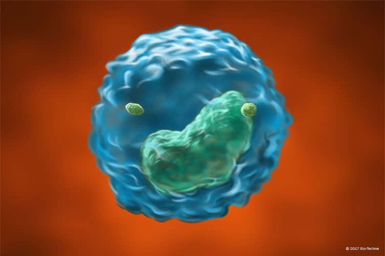
Natural killer (NK) cells are innate lymphoid cells with both intrinsic cytotoxic potential and cytokine-producing capabilities that have been classified as group 1 ILCs. NK cells activation occurs following the detection of abnormalities in target cells, including cells infected with certain bacteria, virally infected cells and cancer. While NK cells share many features with ILC1s including expression of the transcription factor T-bet and activation-induced production of IFN-gamma and TNF-alpha, they also express the transcription factor, eomesodermin (Eomes), along with granzyme B and perforin, which ILC1s do not. Accordingly, NK cells have been suggested to be the innate counterpart of CD8+ T cells.
Mouse Cell Surface Markers
In mice, NK cells are defined as CD3-NK1.1+ or CD3-NKp46+ cells that also typically express Integrin alpha 2/CD49b, Integrin alpha M/CD11b, CD27, T-bet, and Eomes, and lack expression of CD127/IL-7 R alpha. Three subsets of mouse NK cells have been characterized based on the differential expression of Integrin alpha M/CD11b and CD27.
These include:
- CD11dimCD27bright NK cells
- CD11bbrightCD27dim NK cells
- CD11bbrightCD27bright NK cells
CD27
dim NK cells have been found to have lower cytotoxic potential and produce lower levels of cytokines than CD27
bright NK cells.
Human Cell Surface Markers
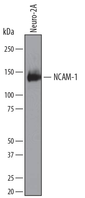
In humans, NK cells are typically defined as CD3-CD56+ cells that are also CD7+CD127-NKp46+T-bet+Eomes+. Different subtypes of human NK cells have been identified that are either CD3-CD56dimCD16+ or CD3-CD56brightCD16-. The CD56dimCD16+ subset of NK cells is predominantly found in the blood and is highly cytotoxic, while the CD56brightCD16- subset is the main subtype found in the lymph nodes and has only weak cytotoxic potential. Some studies have suggested that CD56< sup>brightCD16- subset are immature precursors of mature CD56dimCD16+ NK cells.
NK cell activation markers
In addition to the markers which are commonly used to identify mouse and human NK cells by flow cytometry, NK cells also express multiple cell surface receptors that regulate their activation. These include the human killer immunoglobulin-like receptors (KIRs) and mouse Ly49 family receptors, CD94-NKG2 heterodimeric receptors, NKG2D, natural cytotoxicity receptors (NCRs).
Featured Product

Fluorokines™ for Cell Marker Research
- Direct Detection
- Reduced processing Time
- High Levels of Bioactivity
Explore Our Fluorokines
Featured Pathway

Natural Killer Cell Receptors: Human Target Cell – NK Cell Ligand-Receptor Interactions
Explore NK Cell Pathway

Data Examples
Identification of Human Natural Killer Cells by Flow Cytometry
Detection of CD3- CD56+ CD16-/+ Human Blood Lymphocytes by Flow Cytometry. Human peripheral blood lymphocytes were stained with a PE-conjugated Mouse Anti-Human NCAM-1/CD56 Monoclonal Antibody (R&D Systems, Catalog #
FAB2408P) and a Fluorescein-conjugated Mouse Anti-Human Fc gamma RIIIA/B/CD16 Monoclonal Antibody (R&D Systems, Catalog #
FAB2546F). Cells were gated on CD3- lymphocytes.
R&D Systems KIR Antibodies Are Rigorously Tested for the Selection of Highly Specific Clones
Detection of CD56/NCAM-1+KIR+ Peripheral Blood Mononuclear Cells by Flow Cytometry. Human peripheral blood mononuclear cells (PBMCs) were stained with an APC-conjugated Mouse Anti-Human NCAM-1/CD56 Monoclonal Antibody (R&D Systems, Catalog #
FAB2408A) and a PE-conjugated Mouse Anti-Human KIR/CD158 Monoclonal Antibody (R&D Systems, Catalog #
FAB1848P). The plot is gated on live lymphocytes.
Detection of CD56/NCAM-1+KIR2DL1/S5+ Peripheral Blood Mononuclear Cells by Flow Cytometry. Human peripheral blood mononuclear cells (PBMCs) were stained with a PE-conjugated Mouse Anti-Human NCAM-1/CD56 Monoclonal Antibody (R&D Systems, Catalog #
FAB2408P) and a Fluorescein-conjugated Mouse Anti-Human KIR2DL1/KIR2DS5 Monoclonal Antibody (R&D Systems, Catalog #
FAB1844F). The plot is gated on live lymphocytes.
Detection of CD56/NCAM-1+KIR2DL3+ Peripheral Blood Mononuclear Cells by Flow Cytometry. Human peripheral blood mononuclear cells (PBMCs) were stained with a PE-conjugated Mouse Anti-Human NCAM-1/CD56 Monoclonal Antibody (R&D Systems, Catalog #
FAB2408P) and a Fluorescein-conjugated Mouse Anti-Human KIR2DL3/CD158b2 Monoclonal Antibody (R&D Systems, Catalog #
FAB2014F). The plot is gated on live lymphocytes.
Detection of CD56/NCAM-1+KIR2DS4+ Peripheral Blood Mononuclear Cells by Flow Cytometry. Human peripheral blood mononuclear cells (PBMCs) were stained with a APC-conjugated Mouse Anti-Human NCAM-1/CD56 Monoclonal Antibody (R&D Systems, Catalog #
FAB2408A) and a PE-conjugated Mouse Anti-Human KIR2DS4/CD158i Monoclonal Antibody (R&D Systems, Catalog #
FAB1847P). The plot is gated on live lymphocytes.
Detection of CD56/NCAM-1+KIR3DL2+ Peripheral Blood Mononuclear Cells by Flow Cytometry. Human peripheral blood mononuclear cells (PBMCs) were stained with a APC-conjugated Mouse Anti-Human NCAM-1/CD56 Monoclonal Antibody (R&D Systems, Catalog #
FAB2408A) and a PE-conjugated Mouse Anti-Human KIR3DL2/CD158k Monoclonal Antibody (R&D Systems, Catalog #
FAB2878P). The plot is gated on live lymphocytes.
Identification of Mouse Natural Killer Cells by Flow Cytometry
Detection of CD161/NK1.1 in Mouse Splenocytes by Flow Cytometry Mouse splenocytes were stained with Rat Anti-Mouse NKp46/NCR1 APC-conjugated Monoclonal Antibody (Catalog #
FAB22252A) and either (A) Rat Anti-Mouse CD161/NK1.1 Alexa Fluor® 488-conjugated Monoclonal Antibody (Catalog #
FAB7614G) or (B) Rat IgG2AAlexa Fluor 488 Isotype Control (Catalog #
IC006G). View our protocol for
Staining Membrane-associated Proteins.
Detection of Integrin alpha M/CD11b in Mouse Bone Marrow Cells by Flow Cytometry. Mouse bone marrow cells were stained with (A) Rat Anti-Mouse Integrin aM/CD11b Fluorescein-conjugated Monoclonal Antibody (Catalog #
FAB1124F) or (B) isotype control antibody (Catalog #
IC013F) and Rat anti-Mouse Gr-1/Ly-6G APC-conjugated Antibody (Catalog #
FAB1037A). View our protocol for
Staining Membrane-associated Proteins.
Detection of NKp46/NCR1 in Mouse Splenocytes by Flow Cytometry. Mouse splenocytes were stained with (A) Rat Anti-Mouse NKp46/NCR1 Monoclonal Antibody (Catalog #
MAB22252) or (B) Rat IgG2AIsotype Control Antibody (Catalog #
MAB006) followed by APC-conjugated Anti-Rat IgG Secondary Antibody (Catalog #
F0113) and anti-Mouse NK1.1 Alexa Fluor® 488-conjugated Monoclonal Antibody (Catalog #
FAB76141G). View our protocol for
Staining Membrane-associated Proteins.
