Human/Mouse FoxP3 Antibody Summary
Met1-Leu71
Accession # Q9BZS1
*Small pack size (-SP) is supplied either lyophilized or as a 0.2 µm filtered solution in PBS.
Applications
Please Note: Optimal dilutions should be determined by each laboratory for each application. General Protocols are available in the Technical Information section on our website.
Scientific Data
 View Larger
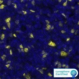
Detection of FOXP3 in Hodgkin's Lymphoma via Multiplex Immunofluorescence staining on COMET™ FOXP3 was detected in immersion fixed paraffin-embedded sections of Hodgkin's Lymphoma using Rabbit Anti-Human FOXP3 Monoclonal Antibody (Catalog # MAB8214) at 20 µg/mL at 37 ° Celsius for 4 minutes. Before incubation with the primary antibody, tissue underwent an all-in-one dewaxing and antigen retrieval preprocessing using PreTreatment Module (PT Module) and Dewax and HIER Buffer H (pH 9). Tissue was stained using the Alexa Fluor™ Plus 647 Goat anti-Rabbit IgG Secondary Antibody at 1:200 at 37 ° Celsius for 2 minutes. (Yellow; Lunaphore Catalog # DR647RB) and counterstained with DAPI (blue; Lunaphore Catalog # DR100). Specific staining was localized to the nucleus. Protocol available in COMET™ Panel Builder.
 View Larger
Detection of FOXP3 in Mouse Thymus via seqIF™ staining on COMET™ FOXP3 Antibody was detected in immersion fixed paraffin-embedded sections of mouse Thymus using Rabbit Anti-Mouse FOXP3 Monoclonal Antibody (Catalog # MAB8214) at 20ug/mL at 37 ° Celsius for 4 minutes. Before incubation with the primary antibody, tissue underwent an all-in-one dewaxing and antigen retrieval preprocessing using PreTreatment Module (PT Module) and Dewax and HIER Buffer H (pH 9; Epredia Catalog # TA-999-DHBH). Tissue was stained using the Alexa Fluor™ Plus 647 Goat anti-Rabbit IgG Secondary Antibody at 1:200 at 37 ° Celsius for 2 minutes. (Yellow; Lunaphore Catalog # DR647RB) and counterstained with DAPI (blue; Lunaphore Catalog # DR100). Specific staining was localized to the nucleus. Protocol available in COMET™ Panel Builder.
 View Larger
Detection of FoxP3 in Human PBMCs stimulated to induce Tregs by Flow Cytometry. Human peripheral blood mononuclear cells (PBMCs) either (A) untreated or (B) stimulated to induce Regulatory T Cells (Tregs) with Recombinant Human TGF-beta 1 (Catalog # 240-B) and Recombinant Human IL-2 (Catalog # 202-IL) for 2 days were stained with Rabbit Anti-Human/Mouse FoxP3 Monoclonal Antibody (Catalog # MAB8214) followed by Phycoerythrin-conjugated Anti-Rabbit IgG Secondary Antibody (Catalog # F0110) and Mouse Anti-Human CD4 APC-conjugated Monoclonal Antibody (Catalog # FAB3791A). Quadrant markers were set based on control antibody staining (Catalog # AB-105-C). To facilitate intracellular staining, cells were fixed and permeabilized with FlowX FoxP3 Fixation & Permeabilization Buffer Kit (Catalog # FC012).
 View Larger
Detection of FoxP3 in Mouse Splenocytes by Flow Cytometry. Mouse splenocytes were stained with Rat Anti-Mouse CD4 APC-conjugated Monoclonal Antibody (Catalog # FAB554A) and either (A) Rabbit Anti-Human/Mouse FoxP3 Monoclonal Antibody (Catalog # MAB8214) or (B) Normal Rabbit IgG Control (Catalog # AB-105-C) followed by Phycoerythrin-conjugated Anti-Rabbit IgG Secondary Antibody (Catalog # F0110). To facilitate intracellular staining, cells were fixed and permeabilized with FlowX FoxP3 Fixation & Permeabilization Buffer Kit (Catalog # FC012).
 View Larger
FoxP3 in HeLa Human Cell Line. FoxP3 was detected in immersion fixed HeLa human cervical epithelial carcinoma cell line using Rabbit Anti-Human/Mouse FoxP3 Monoclonal Antibody (Catalog # MAB8214) at 8 µg/mL for 3 hours at room temperature. Cells were stained using the NorthernLights™ 557-conjugated Anti-Rabbit IgG Secondary Antibody (red, upper panel; Catalog # NL004) and counterstained with DAPI (blue, lower panel). Specific staining was localized to nuclei. View our protocol for Fluorescent ICC Staining of Cells on Coverslips.
 View Larger
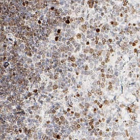
FoxP3 in Human Tonsil. FoxP3 was detected in immersion fixed paraffin-embedded sections of human tonsil using Rabbit Anti-Human/Mouse FoxP3 Monoclonal Antibody (Catalog # MAB8214) at 15 µg/mL overnight at 4 °C. Tissue was stained using the Anti-Rabbit HRP-DAB Cell & Tissue Staining Kit (brown; Catalog # CTS005) and counterstained with hematoxylin (blue). Specific staining was localized to nuclei. View our protocol for Chromogenic IHC Staining of Paraffin-embedded Tissue Sections.
 View Larger
FoxP3 in Human Ovarian Cancer Tissue. FoxP3 was detected in immersion fixed paraffin-embedded sections of human ovarian cancer tissue using Rabbit Anti-Human/Mouse FoxP3 Monoclonal Antibody (Catalog # MAB8214) at 5 µg/mL for 1 hour at room temperature followed by incubation with the Anti-Rabbit IgG VisUCyte™ HRP Polymer Antibody (Catalog # VC003). Tissue was stained using DAB (brown) and counterstained with hematoxylin (blue). Specific staining was localized to nuclei. View our protocol for IHC Staining with VisUCyte HRP Polymer Detection Reagents.
 View Larger
FoxP3 in Human Tonsil Using Dual RNAscope®ISH and IHC. FoxP3 mRNA (red) and protein (green) was detected in formalin-fixed paraffin-embedded tissue sections of human tonsil probed with ACD RNAScope®Probe (Catalog # 418471) followed by immunohistochemistry using R&D Systems Rabbit Anti-Human/Mouse FoxP3 Monoclonal Antibody (Catalog# MAB8214) at 5ug/mL for 1 hour at room temperature followed by incubation with the Anti-Rabbit IgG VisUCyte HRP Polymer Antibody (R&D Systems, Catalog # VC003). Tissue was stained using ACD RNAscope®2.5 HD Duplex Detection Reagents (Catalog # 322500).
 View Larger
Detection of Human FoxP3 by Immunocytochemistry/Immunofluorescence Multiplex fluorescent immunohistochemistry staining. Image collected and cropped by CiteAb from the following publication (https://pubmed.ncbi.nlm.nih.gov/31192136), licensed under a CC-BY license. Not internally tested by R&D Systems.
 View Larger
Detection of FoxP3 in Mouse Thymus. FoxP3 was detected in immersion fixed paraffin-embedded sections of mouse thymus using Rabbit Anti-Human/Mouse FoxP3 Monoclonal Antibody (Catalog # MAB8214) at 15 µg/ml for 1 hour at room temperature followed by incubation with the Anti-Rabbit IgG VisUCyte™ HRP Polymer Antibody (Catalog # VC003). Before incubation with the primary antibody, tissue was subjected to heat-induced epitope retrieval using VisUCyte Antigen Retrieval Reagent-Basic (Catalog # VCTS021). Tissue was stained using DAB (brown) and counterstained with hematoxylin (blue). Specific staining was localized to the nucleus. View our protocol for IHC Staining with VisUCyte HRP Polymer Detection Reagents.
Reconstitution Calculator
Preparation and Storage
- 12 months from date of receipt, -20 to -70 °C as supplied.
- 1 month, 2 to 8 °C under sterile conditions after reconstitution.
- 6 months, -20 to -70 °C under sterile conditions after reconstitution.
Background: FoxP3
Human FoxP3 is a 47 kDa member of the P subclass of the FOX (forkhead box) family of transcription factors. It contains a Leu-rich repeat, a C2H2 zinc finger region, and a C-terminal FKH (fork head), DNA-binding domain. Three isoforms for FoxP3 have been reported. All three isoforms share the sequence used as the immunogen. FoxP3 directly associates with NFAT and NFkB, suppressing their activity in CD4+ T cells. In human, FoxP3 is found in CD4+, CD8+ and CD4+CD25+ T cells. Over the region used for immunization of the amino acid sequence, mouse FoxP3 is 83% to 88% identical to rat, human, canine, and bovine FoxP3.
Product Datasheets
Citations for Human/Mouse FoxP3 Antibody
R&D Systems personnel manually curate a database that contains references using R&D Systems products. The data collected includes not only links to publications in PubMed, but also provides information about sample types, species, and experimental conditions.
19
Citations: Showing 1 - 10
Filter your results:
Filter by:
-
NOD mice, susceptible to pancreatic autoimmunity, demonstrate delayed growth of pancreatic cancer.
Authors: Dooley J, Pasciuto E, Lagou V, Lampi Y, Dresselaers T, Himmelreich U, Liston A
Oncotarget, 2017-09-24;8(46):80167-80174.
-
Microglia Require CD4 T Cells to Complete the Fetal-to-Adult Transition
Authors: Emanuela Pasciuto, Oliver T. Burton, Carlos P. Roca, Vasiliki Lagou, Wenson D. Rajan, Tom Theys et al.
Cell
-
Genome-scale in vivo CRISPR screen identifies RNLS as a target for beta cell protection in type 1 diabetes
Authors: Cai EP, Ishikawa Y, Zhang W et al.
Nat Metab
-
Mesothelial cell-derived antigen-presenting cancer-associated fibroblasts induce expansion of regulatory T cells in pancreatic cancer
Authors: Huocong Huang, Zhaoning Wang, Yuqing Zhang, Rachana N. Pradhan, Debolina Ganguly, Raghav Chandra et al.
Cancer Cell
-
Astrocyte-targeted gene delivery of interleukin 2 specifically increases brain-resident regulatory T cell numbers and protects against pathological neuroinflammation
Authors: Lidia Yshii, Emanuela Pasciuto, Pascal Bielefeld, Loriana Mascali, Pierre Lemaitre, Marika Marino et al.
Nature Immunology
-
Near-infrared photoimmunotherapy targeting human-EGFR in a mouse tumor model simulating current and future clinical trials
Authors: Ryuhei Okada, Aki Furusawa, Daniel W. Vermeer, Fuyuki Inagaki, Hiroaki Wakiyama, Takuya Kato et al.
EBioMedicine
-
Integrated Analysis Highlights the Immunosuppressive Role of TREM2+ Macrophages in Hepatocellular Carcinoma
Authors: Lisha Zhou, Meiling Wang, Hanrui Guo, Jun Hou, Yingna Zhang, Man Li et al.
Frontiers in Immunology
-
PRSS2 remodels the tumor microenvironment via repression of Tsp1 to stimulate tumor growth and progression
Authors: L Sui, S Wang, D Ganguly, TP El Rayes, C Askeland, A Børretzen, D Sim, OJ Halvorsen, G Knutsvik, J Arnes, S Aziz, S Haukaas, WD Foulkes, DR Bielenberg, A Ziemys, V Mittal, RA Brekken, LA Akslen, RS Watnick
Nature Communications, 2022-12-27;13(1):7959.
Species: Mouse
Sample Types: Whole Tissue
Applications: IHC -
Tumor-infiltrated activated B cells suppress liver metastasis of colorectal cancers
Authors: Y Xu, Z Wei, M Feng, D Zhu, S Mei, Z Wu, Q Feng, W Chang, M Ji, C Liu, Y Zhu, L Shen, F Yang, Y Chen, Y Feng, J Xu, D Zhu
Cell Reports, 2022-08-30;40(9):111295.
Species: Mouse
Sample Types: Whole Tissue
Applications: IHC -
VEGFR2 activity on myeloid cells mediates immune suppression in the tumor microenvironment
Authors: Y Zhang, H Huang, M Coleman, A Ziemys, P Gopal, SM Kazmi, RA Brekken
JCI Insight, 2021-12-08;6(23):.
Species: Mouse
Sample Types: Whole Tissue
Applications: IHC -
Dissecting spatial heterogeneity and the immune-evasion mechanism of CTCs by single-cell RNA-seq in hepatocellular carcinoma
Authors: YF Sun, L Wu, SP Liu, MM Jiang, B Hu, KQ Zhou, W Guo, Y Xu, Y Zhong, XR Zhou, ZF Zhang, G Liu, S Liu, YH Shi, Y Ji, M Du, NN Li, GB Li, ZK Zhao, XY Huang, LQ Xu, QC Yu, DH Peng, SJ Qiu, HC Sun, M Dean, XD Wang, WY Chung, AR Dennison, J Zhou, Y Hou, J Fan, XR Yang
Nature Communications, 2021-07-02;12(1):4091.
Species: Human
Sample Types: Whole Tissue
Applications: IHC -
Regulatory T-cell expansion in oral and maxillofacial Langerhans cell histiocytosis
Authors: C Zhang, J Gao, J He, C Liu, X Lv, X Yin, Y Deng, Z Lu, Z Tian
Oral Surg Oral Med Oral Pathol Oral Radiol, 2020-08-08;0(0):.
Species: Human
Sample Types: Whole Tissue
Applications: IHC -
Formation of Human Neuroblastoma in Mouse-Human Neural Crest Chimeras
Authors: MA Cohen, S Zhang, S Sengupta, H Ma, GW Bell, B Horton, B Sharma, RE George, S Spranger, R Jaenisch
Cell Stem Cell, 2020-03-05;0(0):.
Species: Mouse
Sample Types: Tissue
Applications: IHC-P -
Improved Multiplex Immunohistochemistry for Immune Microenvironment Evaluation of Mouse Formalin-Fixed, Paraffin-Embedded Tissues
Authors: N Sorrelle, D Ganguly, ATA Dominguez, Y Zhang, H Huang, LN Dahal, N Burton, A Ziemys, RA Brekken
J. Immunol., 2018-12-03;0(0):.
Species: Mouse
Sample Types: Whole Tissue
Applications: IHC -
CD25 and TGF-? blockade based on predictive integrated immune ratio inhibits tumor growth in pancreatic cancer
Authors: N Pu, G Zhao, H Yin, JA Li, A Nuerxiati, D Wang, X Xu, T Kuang, D Jin, W Lou, W Wu
J Transl Med, 2018-10-25;16(1):294.
Species: Mouse
Sample Types: Whole Tissue
Applications: IHC -
Mice transgenic for human CTLA4-CD28 fusion gene show proliferation and transformation of ATLL-like and AITL-like T cells
Authors: Gyu Jin Lee, Yukyung Jun, Yoon Kyung Jeon, Daekee Lee, Sanghyuk Lee, Jaesang Kim
OncoImmunology
-
Targeting TGFbetaR2-mutant tumors exposes vulnerabilities to stromal TGFbeta blockade in pancreatic cancer
Authors: H Huang, Y Zhang, V Gallegos, N Sorrelle, MM Zaid, J Toombs, W Du, S Wright, M Hagopian, Z Wang, AN Hosein, AA Sathe, C Xing, EJ Koay, KE Driscoll, RA Brekken
EMBO Mol Med, 2019-10-14;0(0):e10515.
-
Senescent Tumor Cells Build a Cytokine Shield in Colorectal Cancer
Authors: Choi YW, Kim YH, Oh SY et al.
Advanced Science
-
RNU (Foxn1 RNU-Nude) Rats Demonstrate an Improved Ability to Regenerate Muscle in a Volumetric Muscle Injury Compared to Sprague Dawley Rats
Authors: Michael J. McClure, Lucas C. Olson, David J. Cohen, Yen Chen Huang, Shirley Zhang, Tri Nguyen et al.
Bioengineering (Basel)
FAQs
-
What detection reagent was used for IHC, to create the Dual RNAscope ISH-IHC image on the datasheet for Catalog # MAB8214?
Green Chromogen from ACD (ACD Catalog # 322550) was ued for IHC detection. See ACD website (https://acdbio.com/store/catalog/product/view/id/22/) for protocol and handling recommendations. Extensive washing with buffers containing PBS is discouraged as it may cause decolorization. It is critical that after staining with green chromogen, slides with tissues are allowed to dry at room temperature prior to using a permanent mounting media.
Reviews for Human/Mouse FoxP3 Antibody
Average Rating: 3 (Based on 1 Review)
Have you used Human/Mouse FoxP3 Antibody?
Submit a review and receive an Amazon gift card.
$25/€18/£15/$25CAN/¥75 Yuan/¥2500 Yen for a review with an image
$10/€7/£6/$10 CAD/¥70 Yuan/¥1110 Yen for a review without an image
Filter by:


