Overview
Neutrophils are granule-containing, polymorphonuclear leukocytes that develop in the bone marrow from myeloid precursors. Neutrophil activation is an essential process that occurs when these cells are exposed to pathogens or inflammatory stimuli. Neutrophils play a central role in the innate immune response by destroying foreign particles either intracellularly in phagosomes or extracellularly by releasing neutrophil extracellular traps (NETs) and promoting acute inflammation.
In humans, neutrophils are the most abundant circulating leukocytes, accounting for 50-70% of white blood cells, while 10-25% of circulating mouse leukocytes are neutrophils. Although neutrophils can be visually identified based on the shape of their nuclei and cytoplasmic granularity, they can also be identified by flow cytometry.
Surface markers are used to identify and classify subsets of neutrophils. These neutrophil markers are important for activation and cellular function of both mature and immature neutrophils.
Mouse Neutrophil Markers
Mouse neutrophils are commonly identified based on the cell surface expression of Ly-6G (NovusBio.com) and CD11b/Integrin alpha M. Since mouse granulocytic myeloid-derived suppressor cells can also express these markers, neutrophils are frequently distinguished from these cells in mice based on their lack of expression of M-CSF R/CD115 and CD244/SLAMF4, along with an absence of immunosuppressive properties.
Human Neutrophil Markers
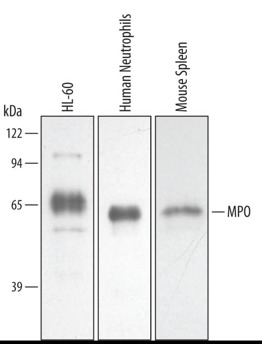
In humans, neutrophils are distinguished from eosinophils and monocytes based on the expression of both CD15, an antigen expressed on the surface of immature neutrophils, and CD16/Fc gamma RIII on human neutrophils, along with the lack of expression of CD14. In addition, CD66b/CEACAM-8, CD11b/Integrin alpha M, CD33, and the cytoplasmic marker, myeloperoxidase, are other common markers that are used to identify human neutrophils.
Data Examples
Identification of Mouse Neutrophils by Flow Cytometry
Detection of Ly-6G (GR-1) in Mouse Bone Marrow Cells by Flow Cytometry. Mouse bone marrow cells were stained with either (A) Rat Anti-Mouse Ly-6g (GR-1) Monoclonal Antibody (Catalog #
MAB10371) or (B) Rat IgG2A Isotype Control (Catalog #
MAB006) followed by Phycoerythrin-conjugated Anti-Rat IgG Secondary Antibody (Catalog #
F0105B) and Rat Anti-Mouse B220/CD45R APC-conjugated Monoclonal Antibody (Catalog #
FAB1217A).View our protocol for
Staining Membrane-associated Proteins.
Detection of Integrin alpha M/CD11b in Mouse Bone Marrow Cells by Flow Cytometry. Mouse bone marrow cells were stained with (A) Rat Anti-Mouse Integrin aM/CD11b Fluorescein-conjugated Monoclonal Antibody (Catalog #
FAB1124F) or (B) isotype control antibody (Catalog #
IC013F) and Rat anti-Mouse Gr-1/Ly-6G APC-conjugated Antibody (Catalog #
FAB1037A). View our protocol for
Staining Membrane-associated Proteins.
Detection of CD45 in Mouse Splenocytes by Flow Cytometry. Mouse splenocytes were stained with Rat Anti-Mouse CD45 APC-conjugated Monoclonal Antibody (Catalog #
FAB114A, filled histogram) or isotype control antibody (Catalog #
IC013A, open histogram). View our protocol for
Staining Membrane-associated Proteins.
Identification of Human Neutrophils by Flow Cytometry
Detection of CD15/Lewis X in Human Blood Granulocytes by Flow Cytometry. Human peripheral blood granulocytes were stained with Mouse Anti-Human CD15/Lewis X Monoclonal Antibody (Catalog #
MAB7368, filled histogram) or Mouse IgM control antibody (open histogram), followed by Phycoerythrin-conjugated Anti-Mouse IgM Secondary Antibody (Catalog #
F0116).
Detection of L-Selectin/CD62 Human PBMC by Flow Cytometry. Human PBMC either (A) resting or (B) treated with 100 ng/ml PMA for 24 hours, were stained with Mouse anti-Human L-Selectin/CD62 monoclonal antibody (Catalog #
MAB728) followed by Allophycocyanin-conjugated anti-Mouse IgG Secondary Antibody (Catalog #
F0101B,) and Mouse anti-Human PE-conjugated CD3 Monoclonal Antibody (Catalog #
FAB100P). Quadrant markers were set based on isotype control antibody staining (Catalog #
MAB003, data not shown). Staining was performed using our
Staining Membrane-Associated Proteins protocol.
Detection of CD45 in Human Blood Lymphocytes by Flow Cytometry. Human peripheral blood lymphocytes were stained with Mouse Anti-Human CD45 Monoclonal Antibody (Catalog #
MAB1430, filled histogram) or isotype control antibody (Catalog #
MAB002, open histogram), followed by Phycoerythrin-conjugated Anti-Mouse IgG Secondary Antibody (Catalog #
F0102B). View our protocol for
Staining Membrane-associated Proteins.
Detection of Integrin beta 2/CD18 in Human Peripheral Blood Lymphocytes by Flow Cytometry. Human peripheral blood Lymphocytes were stained with Mouse Anti-Human Integrin beta 2/CD18 PE-conjugated Monoclonal Antibody (Catalog #
FAB1730P, filled histogram) or isotype control antibody (Catalog #
IC002P, open histogram). View our protocol for
Staining Membrane-associated Proteins.
Detection of Siglec-3/CD33 in HEK293 Human Cell Line Transfected with Human Siglec-3/CD33 and eGFP by Flow Cytometry. HEK293 human embryonic kidney cell line transfected with human Siglec-3/CD33 and eGFP was stained with (A) Rabbit Anti-Human Siglec-3/CD33 Monoclonal Antibody (Catalog #
MAB11373) or (B) Rabbit IgG control antibody (Catalog #
MAB1050) followed by Allophycocyanin-conjugated Anti-Rabbit IgG Secondary Antibody (Catalog #
F0111). View our protocol for
Staining Membrane-associated Proteins.


