Microglia Activation During Neuroinflammation: Overview
Click on one of the stages of microglia activation to see the molecules involved in that process.
Maintenance of
"Resting" Microglia
Maintenance of
"Resting" Microglia
Microglia
Steady-State
Markers
Microglia
Steady-State
Markers
Laminin alpha 3/Laminin-5
Laminin alpha 4
Laminin gamma 1
Laminin S
Laminin-1
Fibronectin
Collagen IV
Collagen IV alpha 1
Hyaluronan
Glypican 1
Glypican 2
Glypican 3
Glypican 5
Glypican 6
Syndecan-1/CD138
Syndecan-2/CD362
Syndecan-3
Syndecan-4
PTP beta/zeta/PTPRZ
Neurocan
Versican
Brevican
Aggrecan
Tenascin C
Tenascin R
Endorepellin/Perlecan
Agrin
(ECM)
(ECM)
Laminin alpha 3/Laminin-5
Laminin alpha 4
Laminin gamma 1
Laminin S
Laminin-1
Fibronectin
Collagen IV
Collagen IV alpha 1
Hyaluronan
Glypican 1
Glypican 2
Glypican 3
Glypican 5
Glypican 6
Syndecan-1/CD138
Syndecan-2/CD362
Syndecan-3
Syndecan-4
PTP beta/zeta/PTPRZ
Neurocan
Versican
Brevican
Aggrecan
Tenascin C
Tenascin R
Endorepellin/Perlecan
Agrin
Injury-Derived
DAMPs
Injury-Derived
DAMPs
DAMPs
DAMPs
Laminin alpha 3/Laminin-5
Laminin alpha 4
Laminin gamma 1
Laminin S
Laminin-1
Fibronectin
Collagen IV
Collagen IV alpha 1
Hyaluronan
Glypican 1
Glypican 2
Glypican 3
Glypican 5
Glypican 6
Syndecan-1/CD138
Syndecan-2/CD362
Syndecan-3
Syndecan-4
PTP beta/zeta/PTPRZ
Neurocan
Versican
Brevican
Aggrecan
Tenascin C
Tenascin R
Endorepellin/Perlecan
Agrin
Microglia
Activation
Microglia
Activation
Disease-Related
DAMPs
Disease-Related
DAMPs
Microglia
Polarization
Microglia
Polarization
IFN-gamma
GM-CSF
IL-1 beta/IL-1F2
IL-6
IFN-gamma
GM-CSF
IL-1 beta/IL-1F2
IL-6
IL-13
IL-13

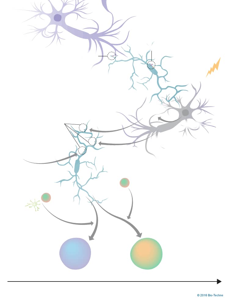
Neuroinflammation, defined as inflammation of nervous tissue, is initiated in response to a variety of endogenous and exogenous sources including invading pathogens, neuronal injury, and toxic compounds. It is characterized by glial cell activation, the release of inflammatory molecules, increased blood-brain barrier permeability, and recruitment of peripheral immune cells into the brain. Neuroinflammation is initiated by microglia, which are the resident immune cells of the central nervous system. Under steady-state conditions, microglia are maintained in a “resting” state through interactions with cell surface and soluble factors from surrounding cells. Microglia become activated following exposure to pathogen-associated molecular patterns (PAMPs) and/or endogenous damage-associated molecular patterns (DAMPs), and removal of the immune-suppressive signals. Activated microglia can acquire different phenotypes depending on cues in its surrounding environment. M1 microglia are initially present following an insult as they promote a proinflammatory response. Over time, the response is shifted to be anti-inflammatory, which is mediated by M2 microglia. In truth, current research has suggested that microglia activation is more complex than originally believed. It has been suggested that there is a range of microglia activation states that span from the M1 to M2 phenotypes, with each phenotype displaying different markers, secreting different compounds, and exhibiting different functions.
To learn more about our products for neuroinflammation, please visit our Neuroinflammation Research Area.
Get Print Copy of this Pathway

