Mouse/Rat Neurocan Antibody Summary
Asp23-Asp637
Accession # NP_031815
Applications
Please Note: Optimal dilutions should be determined by each laboratory for each application. General Protocols are available in the Technical Information section on our website.
Scientific Data
 View Larger
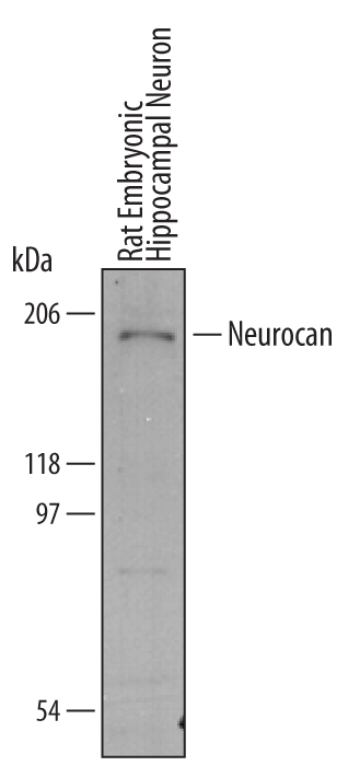
Detection of Mouse/Rat Neurocan by Western Blot. Western blot shows lysates of rat embryonic hippocampal neurons. PVDF membrane was probed with 1 µg/mL of Mouse/Rat Neurocan Antigen Affinity-purified Polyclonal Antibody (Catalog # AF5800) followed by HRP-conjugated Anti-Sheep IgG Secondary Antibody (Catalog # HAF016). A specific band was detected for Neurocan at approximately 200 kDa (as indicated). This experiment was conducted under reducing conditions and using Immunoblot Buffer Group 8.
 View Larger
Neurocan in Mouse Brain. Neurocan was detected in perfusion fixed frozen sections of mouse brain (medulla) using Mouse/Rat Neurocan Antigen Affinity-purified Polyclonal Antibody (Catalog # AF5800) at 15 µg/mL overnight at 4 °C. Tissue was stained using the Anti-Sheep HRP-DAB Cell & Tissue Staining Kit (brown; Catalog # CTS019) and counterstained with hematoxylin (blue). View our protocol for Chromogenic IHC Staining of Frozen Tissue Sections.
Reconstitution Calculator
Preparation and Storage
- 12 months from date of receipt, -20 to -70 °C as supplied.
- 1 month, 2 to 8 °C under sterile conditions after reconstitution.
- 6 months, -20 to -70 °C under sterile conditions after reconstitution.
Background: Neurocan
Neurocan is a 220 kDa nervous tissue-specific chondroitin sulfate proteoglycan member of the aggrecan/versican proteoglycan family (1). Mouse Neurocan is synthesized as a 1268 amino acid (aa) precursor that contains a 22 aa signal sequence and a 1246 aa mature chain. The mature chain contains one Ig-like V-type domain (aa 37-157), two Link domains (aa 159-254 and 258-356), two EGF-like domains (aa 960-996 and 998-1034), one C-type lectin-like domain (aa 1036-1165), one Sushi domain (aa 1165-1224), and five potential sites for N-linked glycosylation. Mature mouse Neurocan is 90% and 66% aa identical to mature rat and human Neurocan, respectively. Neurocan binds with high affinity to the cell adhesion molecules (CAM) Ng-CAM and N-CAM to inhibit Neuronal adhesion and neurite growth (2-3). In the developing rat retina, the expression of Neurocan is regulated both temporally and spatially, which suggests that it may play a role in the differentiation of and neural network formation of the mammalian retina (1). Injury to the CNS leads to permanent loss of function due to the inability of severed nerve fibers to regenerate back to their targets (4). The lack of CNS repair is attributed in part to the extracellular matrix chondroitin sulfate proteoglycans, such as Neurocan, which are produced by activated glial cells post-injury (4).
- Inatani, M. et al. (1999) Invest. Ophthalmol. Vis. Sci. 40:2350.
- Friedlander, D.R. et al. (1994) J. Cell Biol. 125:669.
- Retzler, C. et al. (1996) J. Biol. Chem. 271:27304.
- Quaglia, X. et al. (2008) Brain 131:240.
Product Datasheets
Citations for Mouse/Rat Neurocan Antibody
R&D Systems personnel manually curate a database that contains references using R&D Systems products. The data collected includes not only links to publications in PubMed, but also provides information about sample types, species, and experimental conditions.
12
Citations: Showing 1 - 10
Filter your results:
Filter by:
-
Astrocyte-Secreted Neurocan Controls Inhibitory Synapse Formation and Function
Authors: D Irala, S Wang, K Sakers, L Nagendren, FP Ulloa-Seve, DS Bindu, C Eroglu
bioRxiv : the preprint server for biology, 2023-04-03;0(0):.
Species: Mouse
Sample Types: Tissue Homogenates, Whole Tissue
Applications: IHC, Western Blot -
Distribution and postnatal development of chondroitin sulfate proteoglycans in the perineuronal nets of cholinergic motoneurons innervating extraocular muscles
Authors: A Ritok, P Kiss, A Zaher, E Wolf, L Ducza, T Bacskai, C Matesz, B Gaal
Scientific Reports, 2022-12-14;12(1):21606.
Species: Mouse
Sample Types: Whole Tissue
Applications: IHC -
Brevican and Neurocan Cleavage Products in the Cerebrospinal Fluid - Differential Occurrence in ALS, Epilepsy and Small Vessel Disease
Authors: W Hu beta ler, L Höhn, C Stolz, S Vielhaber, C Garz, FC Schmitt, ED Gundelfing, S Schreiber, CI Seidenbech
Frontiers in Cellular Neuroscience, 2022-04-11;16(0):838432.
Species: Human
Sample Types: Serum
Applications: ELISA Development -
Neurocan is a New Substrate for the ADAMTS12 Metalloprotease: Potential Implications in Neuropathies
Authors: T Fontanil, Y Mohamedi, A Moncada-Pa, T Cobo, JA Vega, JL Cobo, O García-Suá, J Cobo, ÁJ Obaya, S Cal
Cell. Physiol. Biochem., 2019-01-01;52(5):1003-1016.
Species: Mouse
Sample Types: Cell Lysates, Whole Tissue
Applications: IHC-P, Western Blot -
Layer-specific expression of extracellular matrix molecules in the mouse somatosensory and piriform cortices
Authors: H Ueno, S Suemitsu, S Murakami, N Kitamura, K Wani, Y Matsumoto, M Okamoto, T Ishihara
IBRO Rep, 2018-11-28;6(0):1-17.
Species: Mouse
Sample Types: Whole Tissue
Applications: IHC-Fr -
Synaptic coupling of inner ear sensory cells is controlled by brevican-based extracellular matrix baskets resembling perineuronal nets
Authors: M Sonntag, M Blosa, S Schmidt, K Reimann, K Blum, T Eckrich, G Seeger, D Hecker, B Schick, T Arendt, J Engel, M Morawski
BMC Biol., 2018-09-26;16(1):99.
Species: Mouse
Sample Types: Cell Lysates, Whole Tissue
Applications: IHC, Western Blot -
BMP-responsive Protease HtrA1 is Differentially Expressed in Astrocytes and Regulates Astrocytic Development and Injury Response
Authors: J Chen, S Van Gulden, TL McGuire, AC Fleming, C Oka, JA Kessler, CY Peng
J. Neurosci., 2018-02-26;0(0):.
Species: Mouse
Sample Types: Cell Lysates
Applications: Western Blot -
Sensory experience-dependent formation of perineuronal nets and expression of Cat-315 immunoreactive components in the mouse somatosensory cortex
Authors: H Ueno, S Suemitsu, M Okamoto, Y Matsumoto, T Ishihara
Neuroscience, 2017-05-08;0(0):.
Species: Mouse
Sample Types: Whole Tissue
Applications: IHC-Fr -
Protein O-Mannosylation in the Murine Brain: Occurrence of Mono-O-Mannosyl Glycans and Identification of New Substrates
PLoS ONE, 2016-11-03;11(11):e0166119.
Species: Mouse
Sample Types: Whole Tissue
Applications: IHC-Fr -
L-type Calcium Channel Cav1.2 Is Required for Maintenance of Auditory Brainstem Nuclei.
Authors: Ebbers L, Satheesh S, Janz K, Ruttiger L, Blosa M, Hofmann F, Morawski M, Griesemer D, Knipper M, Friauf E, Nothwang H
J Biol Chem, 2015-08-04;290(39):23692-710.
Species: Mouse
Sample Types: Whole Tissue
Applications: IHC -
Neurons and glia modify receptor protein-tyrosine phosphatase zeta (RPTPzeta)/phosphacan with cell-specific O-mannosyl glycans in the developing brain.
Authors: Dwyer C, Katoh T, Tiemeyer M, Matthews R
J Biol Chem, 2015-03-03;290(16):10256-73.
Species: Mouse
Sample Types: Tissue Homogenates
Applications: Western Blot -
Neurocan contributes to the molecular heterogeneity of the perinodal ECM.
Authors: Bekku Y, Oohashi T
Arch. Histol. Cytol., 2010-01-01;73(2):95-102.
Species: Mouse
Sample Types: Tissue Homogenates, Whole Tissue
Applications: IHC-Fr, Western Blot
FAQs
No product specific FAQs exist for this product, however you may
View all Antibody FAQsReviews for Mouse/Rat Neurocan Antibody
There are currently no reviews for this product. Be the first to review Mouse/Rat Neurocan Antibody and earn rewards!
Have you used Mouse/Rat Neurocan Antibody?
Submit a review and receive an Amazon gift card.
$25/€18/£15/$25CAN/¥75 Yuan/¥1250 Yen for a review with an image
$10/€7/£6/$10 CAD/¥70 Yuan/¥1110 Yen for a review without an image

