Mouse/Rat CD47 N-terminal IgV-like Extracellular Domain Antibody Summary
Gln19-Pro158
Accession # NP_034711
Applications
Please Note: Optimal dilutions should be determined by each laboratory for each application. General Protocols are available in the Technical Information section on our website.
Scientific Data
 View Larger
Detection of Mouse CD47 by Western Blot. Western blot shows lysates of mouse ovary tissue and mouse thymus tissue. PVDF membrane was probed with 0.25 µg/mL of Goat Anti-Mouse/Rat CD47 N-terminal IgV-like Extracellular Domain Antigen Affinity-purified Polyclonal Antibody (Catalog # AF1866) followed by HRP-conjugated Anti-Goat IgG Secondary Antibody (HAF019). Specific bands were detected for CD47 at approximately 40-60 kDa (as indicated). This experiment was conducted under reducing conditions and using Immunoblot Buffer Group 1.
 View Larger
Detection of Rat CD47 by Western Blot. Western blot shows lysates of rat placenta tissue. PVDF membrane was probed with 1 µg/mL of Goat Anti-Mouse/Rat CD47 N-terminal IgV-like Extracellular Domain Antigen Affinity-purified Polyclonal Antibody (Catalog # AF1866) followed by HRP-conjugated Anti-Goat IgG Secondary Antibody (HAF017). Specific bands were detected for CD47 at approximately 40-60 kDa (as indicated). This experiment was conducted under reducing conditions and using Immunoblot Buffer Group 1.
 View Larger
Detection of Mouse CD47 by Western Blot CD47 regulates thrombospondin-1, TGF-beta 1, and collagen deposition after injury. a Whole cell lysates from freshly isolated intestinal epithelial cells from Cd47f/f and Cd47 delta IEC mice were subjected to SDS–PAGE and immunoblot for thrombospondin-1/TSP-1, TGF-beta 1, and phosphorylated SMAD2 and SMAD3. Results are representative of three independent experiments. b Representative Masson’s trichrome staining of wounds beds and chronic DSS-colitis colons from Cd47f/f and Cd47 delta IEC mice. Scale bars = 50 μm. Results representative of three independent experiments with 5–7 mice per group. Source data are provided as a Source Data file Image collected and cropped by CiteAb from the following publication (https://pubmed.ncbi.nlm.nih.gov/31676794), licensed under a CC-BY license. Not internally tested by R&D Systems.
 View Larger
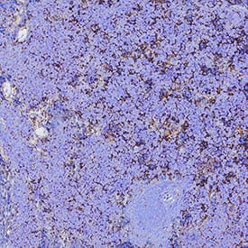
Detection of CD47 in Mouse Spleen. CD47 was detected in immersion fixed paraffin-embedded sections of Mouse Spleen using Goat Anti-Mouse/Rat CD47 N-terminal IgV-like Extracellular Domain Antigen Affinity-purified Polyclonal Antibody (Catalog # AF1866) at 1 µg/mL for 1 hour at room temperature followed by incubation with the Anti-Mouse IgG VisUCyte™ HRP Polymer Antibody (Catalog # VC001). Before incubation with the primary antibody, tissue was subjected to heat-induced epitope retrieval using VisUCyte Antigen Retrieval Reagent-Basic (Catalog # VCTS021). Tissue was stained using DAB (brown) and counterstained with hematoxylin (blue). Specific staining was localized to Cell surface and cytoplasm. View our protocol for IHC Staining with VisUCyte HRP Polymer Detection Reagents.
 View Larger
Detection of Mouse CD47 by Western Blot CD47 associates with beta 1 integrin and promotes focal adhesion formation. a In situ proximity ligation assay utilizing antibodies against CD47 and beta 1 integrin indicating close association between CD47 and beta 1 integrin in the colonic epithelium. Positive PLA signals are shown in green, beta-catenin in magenta and DAPI/nuclei in blue. Crypts are indicated by dashed lines. Arrowheads and asterisks (*) indicate positive PLA signals on IECs and immune cells in the lamina propria, respectively. Strong PLA signals are detected in IECs that are mainly concentrated at the base of the crypts in Cd47f/f colon in contrast to very few PLA signals are observed in crypts IECs in Cd47 delta IEC colons. Scale bars = 20 μm. The specificity of the PLA signals was evaluated in Supplementary figure 6 demonstrating no PLA signals in Cd47−/− colons. b Whole-cell lysates from freshly isolated intestinal epithelial cells from Cd47f/f and Cd47 delta IEC mice were subjected to SDS–PAGE and immunoblot for signaling molecules downstream of beta 1 integrin-dependent cell adhesion, revealing decreased beta 1 integrin protein expression and reduced phosphorylation of SrcY416, FAKY397/Y861, and p130CASY410 in cells from Cd47 delta IEC mice. Results are representative of three independent experiments. c Murine enteroid-derived primary epithelial cell monolayers were scratch-wounded and harvested at the indicated time points, then analyzed by SDS–PAGE and immunoblot. CD47-deficient epithelial cells show a reduction in phosphorylated SrcY416, FAKY397/Y861, and p130CASY410 upon wounding, whereas maintaining reduced beta 1 integrin protein baseline expression. d Epithelial cells immediately adjacent to wounds from Cd47f/f and Cd47 delta IEC mice were imaged by confocal microscopy, showing disrupted basal co-staining for phosphorylated FAKY861 and beta 1 integrin (apical surface indicated by dashed line). Arrows indicate reduced colocalization of phospho-FAKY861 and beta 1 integrin in the absence of CD47 expression. Scale bars = 10 μm. Results are representative of three independent experiments with three mice per treatment group. e Lamellipodia of CD47(−) cells exhibited fewer phosphorylated FAKY861-positive focal adhesions in comparison with CD47-expressing cells (insets). Scale bars = 10 μm. Results are representative of three independent experiments with two independently derived enteroid culture lines. Source data are provided as a Source Data file Image collected and cropped by CiteAb from the following publication (https://pubmed.ncbi.nlm.nih.gov/31676794), licensed under a CC-BY license. Not internally tested by R&D Systems.
 View Larger
Detection of Mouse CD47 by Immunocytochemistry/Immunofluorescence Loss of CD47 in IEC does not induce immune-mediated mucosal damage. a–c Naive Cd47 delta IEC and tamoxifen-treated Cd47ER delta IEC mice housed in pathogen-free conditions were analyzed for intestinal epithelial CD47 expression. Cd47ER delta IEC mice were treated with tamoxifen to induce acute CD47 deletion in intestinal epithelial cells, and analyzed 2 weeks later. a Tissue sections from naive Cd47 delta IEC and tamoxifen-treated Cd47ER delta IEC mice were stained with anti-CD47 antibodies (green) with DAPI counterstain (blue). CD47 expression is absent in the epithelium but retained in the lamina propria and submucosa. Scale bars = 100 μm upper panels, 50 μm lower panels. b IECs were isolated from the terminal ileum of naive mice. Protein lysates were analyzed by SDS–PAGE and immunoblot for CD47. c IECs were isolated from Cd47ER delta IEC mice treated with vehicle (corn oil) or tamoxifen, and analyzed as in b. Results are representative of three independent experiments with 3–5 mice per group. d Paraffin-embedded colon tissue sections from Cd47 delta IEC mice were stained with Hematoxylin and Eosin counterstain for histological examination. Gross mucosal architecture is intact in the absence of epithelial CD47 expression. Scale bars = 50 μm upper panels, 100 μm lower panels. e Colon tissue digests from naive Cd47 delta IEC mice were stained and analyzed by flow cytometry, showing no significant differences in major lamina propria immune cell populations. Points represent individual samples each containing two mice (n = 4 mice per group). Data are means ± SEM and are representative of three independent experiments. Source data are provided as a Source Data file Image collected and cropped by CiteAb from the following publication (https://pubmed.ncbi.nlm.nih.gov/31676794), licensed under a CC-BY license. Not internally tested by R&D Systems.
 View Larger
Detection of Mouse CD47 by Immunocytochemistry/Immunofluorescence IEC-specific deletion of CD47 results in impaired mucosal healing. Utilizing a miniature video endoscope and biopsy scissors, 5–7 wounds were created in the dorsal aspect of the descending colon mucosa of anesthetized mice. Cd47ER delta IEC mice were wounded 2 weeks after tamoxifen treatment. a Digital measurement of wound surface area at 24 and 72 h post wounding revealed a striking impairment in wound closure in Cd47 delta IEC and tamoxifen-treated Cd47ER delta IEC mice. Points represent mean value within all wounds from an individual mouse. Data are representative of two independent experiments with 5–6 mice per group. Data are means ± SEM. ***p < 0.001; one-way ANOVA. b Tissue sections taken from day 3 wounds were stained with the epithelial-specific marker E-Cadherin (green), plus DAPI counterstain (blue). Re-epithelialization of the wound is disorganized in the absence of CD47 (Cd47 delta IEC) compared with control Cd47f/f. c Tissue sections taken from day 3 wounds were stained with the epithelial-specific marker E-Cadherin (green), brush border protein Villin (magenta), and DAPI (blue). Insets of epithelial cells on top of the wound bed show polarized wound-associated epithelial cells (WAE) expressing Villin (arrows) in Cd47f/f wounds while cells are not polarized in Cd47 delta IEC wounds. Scale bars = 100 μm. d Ki67 staining of frozen sections of wounded colon mucosa 3 days post-wounding (red) revealed similar proliferation rates in crypt epithelial cells immediately adjacent to wounds in the absence of epithelial CD47. Sections were counterstained with E-Cadherin (green) and DAPI (blue). Scale bars = 50 μm. Points represent the average number of Ki67-positive cells for four crypts adjacent to wounds for each individual mouse. Data are means ± SEM and are representative of two independent experiments with 4–6 mice per group. Source data are provided as a Source Data file Image collected and cropped by CiteAb from the following publication (https://pubmed.ncbi.nlm.nih.gov/31676794), licensed under a CC-BY license. Not internally tested by R&D Systems.
 View Larger
Detection of Mouse CD47 by Western Blot CD47 associates with beta 1 integrin and promotes focal adhesion formation. a In situ proximity ligation assay utilizing antibodies against CD47 and beta 1 integrin indicating close association between CD47 and beta 1 integrin in the colonic epithelium. Positive PLA signals are shown in green, beta-catenin in magenta and DAPI/nuclei in blue. Crypts are indicated by dashed lines. Arrowheads and asterisks (*) indicate positive PLA signals on IECs and immune cells in the lamina propria, respectively. Strong PLA signals are detected in IECs that are mainly concentrated at the base of the crypts in Cd47f/f colon in contrast to very few PLA signals are observed in crypts IECs in Cd47 delta IEC colons. Scale bars = 20 μm. The specificity of the PLA signals was evaluated in Supplementary figure 6 demonstrating no PLA signals in Cd47−/− colons. b Whole-cell lysates from freshly isolated intestinal epithelial cells from Cd47f/f and Cd47 delta IEC mice were subjected to SDS–PAGE and immunoblot for signaling molecules downstream of beta 1 integrin-dependent cell adhesion, revealing decreased beta 1 integrin protein expression and reduced phosphorylation of SrcY416, FAKY397/Y861, and p130CASY410 in cells from Cd47 delta IEC mice. Results are representative of three independent experiments. c Murine enteroid-derived primary epithelial cell monolayers were scratch-wounded and harvested at the indicated time points, then analyzed by SDS–PAGE and immunoblot. CD47-deficient epithelial cells show a reduction in phosphorylated SrcY416, FAKY397/Y861, and p130CASY410 upon wounding, whereas maintaining reduced beta 1 integrin protein baseline expression. d Epithelial cells immediately adjacent to wounds from Cd47f/f and Cd47 delta IEC mice were imaged by confocal microscopy, showing disrupted basal co-staining for phosphorylated FAKY861 and beta 1 integrin (apical surface indicated by dashed line). Arrows indicate reduced colocalization of phospho-FAKY861 and beta 1 integrin in the absence of CD47 expression. Scale bars = 10 μm. Results are representative of three independent experiments with three mice per treatment group. e Lamellipodia of CD47(−) cells exhibited fewer phosphorylated FAKY861-positive focal adhesions in comparison with CD47-expressing cells (insets). Scale bars = 10 μm. Results are representative of three independent experiments with two independently derived enteroid culture lines. Source data are provided as a Source Data file Image collected and cropped by CiteAb from the following publication (https://pubmed.ncbi.nlm.nih.gov/31676794), licensed under a CC-BY license. Not internally tested by R&D Systems.
 View Larger
Detection of Mouse CD47 by Immunocytochemistry/Immunofluorescence Loss of CD47 in IEC results in impaired recovery from DSS-induced colitis. Age- and sex-matched Cd47 delta IEC and tamoxifen-treated Cd47ER delta IEC mice were treated with three consecutive cycles of 2.5% DSS in drinking water for either 4 days (Cd47 delta IEC) or 3 days (Cd47ER delta IEC), followed by 5 days of water recovery. Cd47ER delta IEC mice were treated with DSS 2 weeks after tamoxifen treatment. a–b Disease activity index scores are represented as an average of scores 0–4 for percent weight loss, stool consistency, and presence of blood in stools. Cyclical treatment of aCd47 delta IEC and b tamoxifen-treated Cd47ER delta IEC mice with 2.5% DSS in drinking water, followed by a plain water recovery period, induced greater DAI scores in the absence of epithelial CD47. Data are representative of two independent experiments with 5–6 mice per group and are expressed as means ± SEM. a *p = 0.02, b *p = 0.036 by two-way ANOVA. c Histological scoring of hematoxylin and eosin (H&E)-stained tissue sections of colonic mucosa: percentage of injury/ulceration represents a ratio of the length of injured/ulcerated areas (≥ 50% crypt loss) relative to the entire colon length, as assessed in Swiss roll mounts of the entire colon. Results indicate greater damage in the absence of epithelial CD47. Points represent individual mice. Data are representative of two independent experiments with 5–6 mice per group and are expressed as means ± SEM. Significance determined by two-tailed Student’s t test. *p = 0.016, ***p = 0.001. d, e Representative H&E staining of colon tissue sections after three cycles of DSS/water revealed extensive crypt destruction in distal colon of Cd47 delta IEC and Cd47ER delta IEC mice. Large areas of ulcerated mucosa with granulocytic infiltrates were present in the mid colon in Cd47 delta IEC and Cd47ER delta IEC mice, in comparison with littermate controls. Scale bars = 100 μm upper panels, 300 μm lower panels. Results are representative of at least two independent experiments with 5–6 mice per group. Source data are provided as a Source Data file Image collected and cropped by CiteAb from the following publication (https://pubmed.ncbi.nlm.nih.gov/31676794), licensed under a CC-BY license. Not internally tested by R&D Systems.
 View Larger
Detection of Mouse CD47 by Immunocytochemistry/Immunofluorescence IEC-specific deletion of CD47 results in impaired mucosal healing. Utilizing a miniature video endoscope and biopsy scissors, 5–7 wounds were created in the dorsal aspect of the descending colon mucosa of anesthetized mice. Cd47ER delta IEC mice were wounded 2 weeks after tamoxifen treatment. a Digital measurement of wound surface area at 24 and 72 h post wounding revealed a striking impairment in wound closure in Cd47 delta IEC and tamoxifen-treated Cd47ER delta IEC mice. Points represent mean value within all wounds from an individual mouse. Data are representative of two independent experiments with 5–6 mice per group. Data are means ± SEM. ***p < 0.001; one-way ANOVA. b Tissue sections taken from day 3 wounds were stained with the epithelial-specific marker E-Cadherin (green), plus DAPI counterstain (blue). Re-epithelialization of the wound is disorganized in the absence of CD47 (Cd47 delta IEC) compared with control Cd47f/f. c Tissue sections taken from day 3 wounds were stained with the epithelial-specific marker E-Cadherin (green), brush border protein Villin (magenta), and DAPI (blue). Insets of epithelial cells on top of the wound bed show polarized wound-associated epithelial cells (WAE) expressing Villin (arrows) in Cd47f/f wounds while cells are not polarized in Cd47 delta IEC wounds. Scale bars = 100 μm. d Ki67 staining of frozen sections of wounded colon mucosa 3 days post-wounding (red) revealed similar proliferation rates in crypt epithelial cells immediately adjacent to wounds in the absence of epithelial CD47. Sections were counterstained with E-Cadherin (green) and DAPI (blue). Scale bars = 50 μm. Points represent the average number of Ki67-positive cells for four crypts adjacent to wounds for each individual mouse. Data are means ± SEM and are representative of two independent experiments with 4–6 mice per group. Source data are provided as a Source Data file Image collected and cropped by CiteAb from the following publication (https://pubmed.ncbi.nlm.nih.gov/31676794), licensed under a CC-BY license. Not internally tested by R&D Systems.
 View Larger
Detection of Mouse CD47 by Immunocytochemistry/Immunofluorescence CD47 associates with beta 1 integrin and promotes focal adhesion formation. a In situ proximity ligation assay utilizing antibodies against CD47 and beta 1 integrin indicating close association between CD47 and beta 1 integrin in the colonic epithelium. Positive PLA signals are shown in green, beta-catenin in magenta and DAPI/nuclei in blue. Crypts are indicated by dashed lines. Arrowheads and asterisks (*) indicate positive PLA signals on IECs and immune cells in the lamina propria, respectively. Strong PLA signals are detected in IECs that are mainly concentrated at the base of the crypts in Cd47f/f colon in contrast to very few PLA signals are observed in crypts IECs in Cd47 delta IEC colons. Scale bars = 20 μm. The specificity of the PLA signals was evaluated in Supplementary figure 6 demonstrating no PLA signals in Cd47−/− colons. b Whole-cell lysates from freshly isolated intestinal epithelial cells from Cd47f/f and Cd47 delta IEC mice were subjected to SDS–PAGE and immunoblot for signaling molecules downstream of beta 1 integrin-dependent cell adhesion, revealing decreased beta 1 integrin protein expression and reduced phosphorylation of SrcY416, FAKY397/Y861, and p130CASY410 in cells from Cd47 delta IEC mice. Results are representative of three independent experiments. c Murine enteroid-derived primary epithelial cell monolayers were scratch-wounded and harvested at the indicated time points, then analyzed by SDS–PAGE and immunoblot. CD47-deficient epithelial cells show a reduction in phosphorylated SrcY416, FAKY397/Y861, and p130CASY410 upon wounding, whereas maintaining reduced beta 1 integrin protein baseline expression. d Epithelial cells immediately adjacent to wounds from Cd47f/f and Cd47 delta IEC mice were imaged by confocal microscopy, showing disrupted basal co-staining for phosphorylated FAKY861 and beta 1 integrin (apical surface indicated by dashed line). Arrows indicate reduced colocalization of phospho-FAKY861 and beta 1 integrin in the absence of CD47 expression. Scale bars = 10 μm. Results are representative of three independent experiments with three mice per treatment group. e Lamellipodia of CD47(−) cells exhibited fewer phosphorylated FAKY861-positive focal adhesions in comparison with CD47-expressing cells (insets). Scale bars = 10 μm. Results are representative of three independent experiments with two independently derived enteroid culture lines. Source data are provided as a Source Data file Image collected and cropped by CiteAb from the following publication (https://pubmed.ncbi.nlm.nih.gov/31676794), licensed under a CC-BY license. Not internally tested by R&D Systems.
 View Larger
Detection of Mouse CD47 by Immunocytochemistry/Immunofluorescence Loss of CD47 in IEC results in impaired recovery from DSS-induced colitis. Age- and sex-matched Cd47 delta IEC and tamoxifen-treated Cd47ER delta IEC mice were treated with three consecutive cycles of 2.5% DSS in drinking water for either 4 days (Cd47 delta IEC) or 3 days (Cd47ER delta IEC), followed by 5 days of water recovery. Cd47ER delta IEC mice were treated with DSS 2 weeks after tamoxifen treatment. a–b Disease activity index scores are represented as an average of scores 0–4 for percent weight loss, stool consistency, and presence of blood in stools. Cyclical treatment of aCd47 delta IEC and b tamoxifen-treated Cd47ER delta IEC mice with 2.5% DSS in drinking water, followed by a plain water recovery period, induced greater DAI scores in the absence of epithelial CD47. Data are representative of two independent experiments with 5–6 mice per group and are expressed as means ± SEM. a *p = 0.02, b *p = 0.036 by two-way ANOVA. c Histological scoring of hematoxylin and eosin (H&E)-stained tissue sections of colonic mucosa: percentage of injury/ulceration represents a ratio of the length of injured/ulcerated areas (≥ 50% crypt loss) relative to the entire colon length, as assessed in Swiss roll mounts of the entire colon. Results indicate greater damage in the absence of epithelial CD47. Points represent individual mice. Data are representative of two independent experiments with 5–6 mice per group and are expressed as means ± SEM. Significance determined by two-tailed Student’s t test. *p = 0.016, ***p = 0.001. d, e Representative H&E staining of colon tissue sections after three cycles of DSS/water revealed extensive crypt destruction in distal colon of Cd47 delta IEC and Cd47ER delta IEC mice. Large areas of ulcerated mucosa with granulocytic infiltrates were present in the mid colon in Cd47 delta IEC and Cd47ER delta IEC mice, in comparison with littermate controls. Scale bars = 100 μm upper panels, 300 μm lower panels. Results are representative of at least two independent experiments with 5–6 mice per group. Source data are provided as a Source Data file Image collected and cropped by CiteAb from the following publication (https://pubmed.ncbi.nlm.nih.gov/31676794), licensed under a CC-BY license. Not internally tested by R&D Systems.
 View Larger
Detection of Mouse CD47 by Immunocytochemistry/Immunofluorescence IEC-specific deletion of CD47 results in impaired mucosal healing. Utilizing a miniature video endoscope and biopsy scissors, 5–7 wounds were created in the dorsal aspect of the descending colon mucosa of anesthetized mice. Cd47ER delta IEC mice were wounded 2 weeks after tamoxifen treatment. a Digital measurement of wound surface area at 24 and 72 h post wounding revealed a striking impairment in wound closure in Cd47 delta IEC and tamoxifen-treated Cd47ER delta IEC mice. Points represent mean value within all wounds from an individual mouse. Data are representative of two independent experiments with 5–6 mice per group. Data are means ± SEM. ***p < 0.001; one-way ANOVA. b Tissue sections taken from day 3 wounds were stained with the epithelial-specific marker E-Cadherin (green), plus DAPI counterstain (blue). Re-epithelialization of the wound is disorganized in the absence of CD47 (Cd47 delta IEC) compared with control Cd47f/f. c Tissue sections taken from day 3 wounds were stained with the epithelial-specific marker E-Cadherin (green), brush border protein Villin (magenta), and DAPI (blue). Insets of epithelial cells on top of the wound bed show polarized wound-associated epithelial cells (WAE) expressing Villin (arrows) in Cd47f/f wounds while cells are not polarized in Cd47 delta IEC wounds. Scale bars = 100 μm. d Ki67 staining of frozen sections of wounded colon mucosa 3 days post-wounding (red) revealed similar proliferation rates in crypt epithelial cells immediately adjacent to wounds in the absence of epithelial CD47. Sections were counterstained with E-Cadherin (green) and DAPI (blue). Scale bars = 50 μm. Points represent the average number of Ki67-positive cells for four crypts adjacent to wounds for each individual mouse. Data are means ± SEM and are representative of two independent experiments with 4–6 mice per group. Source data are provided as a Source Data file Image collected and cropped by CiteAb from the following publication (https://pubmed.ncbi.nlm.nih.gov/31676794), licensed under a CC-BY license. Not internally tested by R&D Systems.
 View Larger
Detection of Mouse CD47 by Immunohistochemistry IEC-specific deletion of CD47 results in impaired mucosal healing. Utilizing a miniature video endoscope and biopsy scissors, 5–7 wounds were created in the dorsal aspect of the descending colon mucosa of anesthetized mice. Cd47ER delta IEC mice were wounded 2 weeks after tamoxifen treatment. a Digital measurement of wound surface area at 24 and 72 h post wounding revealed a striking impairment in wound closure in Cd47 delta IEC and tamoxifen-treated Cd47ER delta IEC mice. Points represent mean value within all wounds from an individual mouse. Data are representative of two independent experiments with 5–6 mice per group. Data are means ± SEM. ***p < 0.001; one-way ANOVA. b Tissue sections taken from day 3 wounds were stained with the epithelial-specific marker E-Cadherin (green), plus DAPI counterstain (blue). Re-epithelialization of the wound is disorganized in the absence of CD47 (Cd47 delta IEC) compared with control Cd47f/f. c Tissue sections taken from day 3 wounds were stained with the epithelial-specific marker E-Cadherin (green), brush border protein Villin (magenta), and DAPI (blue). Insets of epithelial cells on top of the wound bed show polarized wound-associated epithelial cells (WAE) expressing Villin (arrows) in Cd47f/f wounds while cells are not polarized in Cd47 delta IEC wounds. Scale bars = 100 μm. d Ki67 staining of frozen sections of wounded colon mucosa 3 days post-wounding (red) revealed similar proliferation rates in crypt epithelial cells immediately adjacent to wounds in the absence of epithelial CD47. Sections were counterstained with E-Cadherin (green) and DAPI (blue). Scale bars = 50 μm. Points represent the average number of Ki67-positive cells for four crypts adjacent to wounds for each individual mouse. Data are means ± SEM and are representative of two independent experiments with 4–6 mice per group. Source data are provided as a Source Data file Image collected and cropped by CiteAb from the following publication (https://pubmed.ncbi.nlm.nih.gov/31676794), licensed under a CC-BY license. Not internally tested by R&D Systems.
Reconstitution Calculator
Preparation and Storage
- 12 months from date of receipt, -20 to -70 °C as supplied.
- 1 month, 2 to 8 °C under sterile conditions after reconstitution.
- 6 months, -20 to -70 °C under sterile conditions after reconstitution.
Background: CD47
CD47, also known as Integrin‑Associated Protein (IAP) and OA3, is a 40‑60 kDa variably glycosylated atypical member of the immunoglobulin superfamily (1, 2). Mouse CD47 is an integral membrane protein that consists of a 122 amino acid (aa) extracellular domain (ECD) with a single Ig‑like domain, five membrane-spanning regions with short intervening loops, and a 16 aa C‑terminal cytoplasmic tail (3). Alternate splicing of mouse CD47 generates an additional isoform with an insertion of 21 aa following the Ig‑like domain (3). Within the N‑terminal ECD, mouse CD47 shares 63% and 84% aa sequence identity with human and rat CD47, respectively. A portion of the N‑terminal ECD can by shed from smooth muscle cells by MMP-2‑mediated proteolysis (4). The ubiquitously expressed CD47 binds to SIRP family members on macrophages, neutrophils, and T cells (5, 6). These interactions prevent macrophage‑mediated clearance of healthy CD47-expressing cells and promote immune cell transmigration across the vascular endothelium (5‑8). CD47 associates in cis with Fas on T cells and enhances Fas‑mediated apoptosis; its ligation promotes T cell anergy and dampens Th1 immune responses (9‑11). CD47 also associates in cis with Integrins alpha 4 beta 1, alpha V beta 3, alpha 2b beta 3, and alpha 2 beta 1 which can positively or negatively modulate Integrin-mediated function (2, 12). In the vasculature, CD47 binding by Thrombospondin‑1 inhibits the angiogenic and vasorelaxant effects of nitric oxide (2, 13, 14). On dendritic cells and myeloma cells, CD47 ligation by TSP‑1 induces giant cell formation and osteoclast differentiation (15).
- Sarfati, M. et al. (2009) Curr. Drug Targ. 9:842.
- Isenberg, J.S. et al. (2008) Arterioscler. Thromb. Vasc. Biol. 28:615.
- Lindberg, F.P. et al. (1993) J. Cell Biol. 123:485.
- Maile, L.A. et al. (2008) Mol. Endocrinol. 22:1226.
- Oldenborg, P.-A. et al. (2000) Science 288:2051.
- Liu, Y. et al. (2002) J. Biol. Chem. 277:10028.
- Stefanidakis, M. et al. (2008) Blood 112:1280.
- de Vries, H.E. et al. (2002) J. Immunol. 168:5832.
- Manna, P.P. et al. (2005) J. Biol. Chem. 280:29637.
- Avice, M.-N. et al. (2001) J. Immunol. 167:2459.
- Bouguermouh, S. et al. (2008) J. Immunol. 180:8073.
- Barazi, H.O. et al. (2002) J. Biol. Chem. 277:42859.
- Isenberg, J.S. et al. (2009) Nitric Oxide 21:52.
- Isenberg, J.S. et al. (2009) Matrix Biol. 28:110.
- Kukreja, A. et al. (2009) Blood 114:3413.
Product Datasheets
Citations for Mouse/Rat CD47 N-terminal IgV-like Extracellular Domain Antibody
R&D Systems personnel manually curate a database that contains references using R&D Systems products. The data collected includes not only links to publications in PubMed, but also provides information about sample types, species, and experimental conditions.
11
Citations: Showing 1 - 10
Filter your results:
Filter by:
-
EGFR‐Induced and c‐Src‐Mediated CD47 Phosphorylation Inhibits TRIM21‐Dependent Polyubiquitylation and Degradation of CD47 to Promote Tumor Immune Evasion
Authors: Linyong Du, Zhipeng Su, Silu Wang, Ying Meng, Fei Xiao, Daqian Xu et al.
Advanced Science
-
CD47-blocking antibodies restore phagocytosis and prevent atherosclerosis
Nature, 2016-07-20;0(0):.
-
CD47 Activation by Thrombospondin-1 in Lymphatic Endothelial Cells Suppresses Lymphangiogenesis and Promotes Atherosclerosis
Authors: Singla B, Aithbathula RV, Pervaiz N et al.
Arteriosclerosis, thrombosis, and vascular biology
-
Airway epithelial CD47 plays a critical role in inducing influenza virus-mediated bacterial super-infection
Authors: Moon, S;Han, S;Jang, IH;Ryu, J;Rha, MS;Cho, HJ;Yoon, SS;Nam, KT;Kim, CH;Park, MS;Seong, JK;Lee, WJ;Yoon, JH;Chung, YW;Ryu, JH;
Nature communications
Species: Mouse
Sample Types: Cell Lysates, Whole Tissue
Applications: Immunohistochemistry, Western Blot -
Engineering Human Pluripotent Stem Cell Lines to Evade Xenogeneic Transplantation Barriers
Authors: Pizzato, HA;Alonso-Guallart, P;Woods, J;Johannesson, B;Connelly, JP;Fehniger, TA;Atkinson, JP;Pruett-Miller, SM;Monsma, FJ;Bhattacharya, D;
bioRxiv : the preprint server for biology
Species: Mouse
Sample Types: Whole Tissue
Applications: IHC -
Loss of microglial SIRP&alpha promotes synaptic pruning in preclinical models of neurodegeneration
Authors: X Ding, J Wang, M Huang, Z Chen, J Liu, Q Zhang, C Zhang, Y Xiang, K Zen, L Li
Nature Communications, 2021-04-01;12(1):2030.
Species: Mouse
Sample Types: Cell Lysates
Applications: Western Blot -
CD47 prevents the elimination of diseased fibroblasts in scleroderma
Authors: T Lerbs, L Cui, ME King, T Chai, C Muscat, L Chung, R Brown, K Rieger, T Shibata, G Wernig
JCI Insight, 2020-08-20;5(16):.
Species: Mouse
Sample Types: Whole Cells, Whole Tissue
Applications: ICC, IHC -
Neutrophil expressed CD47 regulates CD11b/CD18-dependent neutrophil transepithelial migration in the intestine in vivo
Authors: V Azcutia, M Kelm, AC Luissint, K Boerner, S Flemming, M Quiros, G Newton, A Nusrat, FW Luscinskas, CA Parkos
Mucosal Immunol, 2020-06-19;0(0):.
Species: Mouse
Sample Types: Whole Cells
Applications: Flow Cytometry -
Epithelial CD47 is critical for mucosal repair in the murine intestine in vivo
Authors: M Reed, AC Luissint, V Azcutia, S Fan, MN O'Leary, M Quiros, J Brazil, A Nusrat, CA Parkos
Nat Commun, 2019-11-01;10(1):5004.
Species: Mouse
Sample Types: Cells
Applications: IF, Western Blot -
Radiation-induced loss of cell surface CD47 enhances immune-mediated clearance of human papillomavirus-positive cancer.
Authors: Vermeer D, Spanos W, Vermeer P, Bruns A, Lee K, Lee J
Int J Cancer, 2013-02-12;133(1):120-9.
Species: Human, Mouse
Sample Types: Cell Lysates
Applications: Western Blot -
Macrophage LRP1 (Low-Density Lipoprotein Receptor-Related Protein 1) Is Required for the Effect of CD47 Blockade on Efferocytosis and Atherogenesis—Brief Report
Authors: Paul A. Mueller, Yoko Kojima, Katherine T. Huynh, Richard A. Maldonado, Jianqin Ye, Hagai Tavori et al.
Arteriosclerosis, Thrombosis, and Vascular Biology
FAQs
No product specific FAQs exist for this product, however you may
View all Antibody FAQsReviews for Mouse/Rat CD47 N-terminal IgV-like Extracellular Domain Antibody
Average Rating: 5 (Based on 3 Reviews)
Have you used Mouse/Rat CD47 N-terminal IgV-like Extracellular Domain Antibody?
Submit a review and receive an Amazon gift card.
$25/€18/£15/$25CAN/¥75 Yuan/¥2500 Yen for a review with an image
$10/€7/£6/$10 CAD/¥70 Yuan/¥1110 Yen for a review without an image
Filter by:

CD47 expression is highly variable in PBMC of mice exposed to different inflammatory and anti-inflammatory stimuli


