Human/Mouse GATA-3 Antibody Summary
Pro135-Ser258
Accession # P23771
*Small pack size (-SP) is supplied either lyophilized or as a 0.2 µm filtered solution in PBS.
Applications
Please Note: Optimal dilutions should be determined by each laboratory for each application. General Protocols are available in the Technical Information section on our website.
Scientific Data
 View Larger
Detection of GATA-3 in Human Breast Cancer via Multiplex Immunofluorescence staining on COMET™ GATA-3 was detected in immersion fixed paraffin-embedded sections of human breast cancer using Mouse Anti-Human GATA‑3 Monoclonal Antibody (MAB6330) at 20 µg/mL at 37 ° Celsius for 4 minutes. Before incubation with the primary antibody, tissue underwent an all-in-one dewaxing and antigen retrieval preprocessing using PreTreatment Module (PT Module) and Dewax and HIER Buffer H (pH 9). Tissue was stained using the Alexa Fluor™ 647 Goat anti-Mouse IgG Secondary Antibody at 1:200 at 37 ° Celsius for 2 minutes. (Yellow; Lunaphore Catalog # DR647MS) and counterstained with DAPI (blue; Lunaphore Catalog # DR100). Specific staining was localized to the nucleus with dim cytoplasmic background signal. Protocol available in COMET™ Panel Builder.
 View Larger
Detection of Human and Mouse GATA‑3 by Western Blot. Western blot shows lysates of HeLa human cervical epithelial carcinoma cell line, MCF-7 human breast cancer cell line, Jurkat human acute T cell leukemia cell line, and EL-4 mouse lymphoblast cell line. PVDF Membrane was probed with 0.1 µg/mL of Moue Anti-Human/Mouse GATA-3 Monoclonal Antibody (Catalog # MAB6330) followed by HRP-conjugated Anti-Mouse IgG Secondary Antibody (HAF007). Specific bands were detected for full length (FL) GATA-3 at approximately 52 kDa and the splice form (SF) found in MCF-7 cells at approximately 40 kDa (as indicated). This experiment was conducted under reducing conditions and using Immunoblot Buffer Group 1.
 View Larger
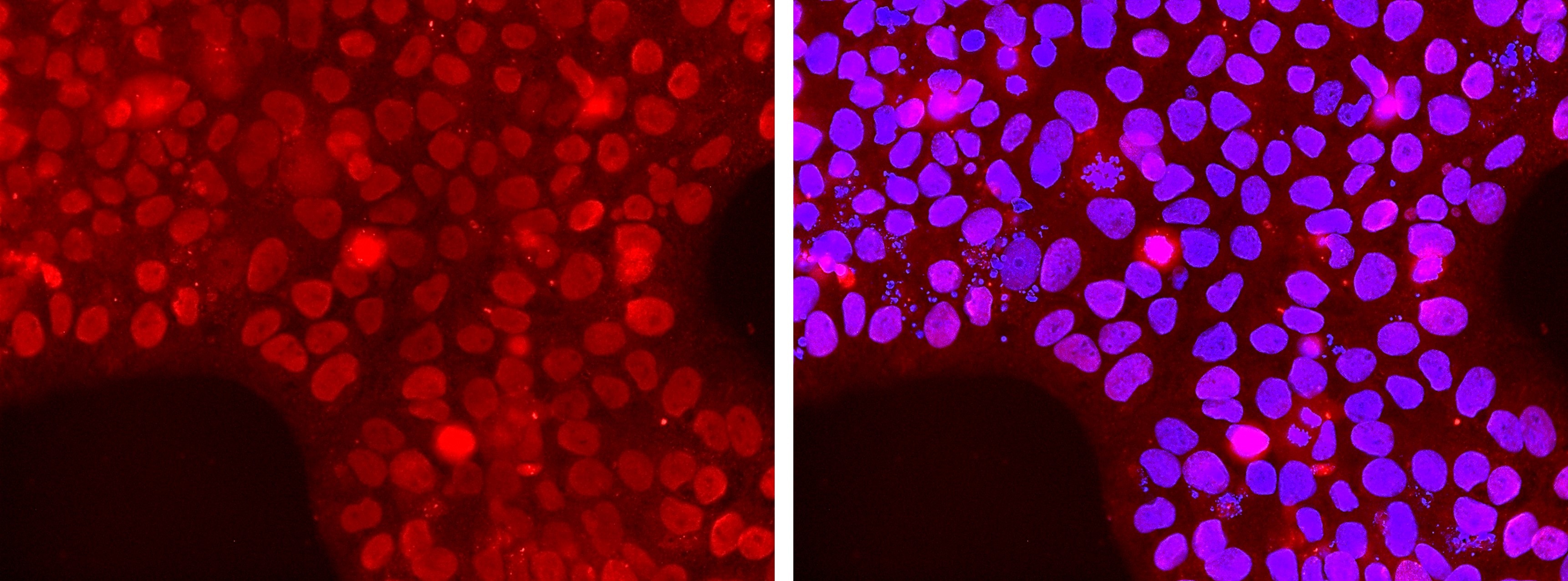
GATA‑3 in MCF‑7 Human Cell Line. GATA-3 was detected in immersion fixed MCF-7 human breast cancer cell line using Mouse Anti-Human/Mouse GATA-3 Monoclonal Antibody (Catalog # MAB6330) at 3 µg/mL for 3 hours at room temperature. Cells were stained using the NorthernLights™ 557-conjugated Anti-Mouse IgG Secondary Antibody (red; NL007) and counterstained with DAPI (blue). Specific staining was localized to nuclei. View our protocol for Fluorescent ICC Staining of Cells on Coverslips.
 View Larger
GATA‑3 in Human Breast Cancer Tissue. GATA-3 was detected in immersion fixed paraffin-embedded sections of human breast cancer tissue using Mouse Anti-Human/Mouse GATA-3 Monoclonal Antibody (Catalog # MAB6330) at 15 µg/mL overnight at 4 °C. Before incubation with the primary antibody, tissue was subjected to heat-induced epitope retrieval using Antigen Retrieval Reagent-Basic (CTS013). Tissue was stained using the Anti-Mouse HRP-DAB Cell & Tissue Staining Kit (brown CTS002) and counterstained with hematoxylin (blue). Specific staining was localized to nuclei. View our protocol for Chromogenic IHC Staining of Paraffin-embedded Tissue Sections.
 View Larger
Detection of Human GATA‑3 by Simple WesternTM. Simple Western lane view shows lysates of Jurkat human acute T cell leukemia cell line and MCF‑7 human breast cancer cell line, loaded at 0.5 mg/mL. Specific bands were detected for GATA‑3 at approximately 53 (splice variant) and 61 kDa (full length) as indicated, using 10 µg/mL of Mouse Anti-Human/Mouse GATA‑3 Monoclonal Antibody (Catalog # MAB6330). This experiment was conducted under reducing conditions and using the 12-230 kDa separation system.
Non-specific interaction with the 230 kDa Simple Western standard may be seen with this antibody.
 View Larger
GATA-3 in Human Breast Cancer Tissue Using Dual RNAscope® ISH and IHC. GATA-3 mRNA (red) and protein (green) was detected in formalin-fixed paraffin-embedded tissue sections of human breast cancer tissue probed with ACD RNAScope® Probe (Catalog # 403551) followed by immunohistochemistry using R&D Systems Mouse Anti-Human/Mouse GATA-3 Monoclonal Antibody (Catalog# MAB6330) at 5 μg/mL for 1 hour at room temperature followed by incubation with the Anti-Mouse IgG VisUCyte HRP Polymer Antibody (VC001). Tissue was stained using ACD RNAscope® 2.5 HD Duplex Detection Reagents (Catalog # 322500).
Reconstitution Calculator
Preparation and Storage
- 12 months from date of receipt, -20 to -70 °C as supplied.
- 1 month, 2 to 8 °C under sterile conditions after reconstitution.
- 6 months, -20 to -70 °C under sterile conditions after reconstitution.
Background: GATA-3
GATA-3 belongs to the GATA family of transcription factors, which bind to the consensus DNA sequence (A/T) GATA (A/G) to control diverse tissue-specific programs of gene expression and morphogenesis. It is widely expressed in mesodermal- and endodermal-derived tissues. GATA-3 has been shown to be an essential regulator for immune cell function, sympathetic neuron development and the maintenance of the differentiated state in epithelial cells.
Product Datasheets
Citations for Human/Mouse GATA-3 Antibody
R&D Systems personnel manually curate a database that contains references using R&D Systems products. The data collected includes not only links to publications in PubMed, but also provides information about sample types, species, and experimental conditions.
16
Citations: Showing 1 - 10
Filter your results:
Filter by:
-
Annexin A1 is a cell-intrinsic metalloregulator of zinc in human ILC2s
Authors: Irie, M;Kabata, H;Sasahara, K;Kurihara, M;Shirasaki, Y;Kamatani, T;Baba, R;Matsusaka, M;Koga, S;Masaki, K;Miyata, J;Araki, Y;Kikawada, T;Kabe, Y;Suematsu, M;Yamagishi, M;Uemura, S;Moro, K;Fukunaga, K;
Cell reports
Species: Human
Sample Types: Cell Lysates
Applications: Western Blot -
Endothelial-to-osteoblast transition in normal mouse bone development
Authors: Song-Chang Lin, Guoyu Yu, Yu-Chen Lee, Jian H. Song, Xingzhi Song, Jianhua Zhang et al.
iScience
-
Analysis of rheumatoid- vs psoriatic arthritis synovial fluid reveals differential macrophage (CCR2) and T helper subsets (STAT3/4 and FOXP3) activation
Authors: F Caso, A Saviano, M Tasso, F Raucci, N Marigliano, S Passavanti, P Frallonard, R Ramonda, V Brancaleon, M Bucci, R Scarpa, L Costa, F Maione
Autoimmunity reviews, 2022-10-01;21(12):103207.
Species: Human
Sample Types: Synovial Fluid
Applications: Western Blot -
Trophectoderm differentiation to invasive syncytiotrophoblast is promoted by endometrial epithelial cells during human embryo implantation
Authors: Peter T Ruane, Terence Garner, Lydia Parsons, Phoebe A Babbington, Ivan Wangsaputra, Susan J Kimber et al.
Human Reproduction
-
Predictive role of endometrial T-bet/GATA3 ratio during mid-luteal phase for live birth in patients undergoing in vitro fertilization: A retrospective observational study
Authors: Y Li, S Yu, C Huang, L Diao, C Chen, W Liu, R Lian, M Mo, C Du, F Liu, Y Zeng
Journal of reproductive immunology, 2021-12-16;149(0):103465.
Species: Human
Sample Types: Whole Tissue
Applications: IHC -
Generation of human blastocyst-like structures from pluripotent stem cells
Authors: Y Fan, Z Min, S Alsolami, Z Ma, E Zhang, W Chen, K Zhong, W Pei, X Kang, P Zhang, Y Wang, Y Zhang, L Zhan, H Zhu, C An, R Li, J Qiao, T Tan, M Li, Y Yu
Cell Discovery, 2021-09-07;7(1):81.
Species: Human
Sample Types: Spheroids
Applications: IHC -
Transplantable human motor networks as a neuron-directed strategy for spinal cord injury
Authors: Zachary T. Olmsted, Cinzia Stigliano, Annalisa Scimemi, Tatiana Wolfe, Jose Cibelli, Philip J. Horner et al.
iScience
-
Cathepsin V suppresses GATA3 protein expression in luminal A breast cancer
Authors: Naphannop Sereesongsaeng, Sara H. McDowell, James F. Burrows, Christopher J. Scott, Roberta E. Burden
Breast Cancer Research
-
Protein O-GlcNAcylation Promotes Trophoblast Differentiation at Implantation
Authors: PT Ruane, CMJ Tan, DJ Adlam, SJ Kimber, DR Brison, JD Aplin, M Westwood
Cells, 2020-10-06;9(10):.
Species: Mouse
Sample Types: Embryo, Whole Cells
Applications: ICC, IHC -
IL-13 is produced by tumor cells in Breast Implant Associated Anaplastic Large Cell Lymphoma: Implications for pathogenesis
Authors: ME Kadin, J Morgan, H Xu, AL Epstein, D Sieber, BA Hubbard, WP Adams, CE Bacchi, JCS Goes, MW Clemens, L Jeffrey Me, RN Miranda
Hum. Pathol., 2018-04-22;0(0):.
Species: Human
Sample Types: Whole Tissue
Applications: IHC-P -
Increased TREM-2 expression on the subsets of CD11c(+) cells in the lungs and lymph nodes during allergic airway inflammation
Authors: SC Hall, DK Agrawal
Sci Rep, 2017-09-19;7(1):11853.
Species: Mouse
Sample Types: Whole Cells
Applications: Flow Cytometry -
ZNF503/Zpo2 drives aggressive breast cancer progression by down-regulation of GATA3 expression
Authors: P Shahi, CY Wang, DA Lawson, EM Slorach, A Lu, Y Yu, MD Lai, H Gonzalez V, Z Werb
Proc. Natl. Acad. Sci. U.S.A, 2017-03-03;0(0):.
Species: Mouse
Sample Types: Cell Lysates
Applications: Western Blot -
Analysis of histological and immunological parameters of metastatic lymph nodes from colon cancer patients reveals that T-helper 1 type immune response is associated with improved overall survival
Authors: Joseph Klausner
Medicine (Baltimore), 2016-11-01;95(45):e5340.
Species: Human
Sample Types: Whole Tissue
Applications: IHC-P -
Methylation of Gata3 protein at Arg-261 regulates transactivation of the Il5 gene in T helper 2 cells.
Authors: Hosokawa H, Kato M, Tohyama H, Tamaki Y, Endo Y, Kimura M, Tumes D, Motohashi S, Matsumoto M, Nakayama K, Tanaka T, Nakayama T
J Biol Chem, 2015-04-10;290(21):13095-103.
Species: Mouse
Sample Types: Cell Lysates
Applications: ChIP -
High-resolution transcriptional and morphogenetic profiling of cells from micropatterned human ESC gastruloid cultures
Authors: KT Minn, YC Fu, S He, S Dietmann, SC George, MA Anastasio, SA Morris, L Solnica-Kr
Elife, 2020-11-18;9(0):.
-
Human Early Syncytiotrophoblasts Are Highly Susceptible to SARS-CoV-2 Infection
Authors: Ruan D, Ye Z, Yuan S et al.
Cell Reports Medicine
FAQs
No product specific FAQs exist for this product, however you may
View all Antibody FAQsReviews for Human/Mouse GATA-3 Antibody
Average Rating: 4 (Based on 1 Review)
Have you used Human/Mouse GATA-3 Antibody?
Submit a review and receive an Amazon gift card.
$25/€18/£15/$25CAN/¥75 Yuan/¥2500 Yen for a review with an image
$10/€7/£6/$10 CAD/¥70 Yuan/¥1110 Yen for a review without an image
Filter by:


