Human L1CAM Antibody Summary
(Ile20Glu1120) & (Arg864Glu1120)
Accession # CAA42508
Applications
Please Note: Optimal dilutions should be determined by each laboratory for each application. General Protocols are available in the Technical Information section on our website.
Scientific Data
 View Larger
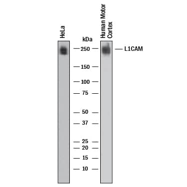
Detection of Human L1CAM by Western Blot. Western blot shows lysates of HeLa human cervical epithelial carcinoma cell line and human brain (motor cortex). PVDF membrane was probed with 2 µg/mL of Rabbit Anti-Human L1CAM Monoclonal Antibody (Catalog # MAB7773) followed by HRP-conjugated Anti-Rabbit IgG Secondary Antibody (HAF008). A specific band was detected for L1CAM at approximately 250 kDa (as indicated). This experiment was conducted under reducing conditions and using Western Blot Buffer Group 1.
 View Larger
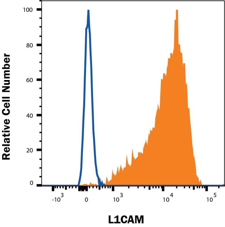
Detection of L1CAM in HeLa Human Cell Line by Flow Cytometry. HeLa human cervical epithelial cell line was stained with Rabbit Anti-Human L1CAM Monoclonal Antibody (Catalog # MAB7773, filled histogram) or Rabbit IgG isotype control antibody (MAB1050, open histogram), followed by Phycoerythrin-conjugated Anti-Rabbit IgG Secondary Antibody (F0110). Staining was performed using our Staining Membrane-associated Proteins protocol.
 View Larger
L1CAM in HeLa Human Cell Line. L1CAM was detected in immersion fixed HeLa human cervical epithelial carcinoma cell line (positive staining) and Daudi human Burkitt's lymphoma cell line (negative staining) using Rabbit Anti-Human L1CAM Monoclonal Antibody (Catalog # MAB7773) at 3 µg/mL for 3 hours at room temperature. Cells were stained using the NorthernLights™ 557-conjugated Anti-Rabbit IgG Secondary Antibody (red; NL004) and counterstained with DAPI (blue). Specific staining was localized to cell membrane. Staining was performed using our protocol for Fluorescent ICC Staining of Non-adherent Cells.
 View Larger
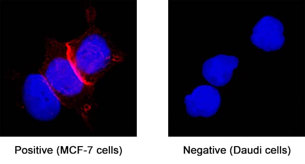
L1CAM in MCF‑7 Human Cell Line. L1CAM was detected in immersion fixed MCF‑7 human breast cancer cell line (positive staining) and Daudi human Burkitt's lymphoma cell line (negative staining) using Rabbit Anti-Human L1CAM Monoclonal Antibody (Catalog # MAB7773) at 3 µg/mL for 3 hours at room temperature. Cells were stained using the NorthernLights™ 557-conjugated Anti-Rabbit IgG Secondary Antibody (red; NL004) and counterstained with DAPI (blue). Specific staining was localized to cell membrane. Staining was performed using our protocol for Fluorescent ICC Staining of Non-adherent Cells.
 View Larger
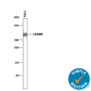
Detection of Human L1CAM by Simple WesternTM. Simple Western lane view shows lysates of HeLa human cervical epithelial carcinoma cell line, loaded at 0.2 mg/mL. A specific band was detected for L1CAM at approximately 338 kDa (as indicated) using 20 µg/mL of Rabbit Anti-Human L1CAM Monoclonal Antibody (Catalog # MAB7773). This experiment was conducted under reducing conditions and using the 66-440 kDa separation system.
Reconstitution Calculator
Preparation and Storage
- 12 months from date of receipt, -20 to -70 °C as supplied.
- 1 month, 2 to 8 °C under sterile conditions after reconstitution.
- 6 months, -20 to -70 °C under sterile conditions after reconstitution.
Background: L1CAM
L1CAM (Neural cell adhesion molecule L1, also known as L1, CD171 and NCAM-L1) is a founding member of the L1 family, Immunoglobulin (Ig) superfamily of molecules (1-4). It was initially described as a 200-230 kDa neural adhesion molecule that likely played a key role in mouse nervous system development (4-6). Subsequent studies have confirmed the adhesive nature of the molecule, and expanded its activities in both neural and nonneural cell types. L1 is now recognized to play a key role in cell migration, adhesion, neurite outgrowth, myelination and neuronal differentiation (1, 7, 8). It does so through a series of cis and trans interactions that involve multiple copartners and target receptors (1, 3, 6, 8). Cells known to express L1 are varied, and include immature oligodendrocytes (9), CD4+ T cells, B cells and monocytes (10), both motor and sensory Schwann cells (11, 12), intestinal epithelial progenitor cells (12), cerebellar granule and Purkinje cells (5, 13, 14), and multiple tumor cells such as melanoma (15) plus pancreatic duct and lung carcinoma cells (16, 17). Human L1 was first identified as a 215 kDa glycoprotein on the surface of SKNAS neuroblastoma cells (18). Subsequent cloning established its precursor as being 1257 amino acids (aa) in length (19, 20). The molecule is a type I transmembrane (TM) protein that contains an 1101 aa extracellular region (aa 20-1120) plus a 114 aa cytoplasmic domain (aa 1144-1257). The extracellular region possesses six C2-type Ig-like domains (aa 35-607) followed by five fibronectin (FN) type III domains (aa 612-1108). As noted, L1 participates in multiple cis and trans interactions, and some of these interaction sites have been mapped to select Ig or FN domains. For instance, Ig-like domains #1, 2 and 6 associate with NP-1, L1 (homotypic binding), and various integrins, respectively (21‑23). The latter interaction is mediated by one (in human) or two (in mouse) RGD motifs (23, 24). Other molecules that heterotypically associate with L1 include NCAM, neurocan, CD24 and EGFR. The cytoplasmic tail contains no kinase motifs, but does possess a FIGQY peptide that interacts with ankyrin, and an RSLE sequence that mediates clathrin-associated endocytosis (1). There are two splice variants, one each in the intracellular and extracellular domains. A deletion of RSLE adversely affects endocytosis, while a Leu substitution for aa 26-31 interfers with numerous heterotypic interactions (25, 26). In general, the full-length L1 molecule is a neuron-associated isoform. L1 is known to undergo proteolysis, either by plasmin or ADAMs. This generates soluble isoforms of varying sizes (140-200 kDa) that retain bioactivity, and which can be incorporated into the surrounding ECM (5, 13, 27-30). The membrane fragments (30-80 kDa) undergo further processing, most importantly by gamma -secretase, to generate a soluble 28 kDa intracellular domain. This domain is SUMOylated, and believed to possess an NLS at Lys1147. Upon presumed entry into the nucleus, L1 is posited to activate L1-responsive genes. Human and mouse L1 precursors share 88% aa sequence identity.
- Maness, P.F. and M. Schachner (2007) Nat. Neurosci. 10:19.
- Wei, C.H. and S.E. Ryu (2012) Exp. Mol. Med. 44:413.
- Faspel, J. and M. Grumet (2003) Front. Biosci. 8:1210.
- Herron, L.R. et al. (2009) Biochem. J. 419:519.
- Rathjen, F.G. and M. Schachner (1984) EMBO. J. 3:1.
- Keifel, H. et al. (2011) Trends Mol. Med. 17:178.
- Dihne, M. et al. (2003) J. Neurosci. 23:6638.
- Kadmon, G. et al. (1998) Dev. Immunol. 6:205.
- Itoh, K. et al. (2000) J. Neurosci. Res. 60:579.
- Jouet, M. et al. (1995) Mol. Brain Res. 30:378.
- He, Q. et al. (2012) Neurosci. Lett. 521:57.
- Thor, G. et al. (1987) EMBO J. 6:2581.
- Sadoul, K. et al. (1988) J. Neurochem. 50:510.
- Hubbe, M. et al. (1993) Eur. J. Immunol. 23:2927.
- Hoja-Lukowicz, D. et al. (2012) Glycoconj J. Apr 29. [Epub ahead of print].
- Geismann, C. et al. (2009) Cancer Res. 69:4517.
- Tischler, V. et al. (2011) Mol. Cancer 10:127.
- Mujoo, K. et al. (1986) J. Biol. Chem. 261:10299.
- Kobayashi, M. et al. (1991) Biochim. Biophys. Acta 1090:238.
- Hlavin, M.L. and V. Lemmon (1991) Genomics 11:416.
- Castellani, V. et al. (2002) EMBO J. 21:6348.
- Zhao, X. and C-H Siu (1995) J. Biol. Chem. 270:29413.
- Oleszewski, M. et al. (1999) J. Biol. Chem. 274:24602.
- Felding-Habermann, B. et al. (1997) J. Cell Biol. 139:1567.
- Kamiguchi, H. et al. (1998) J. Neurosci. 18:5311.
- De Angelis, E. et al. (2001) J. Biol. Chem. 276:32738.
- Montgomery, A.M.P. et al. (1996) J. Cell Biol. 132:475.
- Sadoul, R. et al. (1989) J. Neurochem. 53:1471.
- Reidle, S. et al. (2009) Biochem. J. 420:391.
- Lutz, D. et al. (2012) J. Biol. Chem. 287:17161.
Product Datasheets
FAQs
No product specific FAQs exist for this product, however you may
View all Antibody FAQsReviews for Human L1CAM Antibody
There are currently no reviews for this product. Be the first to review Human L1CAM Antibody and earn rewards!
Have you used Human L1CAM Antibody?
Submit a review and receive an Amazon gift card.
$25/€18/£15/$25CAN/¥75 Yuan/¥2500 Yen for a review with an image
$10/€7/£6/$10 CAD/¥70 Yuan/¥1110 Yen for a review without an image

