Human CXCR7/RDC-1 Antibody Summary
Met1-Lys362
Accession # AAA62370
Applications
Please Note: Optimal dilutions should be determined by each laboratory for each application. General Protocols are available in the Technical Information section on our website.
Scientific Data
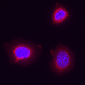 View Larger
CXCR7/RDC‑1 in MCF‑7 Human Cell Line. CXCR7/RDC‑1 was detected in immersion fixed MCF‑7 human breast cancer cell line using Mouse Anti-Human CXCR7/RDC‑1 Monoclonal Antibody (Catalog # MAB42274) at 8 µg/mL for 3 hours at room temperature. Cells were stained using the NorthernLights™ 557-conjugated Anti-Mouse IgG Secondary Antibody (red; NL007) and counterstained with DAPI (blue). Specific staining was localized to cell surface and cytoplasm. Staining was performed using our protocol for Fluorescent ICC Staining of Non-adherent Cells.
 View Larger
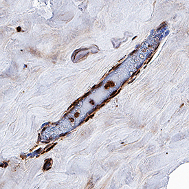
CXCR7/RDC‑1 in Human Breast Cancer Tissue. CXCR7/RDC‑1 was detected in immersion fixed paraffin-embedded sections of human breast cancer tissue using Mouse Anti-Human CXCR7/RDC‑1 Monoclonal Antibody (Catalog # MAB42274) at 0.5 µg/mL for 1 hour at room temperature followed by incubation with the Anti-Mouse IgG VisUCyte™ HRP Polymer Antibody (VC001). Before incubation with the primary antibody, tissue was subjected to heat-induced epitope retrieval using Antigen Retrieval Reagent-Basic (CTS013). Tissue was stained using DAB (brown) and counterstained with hematoxylin (blue). Specific staining was localized to endothelial cells. Staining was performed using our protocol for IHC Staining with VisUCyte HRP Polymer Detection Reagents.
Reconstitution Calculator
Preparation and Storage
- 12 months from date of receipt, -20 to -70 °C as supplied.
- 1 month, 2 to 8 °C under sterile conditions after reconstitution.
- 6 months, -20 to -70 °C under sterile conditions after reconstitution.
Background: CXCR7/RDC-1
The G protein-coupled receptor, RDC1, belongs to a subgroup of chemokine receptors and has been designated CXCR7. CXCR7 can bind with high-affinity to CXCL12/SDF-1 and CXCL11/I-TAC. It is also a co-receptor for several HIV and SIV strains. In their N-termini and extracellular loops 1, 2, and 3, human and mouse CXCR7 share 84%, 100%, 96% and 86% amino acid sequence identity, respectively. Reports of mRNA levels and/or protein expression (as assessed using anti‑CXCR7, clone 9C4) (1, 2) indicate that CXCR7 occurs on a wide variety of tissues and cells including monocytes, B cells, T cells and mature dendritic cells. In contrast, based on ligand binding analysis and receptor level (as assessed using anti‑CXCR7, clone 11G8), surface expression of CXCR7 was reported to be restricted to tumor cells, activated endothelial cells, fetal liver cells, and few other cell types (3). The basis of these inconsistent observations is not known but may be attributed to cell context and the use of different antibodies that may recognize different epitopes.
- Balabanian, K. et al. (2005) J. Biol. Chem. 280:35760.
- Infantino, S. et al. (2006) J. Immunol. 176:2197.
- Burns, J.M. et al. (2006) J. Exp. Med. 203:2201.
Product Datasheets
FAQs
No product specific FAQs exist for this product, however you may
View all Antibody FAQsReviews for Human CXCR7/RDC-1 Antibody
Average Rating: 5 (Based on 1 Review)
Have you used Human CXCR7/RDC-1 Antibody?
Submit a review and receive an Amazon gift card.
$25/€18/£15/$25CAN/¥75 Yuan/¥1250 Yen for a review with an image
$10/€7/£6/$10 CAD/¥70 Yuan/¥1110 Yen for a review without an image
Filter by:


