Mouse Rae-1 Pan Specific Antibody Summary
Leu29-Ser231
Accession # O08604
Applications
Please Note: Optimal dilutions should be determined by each laboratory for each application. General Protocols are available in the Technical Information section on our website.
Scientific Data
 View Larger
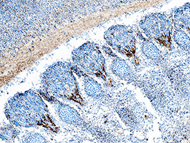
Rae‑1 in Mouse Embryo. Rae-1 was detected in immersion fixed frozen sections of mouse embryo (13 d.p.c.) using Goat Anti-Mouse Rae-1 Pan Specific Antigen Affinity-purified Polyclonal Antibody (Catalog # AF1136) at 5 µg/mL overnight at 4 °C. Tissue was stained using the Anti-Goat HRP-DAB Cell & Tissue Staining Kit (brown; Catalog # CTS008) and counterstained with hematoxylin (blue). Specific staining was localized to axons of primary sensory neurons. View our protocol for Chromogenic IHC Staining of Frozen Tissue Sections.
Reconstitution Calculator
Preparation and Storage
- 12 months from date of receipt, -20 to -70 °C as supplied.
- 1 month, 2 to 8 °C under sterile conditions after reconstitution.
- 6 months, -20 to -70 °C under sterile conditions after reconstitution.
Background: Rae-1
Rae-1 gamma is a member of a family of cell-surface proteins that function as ligands for mouse NKG2D. Other family members are designated Rae-1 alpha, beta, δ and epsilon. Amino acid sequence identity within this family ranges from 88‑95%. The Rae-1 proteins are distantly related to MHC class I proteins, but they possess only the alpha 1 and alpha 2 Ig-like domains, and they have no capacity to bind peptide or interact with beta 2-microglobulin. The genes encoding these proteins are not found within the Major Histocompatibility Complex on mouse chromosome 17, but rather map to mouse chromosome 10. The Rae-1 proteins are anchored to the membrane via a GPI‑linkage. The name of this family derives from the original identification of these proteins as the product of retinoic acid early inducible transcripts. Rae-1 expression is developmentally controlled. Transcripts were observed in the brain/head region of day 10‑14 embryos but disappeared by day 18. Rae-1 transcripts were detected in several transformed cell lines but are absent from most normal adult tissues. All Rae-1 family members bind to mouse NKG2D, an activating receptor expressed on NK cells and some T cell subsets, resulting in the activation of cytolytic activity and/or cytokine production by these effector cells. Ectopic expression of Rae-1 on mouse tumor cell lines resulted in the in vivo rejection of the tumors (1‑6).
- Zou, Z. et al. (1996) J. Biochem (Tokyo) 119:319.
- Diefenbach, A. et al. (2000) Nature Immunol. 1:119.
- Cerwenka, A. et al. (2000) Immunity 12:721.
- Cerwenka, A. et al. (2001) Proc. Natl. Acad. Sci. USA 98:11521.
- Diefenbach, A. et al. (2001) Nature 413:165.
- NKG2D and its Ligands, www.RnDSystems.com.
Product Datasheets
Citations for Mouse Rae-1 Pan Specific Antibody
R&D Systems personnel manually curate a database that contains references using R&D Systems products. The data collected includes not only links to publications in PubMed, but also provides information about sample types, species, and experimental conditions.
8
Citations: Showing 1 - 8
Filter your results:
Filter by:
-
Effects of obesity on NK cells in a mouse model of postmenopausal breast cancer
Authors: J Spielmann, L Mattheis, JS Jung, H Rau beta e, M Gla beta, I Bähr, D Quandt, J Oswald, H Kielstein
Sci Rep, 2020-11-26;10(1):20606.
Species: Mouse
Sample Types: Whole Tissue
Applications: IHC -
Toxicity Induced by a Bispecific T Cell-Redirecting Protein Is Mediated by Both T Cells and Myeloid Cells in Immunocompetent Mice
Authors: C Godbersen-, TA Coupet, Z Grada, SC Zhang, CL Sentman
J. Immunol., 2020-04-15;204(11):2973-2983.
Species: Mouse
Sample Types: Whole Tissue
Applications: IHC -
Cytotoxicity of natural killer cells activated through NKG2D contributes to the development of bronchiolitis obliterans in a murine heterotopic tracheal transplant model
Authors: T Kawakami, K Ito, Y Matsuda, M Noda, A Sakurada, Y Hoshikawa, Y Okada, K Ogasawara
Am. J. Transplant, 2017-04-21;0(0):.
Species: Mouse
Sample Types: Whole Tissue
Applications: IHC-P -
NK1.1(-) CD4(+) NKG2D(+) T cells suppress DSS-induced colitis in mice through production of TGF-?
Authors: X Qian, C Hu, S Han, Z Lin, W Xiao, Y Ding, Y Zhang, L Qian, X Jia, G Zhu, W Gong
J. Cell. Mol. Med, 2017-02-22;0(0):.
Species: Mouse
Sample Types: Whole Tissue
Applications: IHC -
The immunoreceptor NKG2D promotes tumour growth in a model of hepatocellular carcinoma
Authors: S Sheppard, J Guedes, A Mroz, AM Zavitsanou, H Kudo, SM Rothery, P Angelopoul, R Goldin, N Guerra
Nat Commun, 2017-01-27;8(0):13930.
Species: Mouse
Sample Types: Whole Tissue
Applications: IHC -
NKG2D ligand overexpression in lupus nephritis correlates with increased NK cell activity and differentiation in kidneys but not in the periphery.
Authors: Spada R, Rojas J, Perez-Yague S, Mulens V, Cannata-Ortiz P, Bragado R, Barber D
J Leukoc Biol, 2015-01-12;97(3):583-98.
Species: Human
Sample Types: Whole Tissue
Applications: IHC, IHC-P -
CD4(+) NKG2D(+) T cells induce NKG2D down-regulation in natural killer cells in CD86-RAE-1epsilon transgenic mice.
Authors: Lin Z, Wang C, Xia H, Liu W, Xiao W, Qian L, Jia X, Ding Y, Ji M, Gong W
Immunology, 2014-03-01;141(3):401-15.
Species: Mouse
Sample Types: Tissue Homogenates, Whole Tissue
Applications: IHC, Western Blot -
Sustained CTL activation by murine pulmonary epithelial cells promotes the development of COPD-like disease.
Authors: Borchers MT, Wesselkamper SC, Curull V, Ramirez-Sarmiento A, Sanchez-Font A, Garcia-Aymerich J, Coronell C, Lloreta J, Agusti AG, Gea J, Howington JA, Reed MF, Starnes SL, Harris NL, Vitucci M, Eppert BL, Motz GT, Fogel K, McGraw DW, Tichelaar JW, Orozco-Levi M
J. Clin. Invest., 2009-02-09;119(3):636-49.
Species: Mouse
Sample Types: Whole Tissue
Applications: IHC-P
FAQs
No product specific FAQs exist for this product, however you may
View all Antibody FAQsReviews for Mouse Rae-1 Pan Specific Antibody
There are currently no reviews for this product. Be the first to review Mouse Rae-1 Pan Specific Antibody and earn rewards!
Have you used Mouse Rae-1 Pan Specific Antibody?
Submit a review and receive an Amazon gift card.
$25/€18/£15/$25CAN/¥75 Yuan/¥1250 Yen for a review with an image
$10/€7/£6/$10 CAD/¥70 Yuan/¥1110 Yen for a review without an image

