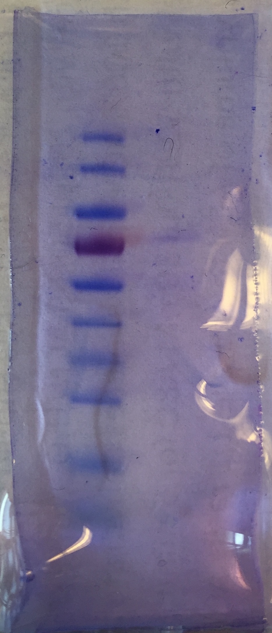
Mouse CX3CL1/Fractalkine Chemokine Domain Antibody Summary
Applications
under non-reducing conditions only
Mouse CX3CL1/Fractalkine Sandwich Immunoassay
Please Note: Optimal dilutions should be determined by each laboratory for each application. General Protocols are available in the Technical Information section on our website.
Scientific Data
 View Larger
Chemotaxis Induced by CX3CL1/Fractalkine and Neutralization by Mouse CX3CL1/Fractalkine Antibody. Recombinant Mouse CX3CL1/Fractalkine (Catalog # 571-MF) chemoattracts the BaF3 mouse pro-B cell line transfected with human CX3CR1 in a dose-dependent manner (orange line). The amount of cells that migrated through to the lower chemotaxis chamber was measured by Resazurin (Catalog # AR002). Chemotaxis elicited by Recombinant Mouse CX3CL1/Fractalkine (30 ng/mL) is neutralized (green line) by increasing concentrations of Rat Anti-Mouse CX3CL1/Fractalkine Chemokine Domain Monoclonal Antibody (Catalog # MAB571). The ND50 is typically 0.75-3.5 µg/mL.
 View Larger
Detection of Mouse CX3CL1/Fractalkine by Immunocytochemistry/Immunofluorescence CX3CL1 expression in the brain during EAE progression. (A) Brain CX3CL1 expression of wild type mice with EAE as determined by quantitative real-time PCR. Gene levels were normalized to GAPDH levels and are displayed as levels relative to naïve mice. Error bars represent the s.e.m. (n = 5 mice per group). (B-M) Brains from naïve wild type mice were harvested and frozen for immunostaining. CX3CL1 expression (red) and DAPI nuclei staining (blue) in (B, F, J) naïve and (C, G, K) Day 10, (D, H, L) Day 14, and (E, I, M) Day 21 post-EAE induced wild type mice. CX3CL1 expression is displayed at the (B-E) choroid plexus, (F-I) in and near the hippocampus, and (J-M) cerebellum. White scale bars represent 50 μm. (N) CX3CL1 expression (relative to non-treated cells) in the CPLacZ-2 mouse choroid plexus cell line after 2 hour treatment with varying concentrations of the A2A adenosine receptor specific adenosine receptor agonist CGS21680. Error bars represent the s.e.m. (O) Lymphocyte migration across a transwell choroid plexus barrier following pretreatment with vehicle treatment alone, CGS21680, or CGS21680 and anti-CX3CL1. Total migration was normalized to the vehicle control (set to 100%). Error bars represent the s.e.m. These results are representative of two separate experiments (n ≤ 3). Image collected and cropped by CiteAb from the following publication (https://jneuroinflammation.biomedcentral.com/articles/10.1186/1742-2094-9-193), licensed under a CC-BY license. Not internally tested by R&D Systems.
 View Larger
Detection of Mouse CX3CL1/Fractalkine by Immunohistochemistry-Frozen CX3CL1 antibody mediated blockade protects mice against EAE and its associated lymphocyte infiltration. Wild type mice were induced to develop EAE and starting at day 8 post induction given daily anti-CX3CL1 antibody or an isotype control treatments (i.p.). (A) EAE disease profile. Error bars represent the s.e.m. (n = 4 mice/group). Significant differences are indicated as determined by two-way ANOVA. EAE scoring data is representative of 2 separate experiments. (B) CD45, CD11b, and F480 stained brain (hippocampal and cerebellum areas) and spinal cord sections from day 28 post-EAE induced mice treated with either anti-CX3CL1 or control antibody. Positively stained cells (red) are shown against a hematoxylin counterstain (blue). Black scale bars represent 50 μm. (C) CD4 and (D) CD8 positive mean cells counts per field at 10x magnification from brain and spinal cord stained frozen brain sections from day 28 post-EAE induced mice treated with either anti-CX3CL1 or control antibody. Error bars represent the standard error of the mean (n ≤ 11). Significant differences (P < 0.05, *) are indicated as determined by the Student’s t-test. Image collected and cropped by CiteAb from the following publication (https://jneuroinflammation.biomedcentral.com/articles/10.1186/1742-2094-9-193), licensed under a CC-BY license. Not internally tested by R&D Systems.
 View Larger
Detection of Mouse CX3CL1/Fractalkine by Immunocytochemistry/Immunofluorescence CX3CL1 expression in the brain during EAE progression. (A) Brain CX3CL1 expression of wild type mice with EAE as determined by quantitative real-time PCR. Gene levels were normalized to GAPDH levels and are displayed as levels relative to naïve mice. Error bars represent the s.e.m. (n = 5 mice per group). (B-M) Brains from naïve wild type mice were harvested and frozen for immunostaining. CX3CL1 expression (red) and DAPI nuclei staining (blue) in (B, F, J) naïve and (C, G, K) Day 10, (D, H, L) Day 14, and (E, I, M) Day 21 post-EAE induced wild type mice. CX3CL1 expression is displayed at the (B-E) choroid plexus, (F-I) in and near the hippocampus, and (J-M) cerebellum. White scale bars represent 50 μm. (N) CX3CL1 expression (relative to non-treated cells) in the CPLacZ-2 mouse choroid plexus cell line after 2 hour treatment with varying concentrations of the A2A adenosine receptor specific adenosine receptor agonist CGS21680. Error bars represent the s.e.m. (O) Lymphocyte migration across a transwell choroid plexus barrier following pretreatment with vehicle treatment alone, CGS21680, or CGS21680 and anti-CX3CL1. Total migration was normalized to the vehicle control (set to 100%). Error bars represent the s.e.m. These results are representative of two separate experiments (n ≤ 3). Image collected and cropped by CiteAb from the following publication (https://jneuroinflammation.biomedcentral.com/articles/10.1186/1742-2094-9-193), licensed under a CC-BY license. Not internally tested by R&D Systems.
 View Larger
Detection of Mouse CX3CL1/Fractalkine by Immunocytochemistry/Immunofluorescence CX3CL1 expression in the brain during EAE progression. (A) Brain CX3CL1 expression of wild type mice with EAE as determined by quantitative real-time PCR. Gene levels were normalized to GAPDH levels and are displayed as levels relative to naïve mice. Error bars represent the s.e.m. (n = 5 mice per group). (B-M) Brains from naïve wild type mice were harvested and frozen for immunostaining. CX3CL1 expression (red) and DAPI nuclei staining (blue) in (B, F, J) naïve and (C, G, K) Day 10, (D, H, L) Day 14, and (E, I, M) Day 21 post-EAE induced wild type mice. CX3CL1 expression is displayed at the (B-E) choroid plexus, (F-I) in and near the hippocampus, and (J-M) cerebellum. White scale bars represent 50 μm. (N) CX3CL1 expression (relative to non-treated cells) in the CPLacZ-2 mouse choroid plexus cell line after 2 hour treatment with varying concentrations of the A2A adenosine receptor specific adenosine receptor agonist CGS21680. Error bars represent the s.e.m. (O) Lymphocyte migration across a transwell choroid plexus barrier following pretreatment with vehicle treatment alone, CGS21680, or CGS21680 and anti-CX3CL1. Total migration was normalized to the vehicle control (set to 100%). Error bars represent the s.e.m. These results are representative of two separate experiments (n ≤ 3). Image collected and cropped by CiteAb from the following publication (https://jneuroinflammation.biomedcentral.com/articles/10.1186/1742-2094-9-193), licensed under a CC-BY license. Not internally tested by R&D Systems.
Reconstitution Calculator
Preparation and Storage
- 12 months from date of receipt, -20 to -70 °C as supplied.
- 1 month, 2 to 8 °C under sterile conditions after reconstitution.
- 6 months, -20 to -70 °C under sterile conditions after reconstitution.
Background: CX3CL1/Fractalkine
CX3CL1, also known as Fractalkine, is a type I membrane protein in which a chemokine domain possessing a unique C-X3-C cysteine motif is tethered on a long mucin-like stalk. It can also be released as a soluble molecule upon proteolysis at a membrane proximal site.
Product Datasheets
Citations for Mouse CX3CL1/Fractalkine Chemokine Domain Antibody
R&D Systems personnel manually curate a database that contains references using R&D Systems products. The data collected includes not only links to publications in PubMed, but also provides information about sample types, species, and experimental conditions.
12
Citations: Showing 1 - 10
Filter your results:
Filter by:
-
The CX3CL1-CX3CR1 chemokine axis can contribute to tumor immune evasion and blockade with a novel CX3CR1 monoclonal antibody enhances response to anti-PD-1 immunotherapy
Authors: Apoorvi Chaudhri, Xia Bu, Yunfei Wang, Michael Gomez, James A. Torchia, Ping Hua et al.
Front Immunol
-
Fractalkine is Involved in Lipopolysaccharide-Induced Podocyte Injury through the Wnt/ beta -Catenin Pathway in an Acute Kidney Injury Mouse Model
Authors: Soulixay Senouthai, Junjie Wang, Dongdong Fu, Yanwu You
Inflammation
-
CX3CL1 Promotes Breast Cancer via Transactivation of the EGF Pathway
Authors: Manuel Tardáguila, Emilia Mira, Miguel A. García-Cabezas, Anna M. Feijoo, Miguel Quintela-Fandino, Iñigo Azcoitia et al.
Cancer Research
-
System analysis based on the cancer–immunity cycle identifies ZNF207 as a novel immunotherapy target for hepatocellular carcinoma
Authors: Xu Wang, Tao Zhou, Xingyi Chen, Yu Wang, Yushi Ding, Haoyang Tu et al.
Journal for ImmunoTherapy of Cancer
-
Inhibition of mPGES-1 attenuates efficient resolution of acute inflammation by enhancing CX3CL1 expression
Authors: P Rappl, S Rösser, P Maul, R Bauer, A Huard, Y Schreiber, D Thomas, G Geisslinge, PJ Jakobsson, A Weigert, B Brüne, T Schmid
Cell Death & Disease, 2021-02-02;12(2):135.
Species: Mouse
Sample Types: In Vivo, Whole Tissue
Applications: IHC, Neutralization -
Prevention of lipopolysaccharide-induced preterm labor by the lack of CX3CL1-CX3CR1 interaction in mice
Authors: M Mizoguchi, Y Ishida, M Nosaka, A Kimura, Y Kuninaka, T Yahata, S Nanjo, S Toujima, S Minami, K Ino, N Mukaida, T Kondo
PLoS ONE, 2018-11-06;13(11):e0207085.
Species: Mouse
Sample Types: In Vivo
Applications: Neutralization -
p16Ink4a and p21Cip1/Waf1 promote tumour growth by enhancing myeloid-derived suppressor cells chemotaxis
Authors: A Okuma, A Hanyu, S Watanabe, E Hara
Nat Commun, 2017-12-12;8(1):2050.
Species: Mouse
Sample Types: In Vivo
Applications: In Vivo -
PGI2 Controls Pulmonary NK Cells That Prevent Airway Sensitization to House Dust Mite Allergen
Authors: Bryan Simons
J. Immunol, 2016-11-28;0(0):.
Species: Mouse
Sample Types:
Applications: Neutralization -
Extracellular adenosine signaling induces CX3CL1 expression in the brain to promote experimental autoimmune encephalomyelitis.
J Neuroinflammation, 2012-08-10;9(0):193.
Species: Mouse
Sample Types: In Vivo, Whole Tissue
Applications: IHC-Fr, Neutralization -
Gene therapy with CX3CL1/Fractalkine induces antitumor immunity to regress effectively mouse hepatocellular carcinoma.
Authors: Tang L, Hu HD, Hu P, Lan YH, Peng ML, Chen M, Ren H
Gene Ther., 2007-06-28;14(16):1226-34.
Species: Mouse
Sample Types: Whole Cells
Applications: ICC -
Regulation of Physical Microglia-Neuron Interactions by Fractalkine Signaling after Status Epilepticus
Authors: Ukpong B. Eyo, Jiyun Peng, Madhuvika Murugan, Mingshu Mo, Almin Lalani, Ping Xie et al.
eNeuro
-
Fibroblast polarization over the myocardial infarction time continuum shifts roles from inflammation to angiogenesis.
Authors: Mouton AJ, Ma Y, Rivera Gonzalez OJ et al.
Basic Res. Cardiol.
FAQs
No product specific FAQs exist for this product, however you may
View all Antibody FAQsReviews for Mouse CX3CL1/Fractalkine Chemokine Domain Antibody
Average Rating: 4 (Based on 1 Review)
Have you used Mouse CX3CL1/Fractalkine Chemokine Domain Antibody?
Submit a review and receive an Amazon gift card.
$25/€18/£15/$25CAN/¥75 Yuan/¥2500 Yen for a review with an image
$10/€7/£6/$10 CAD/¥70 Yuan/¥1110 Yen for a review without an image
Filter by:


