Human UCH-L1/PGP9.5 Antibody Summary
Gln2-Ala223
Accession # P09936
Applications
Please Note: Optimal dilutions should be determined by each laboratory for each application. General Protocols are available in the Technical Information section on our website.
Scientific Data
 View Larger
Detection of Human UCH-L1/PGP9.5 by Western Blot. Western blot shows lysates of human brain (cortex) tissue. PVDF membrane was probed with 2 µg/mL of Mouse Anti-Human UCH-L1/PGP9.5 Monoclonal Antibody (Catalog # MAB6007) followed by HRP-conjugated Anti-Mouse IgG Secondary Antibody (Catalog # HAF007). A specific band was detected for UCH-L1/PGP9.5 at approximately 29 kDa (as indicated). This experiment was conducted under reducing conditions and using Immunoblot Buffer Group 1.
 View Larger
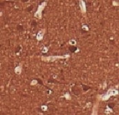
UCH-L1/PGP9.5 in Human Prostate. UCH-L1/PGP9.5 was detected in immersion fixed paraffin-embedded sections of human prostate using Mouse Anti-Human UCH-L1/PGP9.5 Monoclonal Antibody (Catalog # MAB6007) at 15 µg/mL overnight at 4 °C. Before incubation with the primary antibody, tissue was subjected to heat-induced epitope retrieval using Antigen Retrieval Reagent-Basic (Catalog # CTS013). Tissue was stained using the Anti-Mouse HRP-DAB Cell & Tissue Staining Kit (brown; Catalog # CTS002) and counterstained with hematoxylin (blue). Specific staining was localized to cytoplasm of glandular epithelial cells. View our protocol for Chromogenic IHC Staining of Paraffin-embedded Tissue Sections.
 View Larger
Detection of Human and Mouse UCH-L1/PGP9.5 by Simple WesternTM. Simple Western lane view shows lysates of human brain (cortex) tissue and mouse brain (cortex) tissue, loaded at 0.2 mg/mL. A specific band was detected for UCH-L1/PGP9.5 at approximately 32 kDa (as indicated) using 10 µg/mL of Mouse Anti-Human UCH-L1/PGP9.5 Monoclonal Antibody (Catalog # MAB6007). This experiment was conducted under reducing conditions and using the 12-230 kDa separation system.
 View Larger
Western Blot Shows Human UCH-L1/PGP9.5 Specificity by Using Knockout Cell Line. Western blot shows lysates of HEK293T human embryonic kidney parental cell line and UCH-L1/PGP9.5 knockout HEK293T cell line (KO). PVDF membrane was probed with 2 µg/mL of Mouse Anti-Human UCH-L1/PGP9.5 Monoclonal Antibody (Catalog # MAB6007) followed by HRP-conjugated Anti-Mouse IgG Secondary Antibody (Catalog # HAF018). A specific band was detected for UCH-L1/PGP9.5 at approximately 28 kDa (as indicated) in the parental HEK293T cell line, but is not detectable in knockout HEK293T cell line. GAPDH (Catalog # MAB5718) is shown as a loading control. This experiment was conducted under reducing conditions and using Immunoblot Buffer Group 1.
Reconstitution Calculator
Preparation and Storage
- 12 months from date of receipt, -20 to -70 °C as supplied.
- 1 month, 2 to 8 °C under sterile conditions after reconstitution.
- 6 months, -20 to -70 °C under sterile conditions after reconstitution.
Background: UCH-L1/PGP9.5
UCH-L1 (ubiquitin carboxyterminal hydrolase isozyme 1; also PGP9.5) is a 24-27 kDa member of the peptidase C12 family of enzymes. It shows restricted expression, being found in neurons and oocytes. UCH-L1 has dual enzymatic activity. As a monomer, it is a ubiquitin hydrolase that removes ubiquitin from modified proteins; as a homodimer, it acts as a ligase that creates ubiquitin dimers. In neurons, UCH-L1’s most important role appears to be that of generating free ubiquitin. Human UCH-L1 is 223 amino acids (aa) in length. It is O-glycosylated, ubiquitinated, and farnesylated; when farnesylated, it becomes associated with cell membranes. Three potential splice forms are reported. One shows a two aa substitution for aa 12-15, a second contains an alternative start site at Met82, and a third shows the same start site coupled with a deletion of aa 138-153. Full-length human UCH-L1 shares 95% aa identity with mouse UCH-L1.
Product Datasheets
Citations for Human UCH-L1/PGP9.5 Antibody
R&D Systems personnel manually curate a database that contains references using R&D Systems products. The data collected includes not only links to publications in PubMed, but also provides information about sample types, species, and experimental conditions.
3
Citations: Showing 1 - 3
Filter your results:
Filter by:
-
Pivotal Role of Ubiquitin Carboxyl-Terminal Hydrolase L1 (UCHL1) in Uterine Leiomyoma
Authors: T Suzuki, Y Dai, M Ono, J Kojima, T Sasaki, H Fujiwara, N Kuji, H Nishi
Biomolecules, 2023-01-18;13(2):.
Species: Human
Sample Types: Protein, Whole Tissue
Applications: IHC, Western Blot -
UCHL1 and Proteasome in Blood Serum in Relation to Dietary Habits, Concentration of Selected Antioxidant Minerals and Total Antioxidant Status among Patients with Alzheimer's Disease
Authors: S Bogdan, A Pu?cion-Ja, K Klimiuk, K Socha, J Kochanowic, E Gorodkiewi
Journal of Clinical Medicine, 2022-01-14;11(2):.
Species: Human
Sample Types: Serum
Applications: Surface Plasmon Resonance (SPR -
Ubiquitin Carboxyl-Terminal Hydrolase L1 (UCHL1) Promotes Uterine Serous Cancer Cell Proliferation and Cell Cycle Progression
Authors: SY Kwan, CL Au-Yeung, TL Yeung, A Rynne-Vida, KK Wong, JI Risinger, HK Lin, RE Schmandt, MS Yates, SC Mok, KH Lu
Cancers (Basel), 2020-01-02;12(1):.
Species: Human
Sample Types: Whole Cells
Applications: IP
FAQs
No product specific FAQs exist for this product, however you may
View all Antibody FAQsReviews for Human UCH-L1/PGP9.5 Antibody
Average Rating: 4.5 (Based on 2 Reviews)
Have you used Human UCH-L1/PGP9.5 Antibody?
Submit a review and receive an Amazon gift card.
$25/€18/£15/$25CAN/¥75 Yuan/¥2500 Yen for a review with an image
$10/€7/£6/$10 CAD/¥70 Yuan/¥1110 Yen for a review without an image
Filter by:
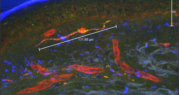
1st PGP9.5 in 1:500, 2nd antibody Cy3
Red staining = laminin + Cy5
+Dapi mounting medium
Collagen autofluorescence was a problem in some of the sections.
Zamboni Fixed, 20% sucr.


