Human SYK (SH2D2 Domain) Antibody Summary
Trp168-Cys259
Accession # P43405
Applications
Please Note: Optimal dilutions should be determined by each laboratory for each application. General Protocols are available in the Technical Information section on our website.
Scientific Data
 antibody by Western Blot.") View Larger
View Larger
Detection of Human and Mouse SYK (SH2D2 Domain) by Western Blot. Western blot shows lysates of EL-4 mouse lymphoblast cell line and Raji human Burkitt's lymphoma cell line. PVDF membrane was probed with 2 µg/mL of Mouse Anti-Human SYK (SH2D2 Domain) Monoclonal Antibody (Catalog # MAB7166) followed by HRP-conjugated Anti-Mouse IgG Secondary Antibody (HAF007). A specific band was detected for SYK (SH2D2 Domain) at approximately 72 kDa (as indicated). This experiment was conducted under reducing conditions and using Immunoblot Buffer Group 1.
 View Larger
Detection of Raji human Burkitt's lymphoma cell line 0.2 by Simple WesternTM. Simple Western lane view shows lysates of Raji human Burkitt's lymphoma cell line, loaded at 0.2 mg/mL. A specific band was detected for SYK at approximately 66 kDa (as indicated) using 20 µg/mL of Mouse Anti-Human SYK (SH2D2 Domain) Monoclonal Antibody (Catalog # MAB7166). This experiment was conducted under reducing conditions and using the 12-230 kDa separation system.
 View Larger
Detection of Mouse SYK by Simple WesternTM. Simple Western lane view shows lysates of EL‑4 mouse lymphoblast cell line, loaded at 0.2 mg/mL. A specific band was detected for SYK at approximately 66 kDa (as indicated) using 20 µg/mL of Mouse Anti-Human SYK (SH2D2 Domain) Monoclonal Antibody (Catalog # MAB7166). This experiment was conducted under reducing conditions and using the 12-230 kDa separation system.
 View Larger
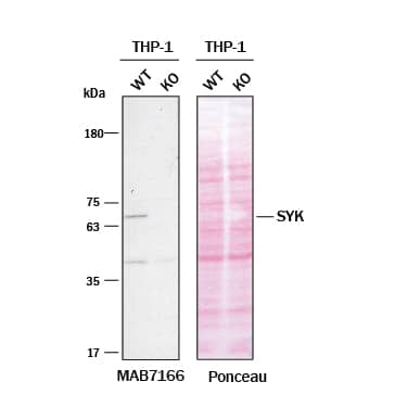
Western Blot Shows Human SYK Specificity Using Knockout Cell Line. Western blot shows lysates of THP‑1 human acute monocytic leukemia cell line and SYK knockout THP-1 cell line (KO). Nitrocellulose membrane was probed with 2 µg/mL of Mouse Anti-Human SYK (SH2D2 Domain) Monoclonal Antibody (Catalog # MAB7166) followed by HRP-conjugated Anti-Mouse IgG Secondary Antibody. A specific band was detected for SYK at approximately 68 kDa (as indicated) in the parental THP-1 cell line, but is not detectable in knockout THP-1 cell line. The Ponceau stained transfer of the blot is shown. This experiment was conducted under reducing conditions. Image, protocol, and testing courtesy of YCharOS Inc. See ycharos.com for additional details.
Reconstitution Calculator
Preparation and Storage
- 12 months from date of receipt, -20 to -70 °C as supplied.
- 1 month, 2 to 8 °C under sterile conditions after reconstitution.
- 6 months, -20 to -70 °C under sterile conditions after reconstitution.
Background: SYK
SYK (spleen tyrosine kinase) is a 70-80 kDa cytoplasmic non-receptor protein tyrosine kinase of the protein kinase superfamily and the SYK/ZAP-70 subfamily of proteins. The 635 amino acid (aa) human SYK contains two SH2 domains (aa 14-106 and aa 168-259), and one protein kinase domain (aa 371-631). A splicing variant produces a second isoform lacking aa 371-631. Within the second SH2 domain (SH2D2), human SYK shares 94% aa sequence identity with mouse and rat SYK. SYK is widely expressed in hematopoietic cells (notably B lymphocytes), where it couples immunoglobulin receptors to downstream events, such as proliferation, differentiation, and phagocytosis.
Product Datasheets
FAQs
No product specific FAQs exist for this product, however you may
View all Antibody FAQsReviews for Human SYK (SH2D2 Domain) Antibody
There are currently no reviews for this product. Be the first to review Human SYK (SH2D2 Domain) Antibody and earn rewards!
Have you used Human SYK (SH2D2 Domain) Antibody?
Submit a review and receive an Amazon gift card.
$25/€18/£15/$25CAN/¥75 Yuan/¥1250 Yen for a review with an image
$10/€7/£6/$10 CAD/¥70 Yuan/¥1110 Yen for a review without an image


