Human p21/CIP1/CDKN1A Antibody Summary
Ser2-Pro164
Accession # P38936
Applications
Please Note: Optimal dilutions should be determined by each laboratory for each application. General Protocols are available in the Technical Information section on our website.
Scientific Data
 View Larger
Detection of Human p21/CIP1/CDKN1A by Western Blot. Western blot shows lysates of MCF-7 human breast cancer cell line untreated (-) or treated (+) with 1 µM camptothecin (CPT) for 16 hours. PVDF membrane was probed with 0.5 µg/mL of Goat Anti-Human p21/CIP1/CDKN1A Antigen Affinity-purified Polyclonal Antibody (Catalog # AF1047), followed by HRP-conjugated Anti-Goat IgG Secondary Antibody (Catalog # HAF017). A specific band was detected for p21/CIP1/CDKN1A at approximately 21 kDa (as indicated). This experiment was conducted under reducing conditions and using Immunoblot Buffer Group 1.
 View Larger
p21/CIP1/CDKN1A in MCF‑7 Human Cell Line. p21/CIP1/CDKN1A was detected in immersion fixed MCF-7 human breast cancer cell line treated with (left panel) or without (right panel) camptothecin using Goat Anti-Human p21/CIP1/CDKN1A Antigen Affinity-purified Polyclonal Antibody (Catalog # AF1047) at 10 µg/mL for 3 hours at room temperature. Cells were stained using the NorthernLights™ 557-conjugated Anti-Goat IgG Secondary Antibody (red; Catalog # NL001) and counterstained with DAPI (blue). Specific staining was localized to nuclei. View our protocol for Fluorescent ICC Staining of Cells on Coverslips.
 View Larger
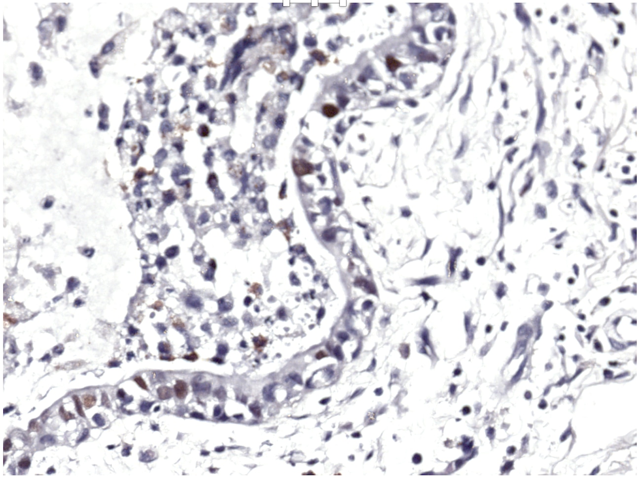
p21/CIP1/CDKN1A in Human Breast Cancer Tissue. p21/CIP1/CDKN1A was detected in immersion fixed paraffin-embedded sections of human breast cancer tissue using 1.7 µg/mL Goat Anti-Human p21/CIP1/CDKN1A Antigen Affinity-purified Polyclonal Antibody (Catalog # AF1047) overnight at 4 °C. Tissue was stained with the Anti-Goat HRP-DAB Cell & Tissue Staining Kit (brown; Catalog # CTS008) and counterstained with hematoxylin (blue). View our protocol for Chromogenic IHC Staining of Paraffin-embedded Tissue Sections.
 View Larger
Detection of p21/CIP1/CDKN1A in MCF‑7 Human Cell Line by Flow Cytometry. MCF-7 human breast cancer cell line was unstimulated (light orange filled histogram) or treated with 1 µM camphtothecin for 16 hours, then stained with Goat Anti-Human p21/CIP1/CDKN1A Antigen Affinity-purified Polyclonal Antibody (Catalog # AF1047, dark orange filled histogram) or isotype control antibody (Catalog # AB-108-C, open histogram), followed by Allophycocyanin-conjugated Anti-Goat IgG Secondary Antibody (Catalog # F0108). To facilitate intracellular staining, cells were fixed with paraformaldehyde and permeabilized with methanol.
 View Larger
Detection of Human p21/CIP1/CDKN1A by Simple WesternTM. Simple Western lane view shows lysates of MCF-7 human breast cancer cell line untreated (-) or treated (+) with 1 µM Camptothecin (CPT) for 16 hours, loaded at 0.2 mg/mL. A specific band was detected for p21/CIP1/CDKN1A at approximately 30 kDa (as indicated) using 5 µg/mL of Goat Anti-Human p21/CIP1/CDKN1A Antigen Affinity-purified Polyclonal Antibody (Catalog # AF1047) followed by 1:50 dilution of HRP-conjugated Anti-Goat IgG Secondary Antibody (Catalog # HAF109). This experiment was conducted under reducing conditions and using the 12-230 kDa separation system.
Reconstitution Calculator
Preparation and Storage
- 12 months from date of receipt, -20 to -70 °C as supplied.
- 1 month, 2 to 8 °C under sterile conditions after reconstitution.
- 6 months, -20 to -70 °C under sterile conditions after reconstitution.
Background: p21/CIP1/CDKN1A
p21CIP1, also called CIP1 (CDK-interacting protein 1) and CDKN1A, is a 21 kDa Cyclin/Cyclin-dependent kinase (Cdk) inhibitor that blocks cell cycle progression from G1 to S phase in the cell cycle. Because p21 is a transcriptional target of the p53 tumor suppressor, p21 expression increases in cells that contain stabilized p53 due to genotoxic stress exposure.
Product Datasheets
Citations for Human p21/CIP1/CDKN1A Antibody
R&D Systems personnel manually curate a database that contains references using R&D Systems products. The data collected includes not only links to publications in PubMed, but also provides information about sample types, species, and experimental conditions.
8
Citations: Showing 1 - 8
Filter your results:
Filter by:
-
Inflammatory responses induced by Helicobacter pylori on the carcinogenesis of gastric epithelial GES‑1 cells
Authors: Jianjun Wang, Yongliang Yao, Qinghui Zhang, Shasha Li, Lijun Tang
International Journal of Oncology
-
The RNA helicase Ddx21 controls Vegfc-driven developmental lymphangiogenesis by balancing endothelial cell ribosome biogenesis and p53 function
Authors: K Koltowska, KS Okuda, M Gloger, M Rondon-Gal, E Mason, J Xuan, S Dudczig, H Chen, H Arnold, R Skoczylas, NI Bower, S Paterson, AK Lagendijk, GJ Baillie, I Leshchiner, C Simons, KA Smith, W Goessling, JK Heath, RB Pearson, E Sanij, S Schulte-Me, BM Hogan
Nature Cell Biology, 2021-11-08;23(11):1136-1147.
Species: Human
Sample Types: Cell Lysates, Whole Cells
Applications: ICC, Western Blot -
Priming with HDAC Inhibitors Sensitizes Ovarian Cancer Cells to Treatment with Cisplatin and HSP90 Inhibitors
Authors: AJ Rodrigues, JJ Bandolik, FK Hansen, T Kurz, A Hamacher, MU Kassack
Int J Mol Sci, 2020-11-05;21(21):.
Species: Human
Sample Types: Cell Lysates
Applications: Western Blot -
Induction of alternative lengthening of telomeres-associated PML bodies by p53/p21 requires HP1 proteins.
Authors: Jiang WQ, Zhong ZH, Nguyen A, Henson JD, Toouli CD, Braithwaite AW, Reddel RR
J. Cell Biol., 2009-05-25;185(5):797-810.
Species: Human
Sample Types: Whole Cells
Applications: ICC -
A high-content cellular senescence screen identifies candidate tumor suppressors, including EPHA3
Authors: Jenni Lahtela, Laura B. Corson, Annabrita Hemmes, Matthew J. Brauer, Sonja Koopal, James Lee et al.
Cell Cycle
-
Cyclin F drives proliferation through SCF-dependent degradation of the retinoblastoma-like tumor suppressor p130/RBL2
Authors: Taylor P Enrico, Wayne Stallaert, Elizaveta T Wick, Peter Ngoi, Xianxi Wang, Seth M Rubin et al.
eLife
-
IL-12-dependent innate immunity arrests endothelial cells in G0-G1 phase by a p21(Cip1/Waf1)-mediated mechanism
Authors: Lucia Napione, Marina Strasly, Claudia Meda, Stefania Mitola, Maria Alvaro, Gabriella Doronzo et al.
Angiogenesis
-
A tissue-bioengineering strategy for modeling rare human kidney diseases in vivo
Authors: JOR Hernandez, X Wang, M Vazquez-Se, M Lopez-Marf, MF Sobral-Rey, A Moran-Horo, M Sundberg, DO Lopez-Cant, CK Probst, GU Ruiz-Espar, K Giannikou, R Abdi, EP Henske, DJ Kwiatkowsk, M Sahin, DR Lemos
Nature Communications, 2021-11-11;12(1):6496.
FAQs
No product specific FAQs exist for this product, however you may
View all Antibody FAQsReviews for Human p21/CIP1/CDKN1A Antibody
Average Rating: 4.3 (Based on 4 Reviews)
Have you used Human p21/CIP1/CDKN1A Antibody?
Submit a review and receive an Amazon gift card.
$25/€18/£15/$25CAN/¥75 Yuan/¥2500 Yen for a review with an image
$10/€7/£6/$10 CAD/¥70 Yuan/¥1110 Yen for a review without an image
Filter by:


