Human/Mouse/Rat TrkB Antibody Summary
Cys32-His430
Accession # Q16620
Applications
Please Note: Optimal dilutions should be determined by each laboratory for each application. General Protocols are available in the Technical Information section on our website.
Scientific Data
 View Larger
Detection of Human, Mouse, and Rat TrkB by Western Blot. Western blot shows lysates of human brain (motor cortex) tissue, mouse brain (cortex) tissue, and rat brain (hippocampus) tissue. PVDF membrane was probed with 2 µg/mL of Mouse Anti-Human/Mouse/Rat TrkB Monoclonal Antibody (Catalog # MAB397) followed by HRP-conjugated Anti-Mouse IgG Secondary Antibody (Catalog # HAF018). Specific bands were detected for TrkB at approximately 95 kDa and 145 kDa (as indicated). This experiment was conducted under reducing conditions and using Immunoblot Buffer Group 1.
 View Larger
TrkB in Human Brain. TrkB was detected in immersion fixed paraffin-embedded sections of human brain (hippocampus) using Mouse Anti-Human/Mouse/Rat TrkB Monoclonal Antibody (Catalog # MAB397) at 25 µg/mL overnight at 4 °C. Before incubation with the primary antibody tissue was subjected to heat-induced epitope retrieval using Antigen Retrieval Reagent-Basic (Catalog # CTS013). Tissue was stained using the Anti-Mouse HRP-DAB Cell & Tissue Staining Kit (brown; Catalog # CTS002) and counterstained with hematoxylin (blue). View our protocol for Chromogenic IHC Staining of Paraffin-embedded Tissue Sections.
 View Larger
Detection of Human TrkB by Western Blot Expression of BDNF and TrkB in CRC cells in vitro.(A): RT-PCR analysis of BDNF and TrkB mRNA levels in CRC cell lines CaCO2 (lane: 1), DLD1 (lane: 2), HT29 (lane: 3), LoVo (lane: 4), and SW480 (lane: 5). GAPDH mRNA levels was used as an internal control. TrkB mRNA levels contain both TrkB.FL and TrkB.T1 mRNAs. (B): Western blotting analysis of BDNF and TrkB protein levels in CRC cell lines CaCO2 (lane: 1), DLD1 (lane: 2), HT29 (lane: 3), LoVo (lane: 4), and SW480 (lane: 5). The full-length TrkB (TrkB.FL, 145 kDa) and the truncated TrkB (TrkB.T1, 95 kDa) were detected. Actin was used as an internal control. Image collected and cropped by CiteAb from the following publication (https://pubmed.ncbi.nlm.nih.gov/24801982), licensed under a CC-BY license. Not internally tested by R&D Systems.
 View Larger
Detection of Human TrkB by Immunocytochemistry/Immunofluorescence BDNF and TrkB protein expression in primary and metastatic colorectal cancer.Representative images of immuno-reactive BDNF and TrkB protein expression in primary CRC and peritoneal metastasis are shown (original magnification: 100×). Anti TrkB antibody (R&D Systems, Foster City, CA, USA) detected both TrkB.FL and TrkB.T1 proteins, which was confirmed by Western blotting analysis. (A): The immunoreactive BDNF protein is located in the cytoplasm of the tumor cells of the primary CRC. (B): The immunoreactive BDNF protein is located in the cytoplasm of the tumor cells in the corresponding peritoneal metastasis. (C): The immunoreactive TrkB protein is located in the nucleus of the tumor cells of the primary CRC. (D): The immunoreactive TrkB protein is located in the nucleus of the tumor cells in the corresponding peritoneal metastasis. These expression patterns for the BDNF and TrkB proteins were confirmed in CRC patients (n = 5) whose primary and peritoneal metastatic nodules were available for immunohistochemistry. Image collected and cropped by CiteAb from the following publication (https://pubmed.ncbi.nlm.nih.gov/24801982), licensed under a CC-BY license. Not internally tested by R&D Systems.
 View Larger
Detection of Human TrkB by Western Blot Expression of BDNF and TrkB in CRC cells in vitro.(A): RT-PCR analysis of BDNF and TrkB mRNA levels in CRC cell lines CaCO2 (lane: 1), DLD1 (lane: 2), HT29 (lane: 3), LoVo (lane: 4), and SW480 (lane: 5). GAPDH mRNA levels was used as an internal control. TrkB mRNA levels contain both TrkB.FL and TrkB.T1 mRNAs. (B): Western blotting analysis of BDNF and TrkB protein levels in CRC cell lines CaCO2 (lane: 1), DLD1 (lane: 2), HT29 (lane: 3), LoVo (lane: 4), and SW480 (lane: 5). The full-length TrkB (TrkB.FL, 145 kDa) and the truncated TrkB (TrkB.T1, 95 kDa) were detected. Actin was used as an internal control. Image collected and cropped by CiteAb from the following publication (https://pubmed.ncbi.nlm.nih.gov/24801982), licensed under a CC-BY license. Not internally tested by R&D Systems.
 View Larger
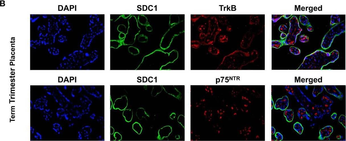
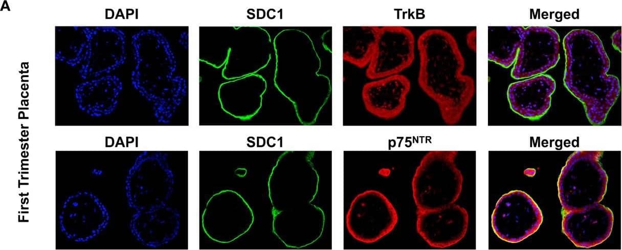
Detection of Human TrkB by Immunohistochemistry Expression of BDNF receptors in human placental tissues. (A) Representative confocal immunofluorescence staining images of showing DAPI (blue) and SDC1 (a syncytiotrophoblast biomarker, green) along with TrkB (red) and p75NTR (red) receptors in the first trimester placenta. (B) Confocal immunofluorescence staining images showing DAPI, SDC1, TrkB, and p75NTR in the term placenta. Image collected and cropped by CiteAb from the following open publication (https://pubmed.ncbi.nlm.nih.gov/34394001), licensed under a CC-BY license. Not internally tested by R&D Systems.
Reconstitution Calculator
Preparation and Storage
- 12 months from date of receipt, -20 to -70 °C as supplied.
- 1 month, 2 to 8 °C under sterile conditions after reconstitution.
- 6 months, -20 to -70 °C under sterile conditions after reconstitution.
Background: TrkB
The neurotrophins, including NGF, BDNF, NT-3, and NT-4/5 constitute a group of structurally related, secreted proteins that play an important role in the development and function of the nervous system. The biological activities of the neurotrophins are mediated by binding to the different members of the Trk family tyrosine kinase receptors. Three Trk family proteins, TrkA, TrkB, and TrkC, exhibiting different ligand specificities, have been identified. TrkA binds NGF, TrkB binds BDNF and NT‑4/5 and TrkC binds NT-3. All Trk family proteins share a conserved complex subdomain organization consisting of a signal peptide, two cysteine-rich domains, a cluster of three leucine-rich motifs, and two immunoglobulin-like domains in the extracellular region, as well as an intracellular region that contains the tyrosine kinase domain. Natural splice variants of the different Trks, including TrkB variants lacking the first cysteine-rich domain, the first and second or all three of the leucine-rich motifs, or the tyrosine kinase domain, have been described. The role of the different extracellular subdomains of TrkB in mediating neurotrophin binding and discrimination is currently being investigated. At the protein sequence level, human and rat TrkB are greater than 90% identical and the proteins exhibit cross-species activity. TrkB is primarily expressed in the nervous system. However, low levels of TrkB expression have also been observed in a wide variety of tissues (pancreas, kidneys, ovary) outside the nervous system.
- Ninkina, N. et al. (1997) J. Biol. Chem. 272:13019.
- Middlemas, D.S. et al. (1991) Mol. Cell Biol. 11:143.
- Soppet, D. et al. (1991) Cell 65:895.
Product Datasheets
Citations for Human/Mouse/Rat TrkB Antibody
R&D Systems personnel manually curate a database that contains references using R&D Systems products. The data collected includes not only links to publications in PubMed, but also provides information about sample types, species, and experimental conditions.
19
Citations: Showing 1 - 10
Filter your results:
Filter by:
-
Synaptic and nonsynaptic localization of protocadherin-gammaC5 in the rat brain.
Authors: Li Y, Serwanski DR, Miralles CP et al.
J Comp Neurol;518(17):3439-63.
-
BDNF inhibits neurodegenerative disease-associated asparaginyl endopeptidase activity via phosphorylation by AKT
Authors: Wang ZH, Wu W, Kang SS et al.
JCI Insight
-
TrkB-containing exosomes promote the transfer of glioblastoma aggressiveness to YKL-40-inactivated glioblastoma cells
Authors: Sandra Pinet, Barbara Bessette, Nicolas Vedrenne, Aurélie Lacroix, Laurence Richard, Marie-Odile Jauberteau et al.
Oncotarget
-
Long‐term NAD+ supplementation prevents the progression of age‐related hearing loss in mice
Authors: Mustafa N. Okur, Burcin Duan Sahbaz, Risako Kimura, Uri Manor, Jaimin Patel, Jae‐Hyeon Park et al.
Aging Cell
-
Akt Phosphorylates NQO1 and Triggers its Degradation, Abolishing its Antioxidative Activities in Parkinson's Disease
Authors: Luo S, Su Kang S, Wang ZH et al.
J. Neurosci.
-
Perfluoroalkyl Substance Exposure and the BDNF Pathway in the Placental Trophoblast
Authors: Melissa J. Marchese, Shuman Li, Bin Liu, Jun J. Zhang, Liping Feng
Front Endocrinol (Lausanne)
-
Cochlear ribbon synapse maturation requires Nlgn1 and Nlgn3
Authors: Ramirez M, Ninoyu Y, Miller C et al.
iScience
-
Dopaminergic neurons derived from human induced pluripotent stem cells survive and integrate into 6-OHDA-lesioned rats.
Authors: Cai J, Yang M, Poremsky E et al.
Stem Cells Dev
-
Defects in the CAPN1 Gene Result in Alterations in Cerebellar Development and Cerebellar Ataxia in Mice and Humans.
Authors: Wang Y, Hersheson J, Lopez D et al.
Cell Rep
-
A TrkB cleavage fragment in hippocampus promotes Depressive-Like behavior in mice
Authors: Wang, J;Yu, H;Li, X;Li, F;Chen, H;Zhang, X;Wang, Y;Xu, R;Gao, F;Wang, J;Liu, P;Shi, Y;Qin, D;Li, Y;Liu, S;Ding, S;Gao, XY;Wang, ZH;
Brain, behavior, and immunity
Species: Mouse
Sample Types: Tissue Homogenates
Applications: Western Blot -
A TrkB agonist prodrug prevents bone loss via inhibiting asparagine endopeptidase and increasing osteoprotegerin
Authors: J Xiong, J Liao, X Liu, Z Zhang, J Adams, R Pacifici, K Ye
Nature Communications, 2022-08-16;13(1):4820.
Species: Mouse
Sample Types: Cell Culture Supernates
Applications: Western Blot -
Deficiency in BDNF/TrkB Neurotrophic Activity Stimulates δ-Secretase by Upregulating C/EBP? in Alzheimer's Disease
Authors: ZH Wang, J Xiang, X Liu, SP Yu, FP Manfredsso, IM Sandoval, S Wu, JZ Wang, K Ye
Cell Rep, 2019-07-16;28(3):655-669.e5.
Species: Mouse, Rat
Sample Types: Cell Lysates, Tissue Homogenates
Applications: Western Blot -
Brain-derived neurotrophic factor (BDNF)-induced tropomyosin-related kinase B (Trk B) signaling is a potential therapeutic target for peritoneal carcinomatosis arising from colorectal cancer.
Authors: Tanaka, Koji, Okugawa, Yoshinag, Toiyama, Yuji, Inoue, Yasuhiro, Saigusa, Susumu, Kawamura, Mikio, Araki, Toshimit, Uchida, Keiichi, Mohri, Yasuhiko, Kusunoki, Masato
PLoS ONE, 2014-05-06;9(5):e96410.
Species: Human
Sample Types: Whole Tissue
Applications: IHC -
Brain-derived neurotrophic factor is increased in serum and skin levels of patients with chronic spontaneous urticaria.
Authors: Rossing K, Novak N, Mommert S, Pfab F, Gehring M, Wedi B, Kapp A, Raap U
Clin. Exp. Allergy, 2011-06-16;41(10):1392-1399.
Species: Human
Sample Types: Whole Tissue
Applications: IHC-P -
Modulation of neurotrophin and neurotrophin receptor expression in nasal mucosa after nasal allergen provocation in allergic rhinitis.
Authors: Raap U, Fokkens W, Bruder M, Hoogsteden H, Kapp A, Braunstahl GJ
Allergy, 2008-02-11;63(4):468-75.
Species: Human
Sample Types: Whole Cells
Applications: ICC -
IL-10 inhibits endothelium-dependent T cell costimulation by up-regulation of ILT3/4 in human vascular endothelial cells.
Authors: Gleissner CA, Zastrow A, Klingenberg R, Kluger MS, Konstandin M, Celik S, Haemmerling S, Shankar V, Giese T, Katus HA, Dengler TJ
Eur. J. Immunol., 2007-01-01;37(1):177-92.
Species: Human
Sample Types: Whole Cells
Applications: Flow Cytometry -
Tissue-Specificity of Antibodies Raised Against TrkB and p75NTR Receptors; Implications for Platelets as Models of Neurodegenerative Diseases
Authors: Fleury S, Boukhatem I, Le Blanc J et al.
Frontiers in Immunology
-
Effects of cerebrolysin on nerve growth factor system in the aging rat brain
Authors: Mikhail Stepanichev, Mikhail Onufriev, Viktor Aniol, Sofia Freiman, Hemma Brandstaetter, Stefan Winter et al.
Restorative Neurology and Neuroscience
-
Differential expression and the anti-apoptotic effect of human placental neurotrophins and their receptors
Authors: K. Fujita, K. Tatsumi, E. Kondoh, Y. Chigusa, H. Mogami, T. Fujii et al.
Placenta
FAQs
No product specific FAQs exist for this product, however you may
View all Antibody FAQsReviews for Human/Mouse/Rat TrkB Antibody
Average Rating: 5 (Based on 1 Review)
Have you used Human/Mouse/Rat TrkB Antibody?
Submit a review and receive an Amazon gift card.
$25/€18/£15/$25CAN/¥75 Yuan/¥2500 Yen for a review with an image
$10/€7/£6/$10 CAD/¥70 Yuan/¥1110 Yen for a review without an image
Filter by:


