Human/Mouse Bcl-2 Antibody Summary
Applications
Please Note: Optimal dilutions should be determined by each laboratory for each application. General Protocols are available in the Technical Information section on our website.
Scientific Data
 View Larger
Detection of Human/Mouse Bcl‑2 by Western Blot. Western blot shows lysates of KG-1 human myeloid leukemia cell line and M-NFS-60 mouse myelogenous leukemia lymphoblast cell line. PVDF membrane was probed with 1 µg/mL of Goat Anti-Human/Mouse Bcl-2 Antigen Affinity-purified Polyclonal Antibody (Catalog # AF810) followed by HRP-conjugated Anti-Goat IgG Secondary Antibody (Catalog # HAF017). A specific band was detected for Bcl-2, at approximately 25 kDa (as indicated). This experiment was conducted under reducing conditions and using Immunoblot Buffer Group 2.
 View Larger
Bcl-2 in Human Breast Cancer Tissue. Bcl‑2 was detected in immersion fixed paraffin-embedded sections of human breast cancer tissue using 15 µg/mL Goat Anti-Human/ Mouse Bcl‑2 Antigen Affinity-purified Polyclonal Antibody (Catalog # AF810) overnight at 4 °C. Tissue was stained (red) and counterstained with hematoxylin (blue). View our protocol for Chromogenic IHC Staining of Paraffin-embedded Tissue Sections.
 View Larger
Immunoprecipitation of Mouse Bcl‑2. Bcl‑2 was immunoprecipitated from lysates (3 x 10⁶cells) of M‑NFS‑60 mouse myelogenous leukemia lymphoblast cell line following incubation with 3 µg Goat Anti-Human/Mouse Bcl‑2 Antigen Affinity-purified Polyclonal Antibody (Catalog # AF810) for 1 hour at 4 °C. Bcl‑2-antibody complexes were absorbed using Protein G (Sigma). Immunoprecipitated Bcl‑2 was detected by Western blot using 1 µg/mL Goat Anti-Human/Mouse Bcl‑2 Antigen Affinity-purified Polyclonal Antibody (Catalog # AF810). View our recommended buffer recipes for immunoprecipitation.
 View Larger
Detection of Human Bcl‑2 by Simple WesternTM. Simple Western lane view shows lysates of KG-1 human acute myelogenous leukemia cell line, loaded at 0.2 mg/mL. A specific band was detected for Bcl-2 at approximately 33 kDa (as indicated) using 50 µg/mL of Goat Anti-Human/Mouse Bcl-2 Antigen Affinity-purified Polyclonal Antibody (Catalog # AF810) followed by 1:50 dilution of HRP-conjugated Anti-Goat IgG Secondary Antibody (Catalog # HAF109). This experiment was conducted under reducing conditions and using the 12-230 kDa separation system.
Reconstitution Calculator
Preparation and Storage
- 12 months from date of receipt, -20 to -70 °C as supplied.
- 1 month, 2 to 8 °C under sterile conditions after reconstitution.
- 6 months, -20 to -70 °C under sterile conditions after reconstitution.
Background: Bcl-2
Bcl-2 is a member of a family of proteins that regulates outer mitochondrial membrane permeability (1, 2). Bcl-2 is an anti-apoptotic member that prevents release of cytochrome c from the mitochondria intermembrane space into the cytosol. Bcl-2 is present on the outer mitochondrial membrane and is also found on other membranes in some cell types. Natural Bcl-2 contains a carboxyl-terminal mitochondria targeting sequence. Recombinant Bcl-2, missing the mitochondrial targeting sequence, maintains its ability to neutralize pro-apoptotic Bcl-2 family members. Neutralization by Bcl-2 appears to be through binding the BH3 region of pro-apoptotic Bcl-2 family members. This activity does not require the mitochondrial targeting sequence.
Product Datasheets
Citations for Human/Mouse Bcl-2 Antibody
R&D Systems personnel manually curate a database that contains references using R&D Systems products. The data collected includes not only links to publications in PubMed, but also provides information about sample types, species, and experimental conditions.
14
Citations: Showing 1 - 10
Filter your results:
Filter by:
-
Interleukin‑22 regulates gastric cancer cell proliferation through regulation of the JNK signaling pathway
Authors: Hao Dong, Fengming Zhu, Shilu Jin, Jing Tian
Experimental and Therapeutic Medicine
-
Targeting Cell Death Mechanism Specifically in Triple Negative Breast Cancer Cell Lines
Authors: Lavinia-Lorena Pruteanu, Cornelia Braicu, Dezső Módos, Maria-Ancuţa Jurj, Lajos-Zsolt Raduly, Oana Zănoagă et al.
International Journal of Molecular Sciences
-
Targeting of H19/cell adhesion molecules circuitry by GSK-J4 epidrug inhibits metastatic progression in prostate cancer
Authors: Pecci, V;Troisi, F;Aiello, A;De Martino, S;Carlino, A;Fiorentino, V;Ripoli, C;Rotili, D;Pierconti, F;Martini, M;Porru, M;Pinto, F;Mai, A;Bassi, PF;Grassi, C;Gaetano, C;Pontecorvi, A;Strigari, L;Farsetti, A;Nanni, S;
Cancer cell international
-
Knockdown of lncRNA CCAT1 Inhibits the Progression of Colorectal Cancer via hsa-miR-4679 Mediating the Downregulation of GNG10
Authors: N Wang, J Li, J He, YG Jing, WD Zhao, WJ Yu, J Wang
Journal of Immunology Research, 2021-12-30;2021(0):8930813.
Species: Human
Sample Types: Tissue Homogenates
Applications: Western Blot -
Lipid storage droplet protein 5 reduces sodium palmitate?induced lipotoxicity in human normal liver cells by regulating lipid metabolism?related factors
Authors: X Ma, F Cheng, K Yuan, K Jiang, T Zhu
Mol Med Rep, 2019-06-06;20(2):879-886.
Species: Human
Sample Types: Cell Lysates
Applications: Western Blot -
B-cell lymphoma 2 is associated with advanced tumor grade and clinical stage, and reduced overall survival in young Chinese patients with colorectal carcinoma
Authors: J Wang, G He, Q Yang, L Bai, B Jian, Q Li, Z Li
Oncol Lett, 2018-04-13;15(6):9009-9016.
Species: Human
Sample Types: Tissue Homogenates, Whole Tissue
Applications: IHC-P, Western Blot -
TCDD promotes lung tumors via attenuation of apoptosis through activation of the Akt and ERK1/2 signaling pathways.
Authors: Chen R, Siao S, Hsu C, Chang C, Chang L, Wu C, Lin P, Wang Y
PLoS ONE, 2014-06-13;9(6):e99586.
Species: Mouse
Sample Types: Whole Tissue
Applications: Bioassay -
Molecular profiling of cervical cancer progression.
Authors: Hagemann T, Bozanovic T, Hooper S, Ljubic A, Slettenaar VI, Wilson JL, Singh N, Gayther SA, Shepherd JH, Van Trappen PO
Br. J. Cancer, 2007-01-29;96(2):321-8.
Species: Human
Sample Types: Whole Tissue
Applications: IHC-P -
The anti-apoptotic effect of Notch-1 requires p56lck-dependent, Akt/PKB-mediated signaling in T cells.
Authors: Sade H, Krishna S, Sarin A
J. Biol. Chem., 2003-10-28;279(4):2937-44.
Species: Human, Mouse
Sample Types: Cell Lysates
Applications: Western Blot -
TGF‑ beta 1 mediates the effects of aspirin on colonic tumor cell proliferation and apoptosis
Authors: Yuyi Wang, Chi Du, Nan Zhang, Mei Li, Yanyang Liu, Maoyuan Zhao et al.
Oncology Letters
-
Securidaca–saponins are natural inhibitors of AKT, MCL-1, and BCL2L1 in cervical cancer cells
Authors: Titus Chukwuemeka Obasi, Cornelia Braicu, Bogdan Cezar Iacob, Ede Bodoki, Ancuta Jurj, Lajos Raduly et al.
Cancer Management and Research
-
Genipin protects against H2O2-induced oxidative damage in�retinal pigment epithelial cells by promoting Nrf2 signaling
Authors: Hailan Zhao, Ruiqing Wang, Mingxia Ye, Lan Zhang
International Journal of Molecular Medicine
-
Influence mechanism of miRNA‑144 on proliferation and apoptosis of osteosarcoma cells
Authors: Xu Zhang, Zhengwei Li, Wei Ji, Xilong Chen, Qiang Gao, Dajin Li et al.
Oncology Letters
-
Protein Synthesis Inhibition Activity of Mesothelin Targeting Immunotoxin LMB-100 Decreases Concentrations of Oncogenic Signaling Molecules and Secreted Growth Factors
Authors: Salma El-Behaedi, Rebekah Landsman, Michael Rudloff, Emily Kolyvas, Rakan Albalawy, Xianyu Zhang et al.
Toxins (Basel)
FAQs
No product specific FAQs exist for this product, however you may
View all Antibody FAQsReviews for Human/Mouse Bcl-2 Antibody
Average Rating: 5 (Based on 2 Reviews)
Have you used Human/Mouse Bcl-2 Antibody?
Submit a review and receive an Amazon gift card.
$25/€18/£15/$25CAN/¥75 Yuan/¥2500 Yen for a review with an image
$10/€7/£6/$10 CAD/¥70 Yuan/¥1110 Yen for a review without an image
Filter by:
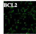
The conjugation of R&D Systems' Bcl-2 antibody for immunofluorescence has been pivotal in advancing our research on apoptosis regulation. Bcl-2 protein's role in cell survival necessitates accurate detection, making the choice of antibody and conjugation method critical.
We selected R&D Systems' antibody for its established reliability and effectiveness in previous studies. Conjugating the antibody to fluorescent dyes enabled precise localization and visualization of Bcl-2 expression in various cellular contexts. The conjugation process was straightforward, and the resulting fluorescence signals were robust and specific, providing clear insights into protein distribution and dynamics within cells.
Validation involved using appropriate controls and optimizing staining protocols to maximize signal-to-noise ratios. The antibody consistently demonstrated high specificity, effectively distinguishing Bcl-2 from non-specific background fluorescence.
In conclusion, R&D Systems' Bcl-2 antibody conjugated for immunofluorescence has been instrumental in our research, offering reliable and detailed visualization of apoptotic mechanisms. Its performance in conjugation and subsequent imaging has enhanced our understanding of Bcl-2's cellular functions, making it a valuable tool for both basic research and potential therapeutic applications


