Human Laminin alpha 3/Laminin-5 Antibody Summary
aa 21-1713
Accession # NP_000218
Applications
Please Note: Optimal dilutions should be determined by each laboratory for each application. General Protocols are available in the Technical Information section on our website.
Scientific Data
 View Larger
Detection of Human Laminin alpha 3/Laminin‑5 by Western Blot. Western blot shows lysates of human lung tissue. PVDF Membrane was probed with 2 µg/mL of Mouse Anti-Human Laminin a3/ Laminin-5 Monoclonal Antibody (Catalog # MAB21441) followed by HRP-conjugated Anti-Mouse IgG Secondary Antibody (Catalog # HAF007). A specific band was detected for Laminin a3/Laminin-5 at approximately 190 kDa (as indicated). This experiment was conducted under non-reducing conditions and using Immunoblot Buffer Group 1.
 View Larger
Laminin alpha 3/Laminin‑5 in NHEK Human Cell Line. Laminin a3/Laminin-5 was detected in immersion fixed NHEK human normal epidermal keratinocytes treated with 10ng/mL Recombinant Human TGF-beta 1 (Catalog # 240-B) for 24 hours using Mouse Anti-Human Laminin a3/Laminin-5 Monoclonal Antibody (Catalog # MAB21441) at 10 µg/mL for 3 hours at room temperature. Cells were stained using the NorthernLights™ 557-conjugated Anti-Mouse IgG Secondary Antibody (red; Catalog # NL007) and counterstained with DAPI (blue). Specific staining was localized to the perinuclear space. View our protocol for Fluorescent ICC Staining of Cells on Coverslips.
 View Larger
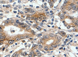
Laminin alpha 3/Laminin‑5 in Human Stomach. Laminin a3/Laminin-5 was detected in immersion fixed paraffin-embedded sections of human stomach using Mouse Anti-Human Laminin a3/Laminin-5 Monoclonal Antibody (Catalog # MAB21441) at 5 µg/mL for 1 hour at room temperature followed by incubation with the Anti-Mouse IgG VisUCyte™ HRP Polymer Antibody (Catalog # VC001). Before incubation with the primary antibody, tissue was subjected to heat-induced epitope retrieval using Antigen Retrieval Reagent-Basic (Catalog # CTS013). Tissue was stained using DAB (brown) and counterstained with hematoxylin (blue). Specific staining was localized to cell surface of epithelial cells in gastric glands. View our protocol for IHC Staining with VisUCyte HRP Polymer Detection Reagents.
 View Larger
Detection of Laminin alpha 3/Laminin-5 in U2OS Human Cell Line by Flow Cytometry. U2OS human osteosarcoma cell line was stained with Mouse Anti-Human Laminin a3/Laminin-5 Monoclonal Antibody (Catalog # MAB21441, filled histogram) or isotype control antibody (Catalog # MAB002, open histogram), followed by Allophycocyanin-conjugated Anti-Mouse IgG Secondary Antibody (Catalog # F0101B). To facilitate intracellular staining, cells were fixed with Flow Cytometry Fixation Buffer (Catalog # FC004) and permeabilized with Flow Cytometry Permeabilization/Wash Buffer I (Catalog # FC005).View our protocol for Staining Intracellular Molecules.
Reconstitution Calculator
Preparation and Storage
- 12 months from date of receipt, -20 to -70 °C as supplied.
- 1 month, 2 to 8 °C under sterile conditions after reconstitution.
- 6 months, -20 to -70 °C under sterile conditions after reconstitution.
Background: Laminin alpha 3/Laminin-5
Laminins are heterotrimeric, noncollagenous glycoproteins composed of alpha, beta, and gamma chains. Through interactions with integrins, dystroglycan and other receptors, laminins contribute to cell differentiation, cell shape and migration, and maintenance of tissue phenotypes and survival. Laminin alpha 3/Laminin-5, also known as epiligrin, includes alpha 3, beta 3, and gamma 2 subunits. It is abundant in transitional epithelium, stratified squamous epithelia, lung mucosa and other epithelial glands and contributes to initiation and maintenance of epithelial cell anchorage to the underlying connective tissue. Within aa 21‑1713 of the alpha 3 subunit, human and mouse share 77% amino acid sequence identity.
Product Datasheets
Citations for Human Laminin alpha 3/Laminin-5 Antibody
R&D Systems personnel manually curate a database that contains references using R&D Systems products. The data collected includes not only links to publications in PubMed, but also provides information about sample types, species, and experimental conditions.
4
Citations: Showing 1 - 4
Filter your results:
Filter by:
-
Paired nicking-mediated COL17A1 reframing for junctional epidermolysis bullosa
Authors: Johannes Bischof, Oliver Patrick March, Bernadette Liemberger, Simone Alexandra Haas, Stefan Hainzl, Igor Petković et al.
Molecular Therapy
-
&alpha2&beta1 integrins spatially restrict Cdc42 activity to stabilise adherens junctions
Authors: JD Howden, M Michael, W Hight-Warb, M Parsons
Bmc Biology, 2021-06-23;19(1):130.
-
5'RNA Trans-Splicing Repair of COL7A1 Mutant Transcripts in Epidermolysis Bullosa
Authors: E Mayr, M Ablinger, T Lettner, EM Murauer, C Guttmann-G, J Piñón Hofb, S Hainzl, M Kaiser, A Klausegger, JW Bauer, U Koller, V Wally
International Journal of Molecular Sciences, 2022-02-02;23(3):.
Species: Human
Sample Types: Whole Tissue
Applications: IHC/IF -
Laminin signals initiate the reciprocal loop that informs breast-specific gene expression and homeostasis by activating NO, p53 and microRNAs
Authors: S Furuta, G Ren, JH Mao, MJ Bissell
Elife, 2018-03-21;7(0):.
Species: Human
Sample Types: Whole Tissue
Applications: IHC-P
FAQs
No product specific FAQs exist for this product, however you may
View all Antibody FAQsReviews for Human Laminin alpha 3/Laminin-5 Antibody
Average Rating: 5 (Based on 3 Reviews)
Have you used Human Laminin alpha 3/Laminin-5 Antibody?
Submit a review and receive an Amazon gift card.
$25/€18/£15/$25CAN/¥75 Yuan/¥2500 Yen for a review with an image
$10/€7/£6/$10 CAD/¥70 Yuan/¥1110 Yen for a review without an image
Filter by:
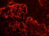
Immunofluorescent staining of frozen human skin.


