Human IRF3 Antibody Summary
Val206-Ser427
Accession # Q14653
Applications
Please Note: Optimal dilutions should be determined by each laboratory for each application. General Protocols are available in the Technical Information section on our website.
Scientific Data
 View Larger
Detection of IRF3 in Daudi Human Cell Line by Flow Cytometry. Daudi human Burkitt's lymphoma cell line was stained with Goat Anti-Human IRF3 Antigen Affinity-purified Polyclonal Antibody (Catalog # AF4019, filled histogram) or isotype control antibody (Catalog # AB-108-C, open histogram), followed by Phycoerythrin-conjugated Anti-Goat IgG Secondary Antibody (Catalog # F0107). To facilitate intracellular staining, cells were fixed with paraformaldehyde and permeabilized with saponin.
 View Larger
Detection of Human IRF3 by Western Blot. Western blot shows lysates of HeLa human cervical epithelial carcinoma cell line, U937 human histiocytic lymphoma cell line, Raji human Burkitt's lymphoma cell line, and Daudi human Burkitt's lymphoma cell line. PVDF membrane was probed with 1 µg/mL of Goat Anti-Human IRF3 Antigen Affinity-purified Polyclonal Antibody (Catalog # AF4019) followed by HRP-conjugated Anti-Goat IgG Secondary Antibody (Catalog # HAF019). A specific band was detected for IRF3 at approximately 50 kDa (as indicated). This experiment was conducted under reducing conditions and using Immunoblot Buffer Group 1.
 View Larger
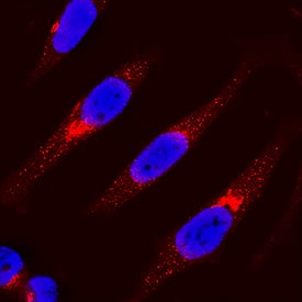
IRF3 in HeLa Human Cell Line. IRF3 was detected in immersion fixed HeLa human cervical epithelial carcinoma cell line using Goat Anti-Human IRF3 Antigen Affinity-purified Polyclonal Antibody (Catalog # AF4019) at 15 µg/mL for 3 hours at room temperature. Cells were stained using the NorthernLights™ 557-conjugated Anti-Goat IgG Secondary Antibody (red; Catalog # NL001) and counterstained with DAPI (blue). Specific staining was localized to cytoplasm. View our protocol for Fluorescent ICC Staining of Cells on Coverslips.
 View Larger
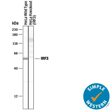
Detection of Human IRF3 by Simple WesternTM. Simple Western lane view shows lysates of HeLa human cervical epithelial carcinoma parental cell line and IRF3 knockout HeLa cell line (KO), loaded at 0.2 mg/mL. A specific band was detected for IRF3 at approximately 57 kDa (as indicated) in the parental HeLa cell line, but is not detectable in knockout HeLa cell line. Goat Anti-Human IRF3 Antigen Affinity-purified Polyclonal Antibody (Catalog # AF4019) was used at 10 µg/mL followed by 1:50 dilution of HRP-conjugated Anti-Goat IgG Secondary Antibody (Catalog # HAF019). This experiment was conducted under reducing conditions and using the 12-230 kDa separation system.
 View Larger
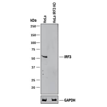
Western Blot Shows Human IRF3 Specificity by Using Knockout Cell Line. Western blot shows lysates of HeLa human cervical epithelial carcinoma parental cell line and IRF3 knockout HeLa cell line (KO). PVDF membrane was probed with 1 µg/mL of Goat Anti-Human IRF3 Antigen Affinity-purified Polyclonal Antibody (Catalog # AF4019) followed by HRP-conjugated Anti-Goat IgG Secondary Antibody (Catalog # HAF017). A specific band was detected for IRF3 at approximately 50 kDa (as indicated) in the parental HeLa cell line, but is not detectable in knockout HeLa cell line. GAPDH (Catalog # AF5718) is shown as a loading control. This experiment was conducted under reducing conditions and using Immunoblot Buffer Group 1.
Reconstitution Calculator
Preparation and Storage
- 12 months from date of receipt, -20 to -70 °C as supplied.
- 1 month, 2 to 8 °C under sterile conditions after reconstitution.
- 6 months, -20 to -70 °C under sterile conditions after reconstitution.
Background: IRF3
Interferon response factor 3 (IRF3) is a 50 kDa, 427 amino acid, member of the IRF family of proteins. Human IRF3 is a transcription factor that binds the interferon-sensitive response element (ISRE) and is activated by Toll-like receptors, TLR3 and TLR4. Viral entry or engagement of TLR3 or TLR4 stimulates IRF3 phosphorylation, nuclear translocation and stimulation of IFN production. There are potential alternate splice forms affecting IRF3 structure, one including a deletion of aa 201‑327, a second with the same deletion and an alternate Met(147) start site, and a third with a 125 aa substitution of the C-terminal 100 aa. Over the region used as immunogen, aa 206‑427, human IRF3 is 76% and 83% identical to the corresponding mouse and porcine proteins, respectively.
Product Datasheets
FAQs
No product specific FAQs exist for this product, however you may
View all Antibody FAQsReviews for Human IRF3 Antibody
There are currently no reviews for this product. Be the first to review Human IRF3 Antibody and earn rewards!
Have you used Human IRF3 Antibody?
Submit a review and receive an Amazon gift card.
$25/€18/£15/$25CAN/¥75 Yuan/¥2500 Yen for a review with an image
$10/€7/£6/$10 CAD/¥70 Yuan/¥1110 Yen for a review without an image


