Human ING1 Antibody Summary
*Small pack size (-SP) is supplied either lyophilized or as a 0.2 µm filtered solution in PBS.
Applications
Please Note: Optimal dilutions should be determined by each laboratory for each application. General Protocols are available in the Technical Information section on our website.
Scientific Data
 View Larger
Detection of Human ING1 by Western Blot. Western blot shows lysates of A549 human lung carcinoma cell line. PVDF membrane was probed with 1 µg/mL of Goat Anti-Human ING1 Antigen Affinity-purified Polyclonal Antibody (Catalog # AF5758) followed by HRP-conjugated Anti-Goat IgG Secondary Antibody (Catalog # HAF019). A specific band was detected for ING1 at approximately 28 kDa (as indicated). This experiment was conducted under reducing conditions and using Immunoblot Buffer Group 8.
.") View Larger
View Larger
ING1 in HeLa Human Cell Line. ING1 was detected in immersion fixed HeLa human cervical epithelial carcinoma cell line using 10 µg/mL Goat Anti-Human ING1 Antigen Affinity-purified Polyclonal Antibody (Catalog # AF5758) for 3 hours at room temperature. Cells were stained with the NorthernLights™ 557-conjugated Anti-Goat IgG Secondary Antibody (red, upper panel; Catalog # NL001) and counterstained with DAPI (blue, lower panel). View our protocol for Chromogenic IHC Staining of Paraffin-embedded Tissue Sections.
 View Larger
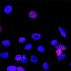
ING1 in A431 Human Cell Line. ING1 was detected in immersion fixed A431 human epithelial carcinoma cell line using Goat Anti-Human ING1 Antigen Affinity-purified Polyclonal Antibody (Catalog # AF5758) at 5 µg/mL for 3 hours at room temperature. Cells were stained using the NorthernLights™ 557-conjugated Anti-Goat IgG Secondary Antibody (red; NL001) and counterstained with DAPI (blue). Specific staining was localized to cell nuclei. Staining was performed using our protocol for Fluorescent ICC Staining of Non-adherent Cells.
.") View Larger
View Larger
ING1 in Human Lung Cancer Tissue. ING1 was detected in immersion fixed paraffin-embedded sections of human lung cancer tissue using 5 µg/mL Goat Anti-Human ING1 Antigen Affinity-purified Polyclonal Antibody (Catalog # AF5758) overnight at 4 °C. Tissue was stained with the Anti-Goat HRP-DAB Cell & Tissue Staining Kit (brown; Catalog # CTS008) and counterstained with hematoxylin (blue). View our protocol for Fluorescent ICC Staining of Cells on Coverslips.
Reconstitution Calculator
Preparation and Storage
Background: ING1
ING1 (Inhibitor of Growth 1; also p33ING1b and ING1A) is a 32 kDa member of the ING family of tumor suppressors. It is very widely expressed, and participates in multiple cell functions. When bound to p53, ING1 inhibits cell growth; when complexed to GADD45, ING1 enhances DNA nucleotide excision repair; and when associated with HAT, ING1 promotes hyperacetylation of histones H3 and H4. Human ING1 is 279 amino acids (aa) in length. It contains a PIP-PBD region (aa 5‑45), an NLS (aa 142‑158), and a plant homeodomain (PHD) (aa 210‑259). Phosphorylation occurs on Ser126 and Ser199. There are at least three isoform variants. p47ING1a contains a 190 aa substitution for aa 1‑46, p27ING1d shows a deletion of aa 2‑45, and p24ING1c has an alternate start site at Met70. Over aa 213‑327, human ING1 shares 82% aa identity with mouse ING1.
Product Datasheets
FAQs
No product specific FAQs exist for this product, however you may
View all Antibody FAQsReviews for Human ING1 Antibody
There are currently no reviews for this product. Be the first to review Human ING1 Antibody and earn rewards!
Have you used Human ING1 Antibody?
Submit a review and receive an Amazon gift card.
$25/€18/£15/$25CAN/¥75 Yuan/¥1250 Yen for a review with an image
$10/€7/£6/$10 CAD/¥70 Yuan/¥1110 Yen for a review without an image

