Human IL-17E/IL-25 Antibody Summary
Tyr33-Gly177
Accession # Q9H293
Applications
This antibody functions as an ELISA capture antibody when paired with Goat Anti-Human IL‑17E/IL‑25 Antigen Affinity-purified Polyclonal Antibody (Catalog # AF1258).
This product is intended for assay development on various assay platforms requiring antibody pairs.
Please Note: Optimal dilutions should be determined by each laboratory for each application. General Protocols are available in the Technical Information section on our website.
Scientific Data
 View Larger
Detection of IL‑17E/IL‑25 in PC‑3 Human Cell Line by Flow Cytometry. PC‑3 human prostate cancer cell line was stained with Human IL‑17E/IL‑25 Monoclonal Antibody (Catalog # MAB1258, filled histogram) or isotype control antibody (Catalog # MAB002, open histogram), followed by Allophycocyanin-conjugated Anti-Mouse IgG F(ab')2Secondary Antibody (Catalog # F0101B). To facilitate intracellular staining, cells were fixed with paraformaldehyde and permeabilized with saponin.
 View Larger
Detection of Human IL-17E/IL-25 by Immunocytochemistry/Immunofluorescence IL-25 IR in neurons, astrocytes and microglia. IFC reveals IL-25 IR (in green) co-localizing with three CNS cell type markers (red), (A) Iba1, a microglial marker, (B) GFAP, an astrocytic marker and (C) NeuN, a neuronal marker. Panel A,B: CA4; Panel C: dentate gyrus. Note that IL-25 IR is also present in the astrocytic endfeet surrounding the bloodvessels (B). The insets are higher power magnifications taken from a representative area, with the added nuclear marker DAPI (blue). Scale bar = 40 μm Image collected and cropped by CiteAb from the following publication (https://pubmed.ncbi.nlm.nih.gov/22935090), licensed under a CC-BY license. Not internally tested by R&D Systems.
 View Larger
Detection of Human IL-17E/IL-25 by Immunocytochemistry/Immunofluorescence Hippocampal expression patterns of IL-25 and CCL4 in Control, mTLE-HS and mTLE + HS patients. Photomicrographs showing typical examples of Il-25 and CCL4 staining in the hippocampal CA1 region (A) and the DG/CA4 region (B). IL-25 immunoreactivity (IR) is evident in cells with neuronal morphology in all three patient groups (A, B); note the increased IL-25 staining in the mTLE + HS hippocampus in small cells. CCL4 IR is low in controls (A, B). Increased CCL4 IR is detected in both mTLE patient groups in the DG-CA4 area (B) and in the CA1 area of mTLE + HS patients (A). Scale bar = 200 μm.The insets are higher power magnifications taken from the same anatomical area Image collected and cropped by CiteAb from the following publication (https://pubmed.ncbi.nlm.nih.gov/22935090), licensed under a CC-BY license. Not internally tested by R&D Systems.
 View Larger
Detection of Human IL-17E/IL-25 by Immunocytochemistry/Immunofluorescence IL-25 IR in neurons, astrocytes and microglia. IFC reveals IL-25 IR (in green) co-localizing with three CNS cell type markers (red), (A) Iba1, a microglial marker, (B) GFAP, an astrocytic marker and (C) NeuN, a neuronal marker. Panel A,B: CA4; Panel C: dentate gyrus. Note that IL-25 IR is also present in the astrocytic endfeet surrounding the bloodvessels (B). The insets are higher power magnifications taken from a representative area, with the added nuclear marker DAPI (blue). Scale bar = 40 μm Image collected and cropped by CiteAb from the following publication (https://pubmed.ncbi.nlm.nih.gov/22935090), licensed under a CC-BY license. Not internally tested by R&D Systems.
 View Larger
Detection of Human IL-17E/IL-25 by Immunocytochemistry/Immunofluorescence IL-25 IR in neurons, astrocytes and microglia. IFC reveals IL-25 IR (in green) co-localizing with three CNS cell type markers (red), (A) Iba1, a microglial marker, (B) GFAP, an astrocytic marker and (C) NeuN, a neuronal marker. Panel A,B: CA4; Panel C: dentate gyrus. Note that IL-25 IR is also present in the astrocytic endfeet surrounding the bloodvessels (B). The insets are higher power magnifications taken from a representative area, with the added nuclear marker DAPI (blue). Scale bar = 40 μm Image collected and cropped by CiteAb from the following publication (https://pubmed.ncbi.nlm.nih.gov/22935090), licensed under a CC-BY license. Not internally tested by R&D Systems.
Reconstitution Calculator
Preparation and Storage
- 12 months from date of receipt, -20 to -70 °C as supplied.
- 1 month, 2 to 8 °C under sterile conditions after reconstitution.
- 6 months, -20 to -70 °C under sterile conditions after reconstitution.
Background: IL-17E/IL-25
The Interleukin 17 (IL-17) family proteins, comprising six members (IL-17, and IL-17B through IL-17F), are secreted, structurally related proteins that share a conserved cysteine-knot fold near the C-terminus, but have considerable sequence divergence at the N-terminus. With the exception of IL-17B, which exists as a non-covalently linked dimer, all IL-17 family members are disulfide-linked dimers. IL-17 family proteins are pro-inflammatory cytokines that induce local cytokine production and are involved in the regulation of immune functions (1, 2).
Human IL-17E cDNA encodes a 177 amino acid (aa) residues precursor protein with a putative 32 aa signal peptide (3). A second isoform of human IL-17E encoding a 161 aa precursor protein also exists (4). The two isoforms differ in their signal peptide sequences. Mature human IL-17E shares 76% aa sequence identity with mature mouse IL-17E. Human IL-17E also shares from 25-36% aa sequence identity with the other human IL-17 family members. IL-17E expression was detected at very low levels by PCR in various peripheral tissues including brain, kidney, lung, prostate, testis, adrenal gland, spinal cord, and trachea (3). IL-17E binds and activates IL-17 B Receptor (IL-17B R) (alternatively known as IL-17 Rh1, IL-17E R, and EVI27) (3), which is expressed in kidney and liver, and at lower levels in brain, testis, and other endocrine tissues. The expression of IL-17B R is up regulated under inflammatory conditions. Ligation of IL-17E to IL-17 RB induces activation of nuclear factor kappa-B and stimulates the production of the pro-inflamatory cytokine IL-8 (3). IL-17 has also been found to promote the expression of the prototypical Th2 genes (4, 5).
- Aggarwal, S. and A.L. Gurney (2002) J. Leukoc. Biol. 71:1.
- Moseley, T.A. et al. (2003) Cytokine & Growth Factor Rev. 14:155.
- Lee, J. et al. (2001) J. Biol. Chem. 276:1660.
- Hurst, S.D. et al. (2002) J. Immunol. 169:443.
- Pan, G. et al. (2001) J. Immunol. 167:6569.
Product Datasheets
Citations for Human IL-17E/IL-25 Antibody
R&D Systems personnel manually curate a database that contains references using R&D Systems products. The data collected includes not only links to publications in PubMed, but also provides information about sample types, species, and experimental conditions.
6
Citations: Showing 1 - 6
Filter your results:
Filter by:
-
IL-25 improves diabetic wound healing through stimulating M2 macrophage polarization and fibroblast activation
Authors: S Li, X Ding, H Zhang, Y Ding, Q Tan
International immunopharmacology, 2022-02-08;106(0):108605.
Species: Mouse
Sample Types: Protein
Applications: Western Blot -
IL-25 or IL-17E Protects against High-Fat Diet-Induced Hepatic Steatosis in Mice Dependent upon IL-13 Activation of STAT6.
Authors: Wang A, Yang Z, Grinchuk V, Smith A, Qin B, Lu N, Wang D, Wang H, Ramalingam T, Wynn T, Urban J, Shea-Donohue T, Zhao A
J Immunol, 2015-09-30;195(10):4771-80.
Species: Human
Sample Types: Whole Tissue
Applications: IHC-P -
Protein expression profiling of inflammatory mediators in human temporal lobe epilepsy reveals co-activation of multiple chemokines and cytokines.
Authors: Kan A, De Jager W, de Wit M, Heijnen C, van Zuiden M, Ferrier C, van Rijen P, Gosselaar P, Hessel E, van Nieuwenhuizen O, de Graan P
J Neuroinflammation, 2012-08-30;9(0):207.
Species: Human
Sample Types: Whole Tissue
Applications: IHC-P -
T-helper cell type 2 (Th2) memory T cell-potentiating cytokine IL-25 has the potential to promote angiogenesis in asthma.
Authors: Corrigan CJ, Wang W, Meng Q, Fang C, Wu H, Reay V, Lv Z, Fan Y, An Y, Wang YH, Liu YJ, Lee TH, Ying S
Proc. Natl. Acad. Sci. U.S.A., 2011-01-04;108(4):1579-84.
Species: Human
Sample Types: Whole Cells
Applications: ICC -
IL-25 in atopic dermatitis: a possible link between inflammation and skin barrier dysfunction?
Authors: Hvid M, Vestergaard C, Kemp K
J. Invest. Dermatol., 2010-09-23;131(1):150-7.
Species: Human
Sample Types: Cell Lysates, Whole Tissue
Applications: IHC-P, Western Blot -
Interleukin-17E, inducible nitric oxide synthase and arginase1 as new biomarkers in the identification of neutrophilic dermatoses
Authors: R. Stalder, N. Brembilla, C. Conrad, N. Yawalkar, A. Navarini, W H Boehncke et al.
Clin Exp Dermatol
FAQs
No product specific FAQs exist for this product, however you may
View all Antibody FAQsReviews for Human IL-17E/IL-25 Antibody
Average Rating: 5 (Based on 1 Review)
Have you used Human IL-17E/IL-25 Antibody?
Submit a review and receive an Amazon gift card.
$25/€18/£15/$25CAN/¥75 Yuan/¥1250 Yen for a review with an image
$10/€7/£6/$10 CAD/¥70 Yuan/¥1110 Yen for a review without an image
Filter by:
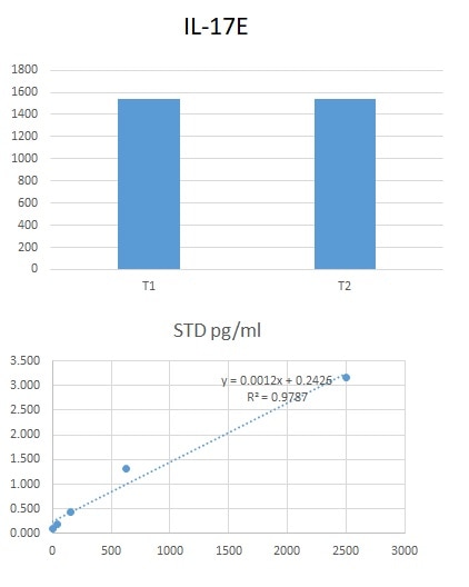
We used this antibody in an in-house ELISA along with pAb (AF1258) and protein (1258-IL-025) to quantify IL-17E in human serum and plasma. This combination detected IL-17E in our samples efficiently.


