Human IL-16 C-terminal Peptide Antibody Summary
*Small pack size (-SP) is supplied either lyophilized or as a 0.2 µm filtered solution in PBS.
Applications
Please Note: Optimal dilutions should be determined by each laboratory for each application. General Protocols are available in the Technical Information section on our website.
Scientific Data
 View Larger
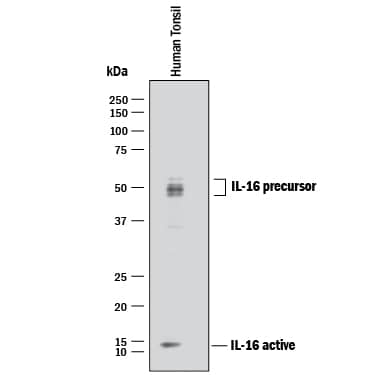
Detection of Human IL‑16 by Western Blot. Western blot shows lysate of human tonsil tissue. PVDF membrane was probed with 1 µg/mL of Goat Anti-Human IL-16 C-terminal Peptide Antigen Affinity-purified Polyclonal Antibody (Catalog # AF-316-PB) followed by HRP-conjugated Anti-Goat IgG Secondary Antibody (Catalog # HAF017). Specific bands were detected for IL-16 at approximately 14 kDa (active) and 45-55 kDa (precursor), as indicated. This experiment was conducted under reducing conditions and using Immunoblot Buffer Group 1.
 View Larger
IL‑16 in Human PBMCs. IL‑16 was detected in immersion fixed LPS-stimulated human peripheral blood mononuclear cells (PBMCs) using 10 µg/mL Human IL‑16 C-terminal Peptide Antigen Affinity-purified Polyclonal Antibody (Catalog # AF‑316‑PB) for 3 hours at room temperature. Cells were stained with the NorthernLights™ 557-conjugated Anti-Goat IgG Secondary Antibody (red; Catalog # NL001) and counterstained with DAPI (blue). View our protocol for Fluorescent ICC Staining of Non-adherent Cells.
 View Larger
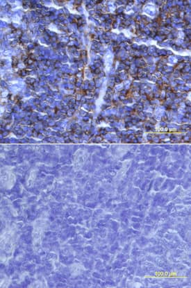
IL-16 in Human Tonsil. IL-16 was detected in immersion fixed paraffin-embedded sections of human tonsil using 15 µg/mL Human IL-16 Antigen Affinity-purified Polyclonal Antibody (Catalog # AF-316-PB) overnight at 4 °C. Tissue was stained with the Anti-Goat HRP-DAB Cell & Tissue Staining Kit (brown; Catalog # CTS008) and counterstained with hematoxylin (blue). Lower panel shows a lack of labeling if primary antibodies are omitted and tissue is stained only with secondary antibody followed by incubation with detection reagents. View our protocol for Chromogenic IHC Staining of Paraffin-embedded Tissue Sections.
Reconstitution Calculator
Preparation and Storage
Background: IL-16
Interleukin 16, also named lymphocyte chemoattractant factor (LCF), was originally identified as a CD8+ T-cell-derived chemoattractant for CD4+ cells. The biologically active form of IL-16 was originally proposed to be a homotetramer of 14 kDa chains containing 130 amino acid residue subunits. The complete pro-IL-16 cDNA was subsequently cloned and shown to encode a 631 amino acid residue hydrophilic protein that lacked a signal peptide. The original 130 amino acid residue polypeptide is now believed to have been derived from the C terminus of the precursor. IL-16 precursor protein has been detected in the lysates of various cells including mitogen stimulated PBMCs. The biologically active and secreted natural IL-16 is assumed to be a proteolytic cleavage product of pro-IL-16 generated by proteases present in or on activated CD8+ cells. A likely cleavage site was proposed to be at aspartate residue 510. This would yield a 121 amino acid residue protein, smaller than the 130 aa residue protein first described. The expression of IL-16 precursor mRNA has been detected in various tissues including spleen, thymus, lymph nodes, peripheral leukocytes, bone marrow and cerebellum. The gene for IL-16 precursor has been localized to chromosome 15. The biological activities ascribed to IL-16 are reported to be dependent on the cell surface expression of CD4, suggesting that IL-16 is a CD4 ligand. Besides its chemotactic properties, IL-16 has also been shown to suppress HIV-1 replication in vitro. Recombinant E. coli-derived IL-16 produced at R&D Systems is present mostly as a monomer, exhibits chemotactic activity for lymphocytes at high concentrations, lacks chemotactic activites for monocytes, and binds the extracellular domain of CD4 with low affinity.
- Cruikshank, W.W. et al. (1994) Proc. Natl. Acad. Sci. USA 91:5109.
- Baier, M. et al. (1997) Proc. Natl. Acad. Sci. USA 94:5273.
- Zhou, A. et al. (1997) Nature Medicine 3:659.
- Bazan, J.F. and T.J. Schall (1996) Nature 381:29.
Product Datasheets
Citation for Human IL-16 C-terminal Peptide Antibody
R&D Systems personnel manually curate a database that contains references using R&D Systems products. The data collected includes not only links to publications in PubMed, but also provides information about sample types, species, and experimental conditions.
1 Citation: Showing 1 - 1
-
Chemoattractant factors in breast milk from allergic and nonallergic mothers.
Authors: Bottcher MF, Jenmalm MC, Bjorksten B, Garofalo RP
Pediatr. Res., 2000-05-01;47(5):592-7.
Species: Human
Sample Types: Serum
Applications: ELISA Development
FAQs
No product specific FAQs exist for this product, however you may
View all Antibody FAQsReviews for Human IL-16 C-terminal Peptide Antibody
Average Rating: 5 (Based on 2 Reviews)
Have you used Human IL-16 C-terminal Peptide Antibody?
Submit a review and receive an Amazon gift card.
$25/€18/£15/$25CAN/¥75 Yuan/¥1250 Yen for a review with an image
$10/€7/£6/$10 CAD/¥70 Yuan/¥1110 Yen for a review without an image
Filter by:
A sandwich ELISA was made with MAB316 as the capture and biotinylated AF-316 as the detection using 316-IL as the immunoassay calibrator.


