Human Glypican 3 Antibody Summary
Gln25-Val558
Accession # P51654
Applications
This antibody functions as an ELISA detection antibody when paired with Mouse Anti-Human Glypican 3 Monoclonal Antibody (Catalog # MAB21191).
This product is intended for assay development on various assay platforms requiring antibody pairs. We recommend the Human Glypican 3 DuoSet ELISA Kit (Catalog # DY2119) for convenient development of a sandwich ELISA or the Human Glypican 3 Quantikine ELISA Kit (Catalog # DGLY30) for a complete optimized ELISA.
Please Note: Optimal dilutions should be determined by each laboratory for each application. General Protocols are available in the Technical Information section on our website.
Scientific Data
 View Larger
Detection of Human Glypican 3 by Western Blot. Western blot shows lysates of HepG2 human hepatocellular carcinoma cell line. PVDF membrane was probed with 2 µg/mL of Sheep Anti-Human Glypican 3 Antigen Affinity-purified Polyclonal Antibody (Catalog # AF2119) followed by HRP-conjugated Anti-Sheep IgG Secondary Antibody (Catalog # HAF016). A specific band was detected for Glypican 3 at approximately 75 kDa (as indicated). This experiment was conducted under reducing conditions and using Immunoblot Buffer Group 1.
.") View Larger
View Larger
Glypican 3 in HepG2 Human Cell Line. Glypican 3 was detected in immersion fixed HepG2 human hepatocellular carcinoma cell line using Sheep Anti-Human Glypican 3 Antigen Affinity-purified Polyclonal Antibody (Catalog # AF2119) at 1.7 µg/mL for 3 hours at room temperature. Cells were stained using the NorthernLights™ 557-conjugated Anti-Sheep IgG Secondary Antibody (red; Catalog # NL010) and counterstained with DAPI (blue). Specific staining was localized to cytoplasm and cell membranes. View our protocol for Fluorescent ICC Staining of Cells on Coverslips.
.") View Larger
View Larger
Glypican 3 in Human Breast. Glypican 3 was detected in immersion fixed paraffin-embedded sections of human breast using 5 µg/mL Sheep Anti-Human Glypican 3 Antigen Affinity-purified Polyclonal Antibody (Catalog # AF2119) overnight at 4 °C. Tissue was stained with the Anti-Sheep HRP-DAB Cell & Tissue Staining Kit (brown; Catalog # CTS019) and counterstained with hematoxylin (blue). View our protocol for Chromogenic IHC Staining of Paraffin-embedded Tissue Sections.
.") View Larger
View Larger
Glypican 3 in Human Liver Cancer Tissue. Glypican 3 was detected in immersion fixed paraffin-embedded sections of human liver cancer tissue using Sheep Anti-Human Glypican 3 Antigen Affinity-purified Polyclonal Antibody (Catalog # AF2119) at 10 µg/mL overnight at 4 °C. Tissue was stained using the Anti-Sheep HRP-DAB Cell & Tissue Staining Kit (brown; Catalog # CTS019) and counterstained with hematoxylin (blue). Lower panel shows a lack of labeling if primary antibodies are omitted and tissue is stained only with secondary antibody followed by incubation with detection reagents. View our protocol for Chromogenic IHC Staining of Paraffin-embedded Tissue Sections.
 View Larger
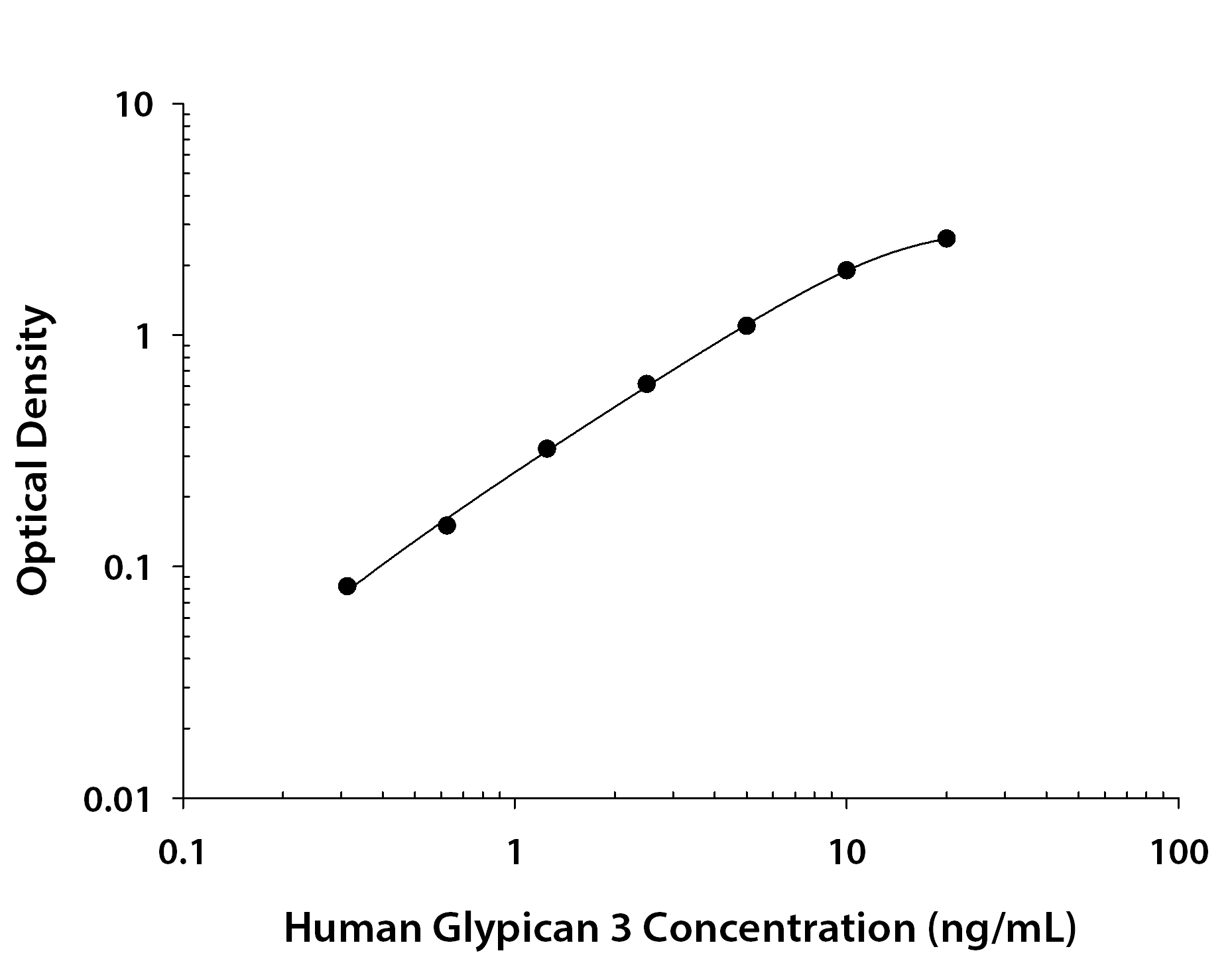
Human Glypican 3 ELISA Standard Curve. Recombinant Human Glypican 3 protein was serially diluted 2-fold and captured by Mouse Anti-Human Glypican 3 Monoclonal Antibody (MAB21191) coated on a Clear Polystyrene Microplate (DY990). Sheep Anti-Human Glypican 3 Antigen Affinity-purified Polyclonal Antibody (Catalog # AF2119) was biotinylated and incubated with the protein captured on the plate. Detection of the standard curve was achieved by incubating Streptavidin-HRP (DY998) followed by Substrate Solution (DY999) and stopping the enzymatic reaction with Stop Solution (DY994).
Reconstitution Calculator
Preparation and Storage
- 12 months from date of receipt, -20 to -70 °C as supplied.
- 1 month, 2 to 8 °C under sterile conditions after reconstitution.
- 6 months, -20 to -70 °C under sterile conditions after reconstitution.
Background: Glypican 3
Glypicans (GPC) are a family of heparan sulfate proteoglycans that are attached to the cell surface by a glycosylphosphatidylinositol (GPI) anchor. Six members of this family have been identified in mammals (GPC1-GPC6). All glypican core proteins contain an N-terminal signal peptide, a large globular cysteine-rich domain (CRD) with 14 invariant cysteine residues, a stalk-like region containing the heparan sulfate attachment sites, and a C-terminal GPI attachment site. While glypican proteins do not share strong amino acid sequence identity (they range from 17-63%), the conserved cysteine residues in their CRDs suggests similarity in their three‑dimensional structure (1, 2).
Mutations in GPC3 cause a rare disorder in humans, Simpson-Golabi-Behmel Syndrome, which is characterized by pre and postnatal overgrowth of multiple tissues and organs and an increased risk for developing embryonic tumors (3). These features are also present in the mouse knock-out of GPC3 indicating that GPC3 regulates cell survival and inhibits cell proliferation during development (4). Glypican 3 has been implicated in regulating many different signaling pathways including: IGF, FGF, BMP, and Wnt. An endoproteolytic processing of GPC3 by proprotein convertases is required for the modulation of Wnt signaling (5). Direct interaction with FGF-basic has been observed and is mediated by the heparan sulfate chains (6).
- Filmus, J. and S.B. Selleck (2001) J. Clinical Invest. 108:497.
- De Cat, B and G. David (2001) Seminars in Cell & Dev. Biol. 12:117.
- Pilia, G. et al. (1996) Nat. Genet. 12: 241.
- Cano-Gauci, D.F. et al. (1999) J. Cell Biol. 146: 255.
- De Cat, B. et al. (2003) J. Cell Biol. 163:625.
- Song, H.H. et al. (1997) J. Biol. Chem. 272:7574.
Product Datasheets
Citations for Human Glypican 3 Antibody
R&D Systems personnel manually curate a database that contains references using R&D Systems products. The data collected includes not only links to publications in PubMed, but also provides information about sample types, species, and experimental conditions.
6
Citations: Showing 1 - 6
Filter your results:
Filter by:
-
Purification of HCC-specific extracellular vesicles on nanosubstrates for early HCC detection by digital scoring
Authors: N Sun, YT Lee, RY Zhang, R Kao, PC Teng, Y Yang, P Yang, JJ Wang, M Smalley, PJ Chen, M Kim, SJ Chou, L Bao, J Wang, X Zhang, D Qi, J Palomique, N Nissen, SB Han, S Sadeghi, RS Finn, S Saab, RW Busuttil, D Markovic, D Elashoff, HH Yu, H Li, AP Heaney, E Posadas, S You, JD Yang, R Pei, VG Agopian, HR Tseng, Y Zhu
Nat Commun, 2020-09-07;11(1):4489.
Species: Human
Sample Types: Plasma
Applications: ELISA Capture -
Bone Marrow-Derived Mesenchymal Stem Cell Potential Regression of Dysplasia Associating Experimental Liver Fibrosis in Albino Rats
Authors: YH Khalifa, GM Mourad, WM Stephanos, SA Omar, RA Mehanna
Biomed Res Int, 2019-11-06;2019(0):5376165.
Species: Rat
Sample Types: Whole Tissue
Applications: IHC-P -
Expression of Glypican 3 is an Independent Prognostic Biomarker in Primary Gastro-Esophageal Adenocarcinoma and Corresponding Serum Exosomes
Authors: M Rahbari, M Pecqueux, D Aust, H Stephan, O Tiebel, A Chatzigeor, T Tonn, F Baenke, V Rao, N Ziegler, H Greif, K Lin, J Weitz, NN Rahbari, C Kahlert
J Clin Med, 2019-05-16;8(5):.
Species: Human
Sample Types: Exosome Lysates, Exosomes
Applications: Flow Cytometry, Western Blot -
Phase I trial of a glypican-3-derived peptide vaccine for advanced hepatocellular carcinoma: immunologic evidence and potential for improving overall survival.
Clin. Cancer Res., 2012-05-10;18(13):3686-96.
Species: Human
Sample Types: Serum
Applications: ELISA Development -
The oncofetal protein glypican-3 is a novel marker of hepatic progenitor/oval cells.
Authors: Grozdanov PN, Yovchev MI, Dabeva MD
Lab. Invest., 2006-10-02;86(12):1272-84.
Species: Rat
Sample Types: Whole Tissue
Applications: IHC-Fr -
Bone-specific heparan sulfates induce osteoblast growth arrest and downregulation of retinoblastoma protein.
Authors: Manton KJ, Sadasivam M, Cool SM, Nurcombe V
J. Cell. Physiol., 2006-10-01;209(1):219-29.
Species: Human
Sample Types: Cell Lysates
Applications: Western Blot
FAQs
No product specific FAQs exist for this product, however you may
View all Antibody FAQsReviews for Human Glypican 3 Antibody
There are currently no reviews for this product. Be the first to review Human Glypican 3 Antibody and earn rewards!
Have you used Human Glypican 3 Antibody?
Submit a review and receive an Amazon gift card.
$25/€18/£15/$25CAN/¥75 Yuan/¥1250 Yen for a review with an image
$10/€7/£6/$10 CAD/¥70 Yuan/¥1110 Yen for a review without an image


