Human FGFR3 Antibody
Human FGFR3 Antibody Summary
Applications
Please Note: Optimal dilutions should be determined by each laboratory for each application. General Protocols are available in the Technical Information section on our website.
Scientific Data
 View Larger
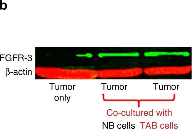
Detection of Mouse FGFR3 by Western Blot TAB cells modulate melanoma cells to express FGFR-3 and its ligand FGF-2 for tumor stroma-tumor cell cross-talk. a WM3749 co-cultured (72 h) with TAB cells (red bar) show increased FGFR-3 mRNA expression when compared with tumors only (open bar) or co-cultured with NB cells (blue bar). b Lysates obtained from pools of melanomas co-cultured (72–120 h) with NB- or TAB cells were probed in western blot with anti-FGFR-3 antibody (left panel), results expressed as relative intensity after beta -actin normalization (right panel). c Melanoma cells co-cultured with TAB cells (72 h) show increased phospho-FGFR-3 expression (right panel; immunofluorescence assays) when compared with melanoma cells alone (left panel) or melanoma cells co-cultured with NB cells (middle panel), scale bars: 40 μm, images captured by Nikon inverted microscope. d Melanoma cells co-cultured with TAB cells (red bar) show increased FGF-2 mRNA expression when compared with melanoma cells alone (open bar) or melanoma cells co-cultured with NB cells (blue bar). e 451Lu and WM989treated with IGF-1 (25 ng/ml/daily for 5 days; red bar) show increased FGFR-3 expression when compared with untreated controls (blue bar), flow cytometry results expressed as net % expression of control antibody. IGF-1 treated melanoma cells (red bars) show higher expression of FGFR-3 compared with untreated cells (blue bars). Bar represents mean + SD of replicate samples. f NB cells treated with FGF-2 (10 ng/ml/daily for 4 days; red bar) show high IGF-1 mRNA expression relative to untreated NB cells (blue bar). g 451Lu and WM989 co-cultured (72 h) with TAB cells in the presence of an anti-IGF-1 neutralizing antibody (10 μg/ml) show decreased FGFR-3 mRNA expression in tumor cells (blue bar) when compared with controls (red bar). h TAB cells co-cultured (72 h) with 451Lu and WM989 in the presence of an anti-FGF-2 neutralizing antibody (1 μg/ml) show decreased IGF-1 mRNA expression in B cells (blue bar) when compared with controls (red bar).Experiments in a, d and f–h were performed using qPCR. In Figures a, d–h, bars represent mean + SE of duplicate samples and are representative of at least two independent experiments. i Summary of cross-talk between melanoma and B cells: FGF-2 is constitutively expressed by tumor cells, released into the microenvironment to bind FGFR-3 on the B cells, activated B cells express increased levels of pro-inflammatory cytokines. IGF-1 released by TAB cells modulates tumor cells to increase their growth, heterogeneity and therapy resistance Image collected and cropped by CiteAb from the following publication (https://pubmed.ncbi.nlm.nih.gov/28928360), licensed under a CC-BY license. Not internally tested by R&D Systems.
Reconstitution Calculator
Preparation and Storage
- 12 months from date of receipt, -20 to -70 °C as supplied.
- 1 month, 2 to 8 °C under sterile conditions after reconstitution.
- 6 months, -20 to -70 °C under sterile conditions after reconstitution.
Background: FGFR3
Fibroblast Growth Factor Receptor 3 (FGF R3) is a type I transmembrane tyrosine kinase receptor that binds FGF ligands along with heparin or heparin sulfate proteoglycans as co‑factors. A segment of the membrane proximal Ig-like domain can be encoded by two different exons resulting in (IIIb) or (IIIc) isoforms. The IIIb or IIIc isoforms recognize FGF -1, -2, -4, -8b, -8e, -8f, -9, and -17b. FGF R3 plays a role in skeletal, brain, lung, intestine, kidney, and skin development.
Product Datasheets
Citations for Human FGFR3 Antibody
R&D Systems personnel manually curate a database that contains references using R&D Systems products. The data collected includes not only links to publications in PubMed, but also provides information about sample types, species, and experimental conditions.
4
Citations: Showing 1 - 4
Filter your results:
Filter by:
-
Effect of Expansion Media on Functional Characteristics of Bone Marrow-Derived Mesenchymal Stromal Cells
Authors: Jakl, V;Popp, T;Haupt, J;Port, M;Roesler, R;Wiese, S;Friemert, B;Rojewski, MT;Schrezenmeier, H;
Cells
Species: Human
Sample Types: Whole Cells
Applications: Flow Cytometry -
A reference collection of patient-derived cell line and xenograft models of proneural, classical and mesenchymal glioblastoma
Authors: BW Stringer, BW Day, RCJ D'Souza, PR Jamieson, KS Ensbey, ZC Bruce, YC Lim, K Goasdoué, C Offenhäuse, S Akgül, S Allan, T Robertson, P Lucas, G Tollesson, S Campbell, C Winter, H Do, A Dobrovic, PL Inglis, RL Jeffree, TG Johns, AW Boyd
Sci Rep, 2019-03-20;9(1):4902.
Species: Human
Sample Types: Whole Cells
Applications: Flow Cytometry -
Tumor-associated B-cells induce tumor heterogeneity and therapy resistance
Authors: R Somasundar, G Zhang, M Fukunaga-K, M Perego, C Krepler, X Xu, C Wagner, D Hristova, J Zhang, T Tian, Z Wei, Q Liu, K Garg, J Griss, R Hards, M Maurer, C Hafner, M Mayerhöfer, G Karanikas, A Jalili, V Bauer-Pohl, F Weihsengru, K Rappersber, J Koller, R Lang, C Hudgens, G Chen, M Tetzlaff, L Wu, DT Frederick, RA Scolyer, GV Long, M Damle, C Ellingswor, L Grinman, H Choi, BJ Gavin, M Dunagin, A Raj, N Scholler, L Gross, M Beqiri, K Bennett, I Watson, H Schaider, MA Davies, J Wargo, BJ Czerniecki, L Schuchter, D Herlyn, K Flaherty, M Herlyn, SN Wagner
Nat Commun, 2017-09-19;8(1):607.
Species: Human
Sample Types: Cell Lysates
Applications: Western Blot -
Diagnostic evaluation of t(4;14) in multiple myeloma and evidence for clonal evolution.
Authors: Stewart AK, Chang H, Trudel S
Leukemia, 2007-06-14;21(11):2358-9.
Species: Human
Sample Types: Whole Cells
Applications: Flow Cytometry
FAQs
-
The immunogen for Human FGFR3 Antibody, Catalog # MAB766 is described as "Pool of NS0-derived recombinant human FGF R3 alpha (IIIb) and Sf21-derived FGF R3 alpha (IIIc)". What does this mean?
To generate Catalog # MAB766, we used a pool of two proteins (NS0-derived recombinant human FGF R3 alpha (IIIb) and Sf21-derived FGF R3 alpha (IIIc)) as the immunogen. This antibody detects the IIIb and IIIc isoforms of human FGF R3 in direct ELISAs and Western blots.
Reviews for Human FGFR3 Antibody
Average Rating: 4 (Based on 1 Review)
Have you used Human FGFR3 Antibody?
Submit a review and receive an Amazon gift card.
$25/€18/£15/$25CAN/¥75 Yuan/¥2500 Yen for a review with an image
$10/€7/£6/$10 CAD/¥70 Yuan/¥1110 Yen for a review without an image
Filter by:
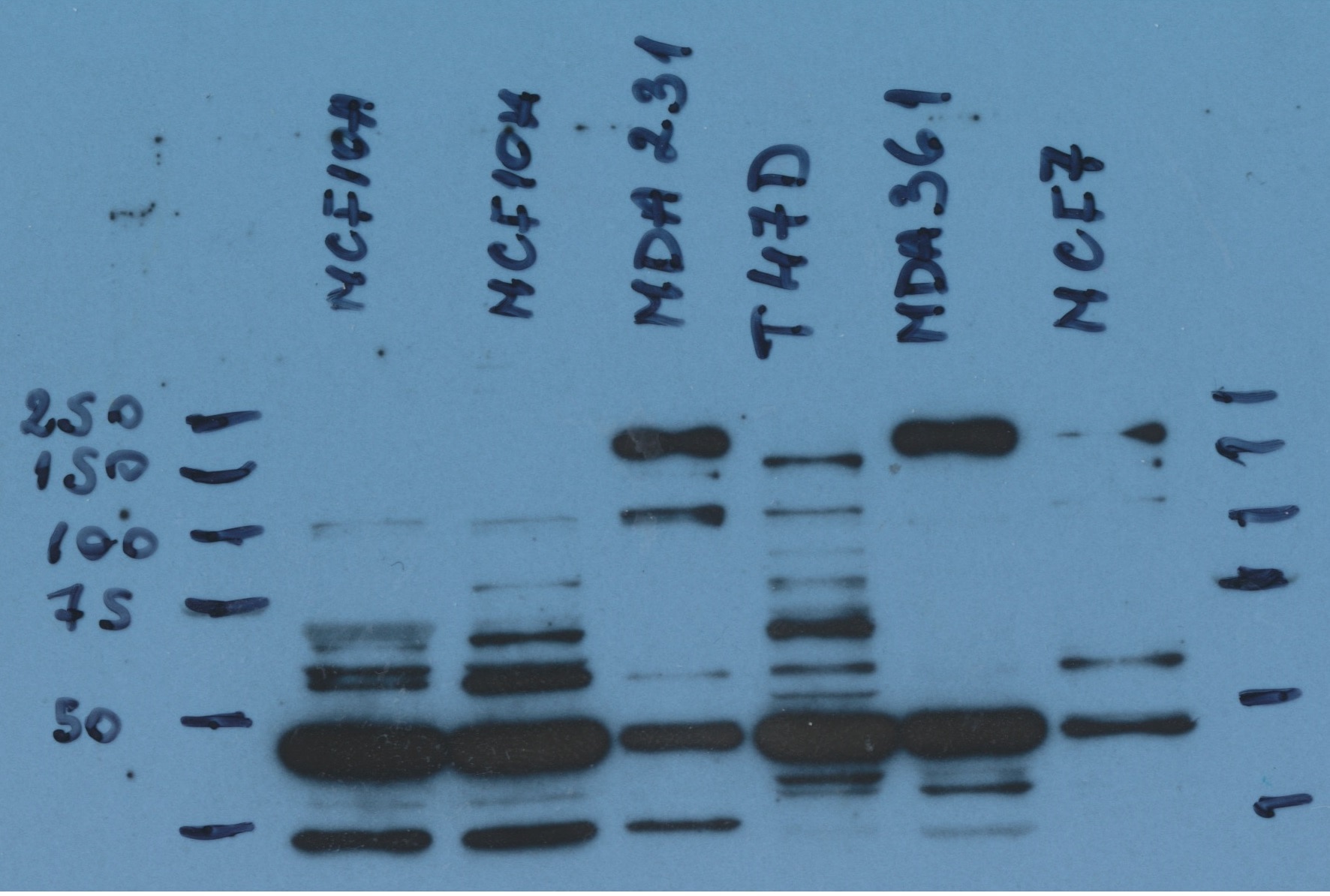
Analysis expression of the protein in breast cancer cell lines


