Recombinant Human CD19 Protein, Atto 647N Conjugate
Recombinant Human CD19 Protein, Atto 647N Conjugate Summary
Learn more about FluorokinesTM Fluorescent-Labeled ProteinsProduct Specifications
| Human CD19 (Glu21-Lys291) Accession # P15391 | IEGRMD | Human IgG1 (Pro100-Lys330) |
| N-terminus | C-terminus | |
Analysis
Product Datasheets
ATM9269
| Formulation | Supplied as a 0.2 μm filtered solution in PBS with BSA as a carrier protein. |
| Shipping | The product is shipped with dry ice or equivalent. Upon receipt, store it immediately at the temperature recommended below. |
| Stability & Storage: | Protect from light. Use a manual defrost freezer and avoid repeated freeze-thaw cycles.
|
Scientific Data
 View Larger
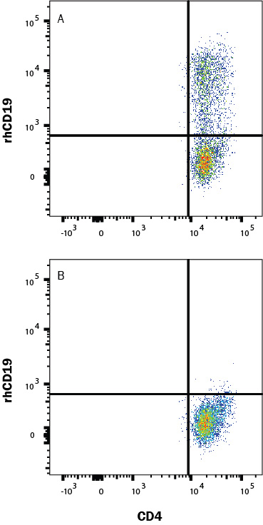
Human PBMC CD4+ and CD8+ T cells were either (A) transduced or (B) not transduced (negative control) with a Human CD19 CAR and then cultured for 11 days. Cells were washed twice with 2mL 1x PBS then stained with a viability dye for 15-30 min at room temp in the dark. Human block (1-001-1) was added during the viability stain to block Fc receptors. Cells were washed twice with Flow Cytometry Staining Buffer (FC001) then stained with Human CD4 PE-Cy7-conjugated Antibody and rhCD19 Atto 647N-conjugated protein (ATM9269) for 25 min at room temp in the dark. Staining was performed using our staining membrane proteins protocol.
 View Larger
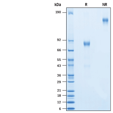
2 μg/lane of Recombinant Human CD19 Fc Chimera Atto 647N (ATM9269) was resolved with SDS-PAGE under reducing (R) and non-reducing (NR) conditions and visualized by Coomassie® Blue staining, showing bands at 77-92 kDa and 154-184 kDa, respectively.
Reconstitution Calculator
Background: CD19
CD19 is a 95 kDa transmembrane glycoprotein that plays a central role in B cell activation and humoral immune responses (1, 2). Mature human CD19 consists of a 272 amino acid (aa) extracellular domain (ECD) with two Ig-like domains, a 22 aa transmembrane segment, and a 243 aa cytoplasmic domain (3). Within the ECD, human CD19 shares 57% amino acid sequence identity with mouse and rat CD19. CD19 is expressed throughout B cell development from pre-B cells to mature B cells, and it is commonly used as a B cell lineage marker (1). It is required for the responsiveness of mature B cell to antigen stimulation, germinal center development, and antibody affinity maturation (4, 5). CD19 associates with the B cell antigen receptor (BCR), CD81, CD38, CD21, CD22, and IFITM1/CD225/Leu13, (6-9). These associations enable CD19 to amplify B cell signaling (7, 9-12) and reduce the threshold for antigen stimulation through the BCR (13). CD19 polymorphisms and up-regulation can lead to the development of autoimmunity by promoting autoantibody production (2).
- Wang, K. et al. (2012) Exp. Hematol. Oncol. 1:36.
- Tedder, T.F. et al. (1997) Immunity 6:107.
- Tedder, T.F. and C.M. Isaacs (1989) J. Immunol. 143:712.
- Rickert, R.C. et al. (1995) Nature 376:352.
- Engel, P. et al. (1995) Immunity 3:39.
- Vences-Catalan, F. et al. (2012) J. Immunol. 137:48.
- Deaglio, S. et al. (2007) Blood 109:5390.
- Bradbury, L. et al. (1992) J. Immunol. 149:2841.
- Krop, I. et al. (1996) J. Immunol. 157:48.
- O'Rourke, L.M. et al. (1998) Immunity 8:635.
- Buhl, A.M. et al. (1997) J. Exp. Med. 186:1897.
- Sato, S. et al. (1995) Proc. Natl. Acad. Sci. USA 92:11558.
- Carter, R.H. and D.T. Fearon (1992) Science 256:105.
FAQs
-
Do you have a protocol for use of Recombinant Human CD19 Protein, Atto 647N Conjugate, Catalog # ATM9269, in Flow Cytometry?
Catalog # ATM9269 works well using our general Flow Cytometry Staining Membrane Associated Proteins protocol. This protocol can be found at this web address. https://www.rndsystems.com/resources/protocols/flow-cytometry-protocol-staining-membrane-associated-proteins-suspended-cells
Reviews for Recombinant Human CD19 Protein, Atto 647N Conjugate
There are currently no reviews for this product. Be the first to review Recombinant Human CD19 Protein, Atto 647N Conjugate and earn rewards!
Have you used Recombinant Human CD19 Protein, Atto 647N Conjugate?
Submit a review and receive an Amazon gift card.
$25/€18/£15/$25CAN/¥75 Yuan/¥1250 Yen for a review with an image
$10/€7/£6/$10 CAD/¥70 Yuan/¥1110 Yen for a review without an image


