Mouse LYVE-1 Biotinylated Antibody Summary
Ala24-Thr234
Accession # Q8BHC0
Applications
Please Note: Optimal dilutions should be determined by each laboratory for each application. General Protocols are available in the Technical Information section on our website.
Scientific Data
 View Larger
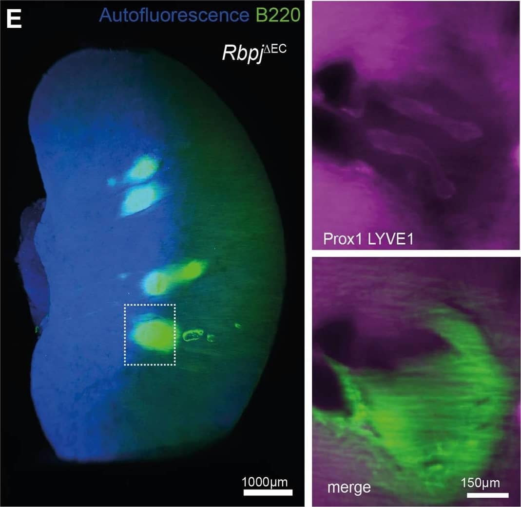
Detection of Mouse LYVE-1 by Immunohistochemistry TLS formation in models of heart failure, conditional lymphatic-EC deletion of Rbpj or kidney ischemia reperfusion. D PAS staining of representative, paraffin-embedded kidney sections. Quantification of infiltrated area [in mm2] per transversal kidney cross-section (sum of all infiltrated areas per section), N = 8 biological replicates per group, Mann–Whitney test, two-tailed, p = 0.51. Graph: Scatter dot blot, Mean, SD (whiskers). ERbpj delta EC Whole kidney staining for B220 (TLS)&Prox1/Lyve1 for lymphatic collecting vessels, light sheet microscopy, ventral view, 3D reconstruction via IMARIS software; scale bar: left image 1000 µm; insets are magnifications of boxed detail, scale bar 150 µm. Exemplary image, kidneys from N = 3 mice stained. Image collected & cropped by CiteAb from the following open publication (https://pubmed.ncbi.nlm.nih.gov/35440634), licensed under a CC-BY license. Not internally tested by R&D Systems.
 View Larger
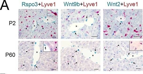
Detection of Mouse LYVE-1 by Immunohistochemistry Lack of Wnt ligand secretion from LSECs impairs adult zonation maintenance.(A) Double in situ hybridization for Rspo3 (green), Wnt9b (green)&Wnt2 (green) showing a few LSCEs (Lyve1+, red) expressing those transcripts (blue arrows&inset) in P2 livers. Only Wnt2 transcripts are detected in some LSECs (inset) in P60 livers. Arrows indicate central vein endothelial cells&arrowheads indicate LSECs. Scale bars: 25 μm. Each image is representative of 3 individual mice (n = 3). Image collected & cropped by CiteAb from the following open publication (https://pubmed.ncbi.nlm.nih.gov/32154783), licensed under a CC-BY license. Not internally tested by R&D Systems.
 View Larger
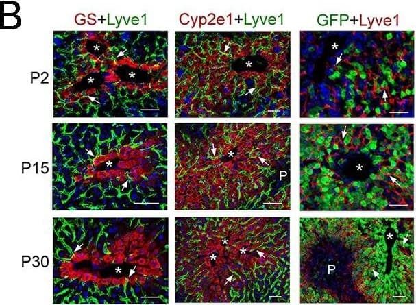
Detection of Mouse LYVE-1 by Immunohistochemistry Lack of Wnt ligand secretion from LSECs impairs adult zonation maintenance. (B) Double-immunofluorescence results show that hepatic Zones 3 (GS+)&2/3 (Cyp2e1+) are densely irrigated by the hepatic sinusoids (Lyve1+, arrows) in P2, P15&P30 wildtype livers. The sinusoidal vasculature (arrows) is also in direct contact with claudin-2/GFP+ hepatocytes in P2, P15&P30 Cldn2-EGFP livers. Scale bars: 50 μm. Each image is representative of 2–4 individual mice (n = 2–4). Image collected & cropped by CiteAb from the following open publication (https://pubmed.ncbi.nlm.nih.gov/32154783), licensed under a CC-BY license. Not internally tested by R&D Systems.
 View Larger
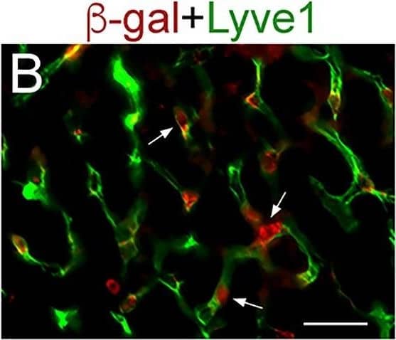
Detection of Mouse LYVE-1 by Immunohistochemistry Lineage tracing results demonstrate selective beta -gal expression in LSECs in Lyve1-Cre;ROSA-LacZ livers.(A, B) Immunofluorescence results showing expression of the lineage tracer beta -gal in PECAM+ endothelial cells connecting to the central vein (A, yellow arrows) and in parenchymal PECAM+/Lyve1+ LSECs (white arrows in (A and B). beta -gal is not expressed in PECAM+ central vein endothelial cells (A, arrowhead; the central vein is surrounded by GS+ hepatocytes). Images are representative of 2 individual livers. Scale bars: 25 μm. Image collected and cropped by CiteAb from the following open publication (https://pubmed.ncbi.nlm.nih.gov/32154783), licensed under a CC-BY license. Not internally tested by R&D Systems.
 View Larger
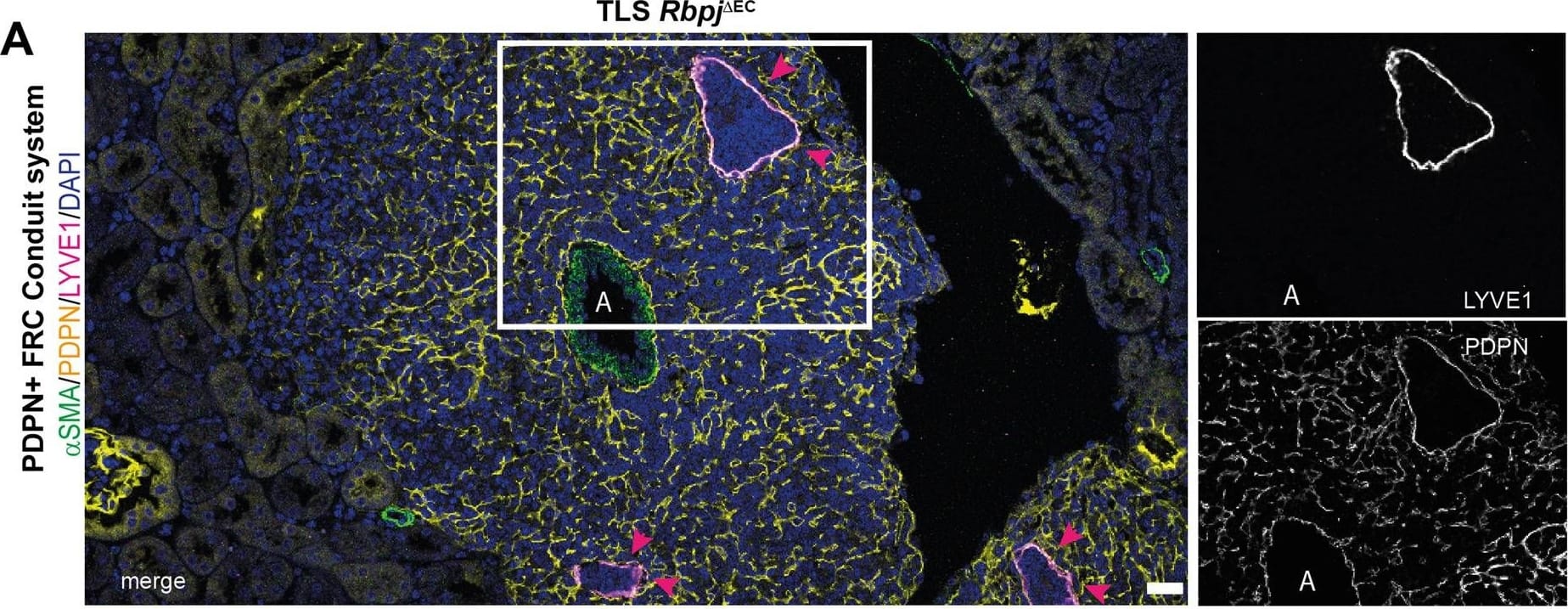
Detection of Mouse LYVE-1 by Immunohistochemistry Molecular, cellular and structural composition of periarterial TLS.A–I Immunofluorescence staining and confocal laser scanning microscopy of representative Rbpj delta EC kidney samples, merged and single channels as indicated. “A” indicates artery. Optical Magnification 200x; different scan areas (see scale bars). Scale: solid bar 50 µm, dotted bar 10 µm (2E). Each micrograph is representative of at least 4 biological replicates. J Whole kidney mRNA expression, relative fold change to control gene Rps9, n = 10/group. Graphs: Scatter dot blot, mean, SD (whiskers). Mann–Whitney test, two-tailed, Exact p-values: Cxcl13, p = 0.0003; Cxcl12, p = 0.393; Cxcr5, p = 0.0433; Cxcr4, p = 0.0288; Ccl19, p = 0.0052; Baff, p = 0.0007; Rankl, p = 0.0005. Source data are provided as a Source Data file. Image collected and cropped by CiteAb from the following open publication (https://pubmed.ncbi.nlm.nih.gov/35440634), licensed under a CC-BY license. Not internally tested by R&D Systems.
 View Larger
Detection of Mouse LYVE-1 by Immunohistochemistry Lyve1-Cre;Wlsf/f mice are refractory to CCl4-induced hepatotoxicity.(A) (Left) Schematic of CCl4 administration&tissue harvesting using Cldn2-EGFP mice. (Right) Double-immunofluorescence results show that claudin-2/GFP+ hepatocytes (arrows) are in close proximity to the sinusoidal endothelium (Lyve1+, arrows) throughout the recovery period that follows CCl4 acute injury. (B) GS (Zone 3), Cyp2e1 (Zone 2/3)&E-cadherin (Zone 1) expression in control&Lyve1-Cre;Wlsf/f livers without CCl4 treatment. Image collected & cropped by CiteAb from the following open publication (https://pubmed.ncbi.nlm.nih.gov/32154783), licensed under a CC-BY license. Not internally tested by R&D Systems.
Reconstitution Calculator
Preparation and Storage
- 12 months from date of receipt, -20 to -70 °C as supplied.
- 1 month, 2 to 8 °C under sterile conditions after reconstitution.
- 6 months, -20 to -70 °C under sterile conditions after reconstitution.
Background: LYVE-1
Lymphatic vessel endothelial hyaluronan (HA) receptor-1 (LYVE-1) is a recently identified receptor of HA, a linear high molecular weight polymer composed of alternating units of D-glucuronic acid and N-acetyl-D-glucosamine. HA is found in the extracellular matrix of most animal tissues and in body fluids. It modulates cell behavior and functions during tissue remodeling, development, homeostasis, and disease (1). The turnover of HA (several grams/day in humans) occurs primarily in the lymphatics and liver, the two major clearance systems that catabolize approximately 85% and 15% of HA, respectively (1-3). LYVE-1 shares 41% homology with the other known HA receptor, CD44 (4). The homology between the two proteins increases to 61% within the HA binding domain. The HA binding domain, known as the link module, is a common structural motif found in other HA binding proteins such as link protein, aggrecan and versican (1, 5). Human and mouse LYVE-1 share 69% amino acid sequence identity.
LYVE-1 is primarily expressed on both the luminal and abluminal surfaces of lymphatic vessels (4, 5). In addition, LYVE-1 is also present in normal hepatic blood sinusoidal endothelial cells (6). LYVE-1 mediates the endocytosis of HA and may transport HA from tissue to lymph by transcytosis, delivering HA to lymphatic capillaries for removal and degradation in the regional lymph nodes (5, 7, 8). Because of its restricted expression patterns, LYVE-1, along with other lymphatic proteins such as VEGF R3, podoplanin and the homeobox protein propero-related (Prox-1), constitute a set of markers useful for distinguishing between lymphatic and blood microvasculature (4, 5, 9-11).
- Knudson, C.B. and W. Knudson (1993) FASEB J. 7:1233.
- Evered, D. and J. Whelan (1989) Ciba Found. Symp. 143:1.
- Laurent, T.C. and J.R.F. Fraser (1992) FASEB J. 6:2397.
- Banerji, S. et al. (1999) J. Cell Biol. 144:789.
- Prevo, R. et al. (2001) J. Biol. Chem. 276:19420.
- Carreira, C.M. et al. (2001) Cancer Research 61:8079.
- Jackson, D.J. et al. (2001) Trends Immunol. 22:317.
- Zhou, B. et al. (2000) J. Biol. Chem. 275:37733.
- Achen, M. et al. (1998) Proc. Natl. Acad. Sci. USA 95:548.
- Breiteneder-Gellef, S. et al. (1999) Am. J. Pathol. 154:385.
- Wiggle, J.T. and G. Oliver (1999) Cell 98:769.
Product Datasheets
Citations for Mouse LYVE-1 Biotinylated Antibody
R&D Systems personnel manually curate a database that contains references using R&D Systems products. The data collected includes not only links to publications in PubMed, but also provides information about sample types, species, and experimental conditions.
13
Citations: Showing 1 - 10
Filter your results:
Filter by:
-
Thrombospondin 1 missense alleles induce extracellular matrix protein aggregation and TM dysfunction in congenital glaucoma
Authors: Fu H, Siggs OM, Knight LS et al.
The Journal of clinical investigation
-
Micro- and Macro-Anatomical Frameworks of Lymph Nodes Indispensable for the Lymphatic System Filtering Function
Authors: Madoka Ozawa, Shihori Nakajima, Daichi Kobayashi, Koichi Tomii, Nan-Jun Li, Tomoya Watarai et al.
Frontiers in Cell and Developmental Biology
-
The phosphatase PTEN links platelets with immune regulatory functions of mouse T follicular helper cells
Authors: X Chen, Y Xu, Q Chen, H Zhang, Y Zeng, Y Geng, L Shen, F Li, L Chen, GQ Chen, C Huang, J Liu
Nature Communications, 2022-05-19;13(1):2762.
Species: Mouse
Sample Types: Whole Tissue
Applications: IHC -
Gremlin 1+ fibroblastic niche maintains dendritic cell homeostasis in lymphoid tissues
Authors: VN Kapoor, S Müller, S Keerthivas, M Brown, C Chalouni, EE Storm, A Castiglion, R Lane, M Nitschke, CX Dominguez, JL Astarita, AT Krishnamur, CB Carbone, Y Senbabaogl, AW Wang, X Wu, V Cremasco, M Roose-Girm, L Tam, J Doerr, MZ Chen, WP Lee, Z Modrusan, YA Yang, R Bourgon, W Sandoval, AS Shaw, FJ de Sauvage, I Mellman, C Moussion, SJ Turley
Nature Immunology, 2021-04-26;22(5):571-585.
Species: Mouse
Sample Types: Whole Cells
Applications: Flow Cytometry, ICC -
Tissue-resident macrophages promote extracellular matrix homeostasis in the mammary gland stroma of nulliparous mice
Authors: Ying Wang, Thomas S Chaffee, Rebecca S LaRue, Danielle N Huggins, Patrice M Witschen, Ayman M Ibrahim et al.
eLife
-
Microtubules control cellular shape and coherence in amoeboid migrating cells
Authors: Aglaja Kopf, Jörg Renkawitz, Robert Hauschild, Irute Girkontaite, Kerry Tedford, Jack Merrin et al.
Journal of Cell Biology
-
Metabolic and non-metabolic liver zonation is established non-synchronously and requires sinusoidal Wnts
Authors: Ruihua Ma, Angelica S Martínez-Ramírez, Thomas L Borders, Fanding Gao, Beatriz Sosa-Pineda
eLife
-
Essential Role of Canonical NF-kappa B Activity in the Development of Stromal Cell Subsets in Secondary Lymphoid Organs
Authors: Dana Bogdanova, Arata Takeuchi, Madoka Ozawa, Yasuhiro Kanda, M. Azizur Rahman, Burkhard Ludewig et al.
The Journal of Immunology
-
A Distinct Subset of Fibroblastic Stromal Cells Constitutes the Cortex-Medulla Boundary Subcompartment of the Lymph Node
Authors: Arata Takeuchi, Madoka Ozawa, Yasuhiro Kanda, Mina Kozai, Izumi Ohigashi, Yoichi Kurosawa et al.
Frontiers in Immunology
-
CHD4-regulated plasmin activation impacts lymphovenous hemostasis and hepatic vascular integrity
J Clin Invest, 2016-05-03;0(0):.
Species: Mouse
Sample Types: Whole Tissue
Applications: IHC-P -
Pathological lymphangiogenesis is modulated by galectin-8-dependent crosstalk between podoplanin and integrin-associated VEGFR-3
Authors: WS Chen, Z Cao, S Sugaya, MJ Lopez, VG Sendra, N Laver, H Leffler, UJ Nilsson, J Fu, J Song, L Xia, P Hamrah, N Panjwani
Nat Commun, 2016-04-12;7(0):11302.
Species: Mouse
Sample Types: Whole Tissue
Applications: IHC -
Endothelial cell O-glycan deficiency causes blood/lymphatic misconnections and consequent fatty liver disease in mice.
Authors: Fu J, Gerhardt H, McDaniel JM, Xia B, Liu X, Ivanciu L, Ny A, Hermans K, Silasi-Mansat R, McGee S, Nye E, Ju T, Ramirez MI, Carmeliet P, Cummings RD, Lupu F, Xia L
J. Clin. Invest., 2008-10-16;118(11):3725-37.
Species: Mouse
Sample Types: Whole Tissue
Applications: IHC-Fr, IHC-P -
Lenalidomide inhibits lymphangiogenesis in preclinical models of mantle cell lymphoma.
Authors: Song K, Herzog B, Sheng M, Fu J, McDaniel J, Chen H, Ruan J, Xia L
Cancer Res, 2013-10-24;73(24):7254-64.
FAQs
No product specific FAQs exist for this product, however you may
View all Antibody FAQsReviews for Mouse LYVE-1 Biotinylated Antibody
There are currently no reviews for this product. Be the first to review Mouse LYVE-1 Biotinylated Antibody and earn rewards!
Have you used Mouse LYVE-1 Biotinylated Antibody?
Submit a review and receive an Amazon gift card.
$25/€18/£15/$25CAN/¥75 Yuan/¥2500 Yen for a review with an image
$10/€7/£6/$10 CAD/¥70 Yuan/¥1110 Yen for a review without an image
