Mouse CD69 Antibody Summary
Gly64-Arg199
Accession # P37217
*Small pack size (-SP) is supplied either lyophilized or as a 0.2 µm filtered solution in PBS.
Applications
Please Note: Optimal dilutions should be determined by each laboratory for each application. General Protocols are available in the Technical Information section on our website.
Scientific Data
 View Larger
Detection of CD69 in mouse splenocytes treated with 50ng/mL PMA and 200ng/mL Ionomycin overnight vs unstimulated mouse splenocytes by Flow Cytometry Mouse splenocytes treated with 50ng/mL PMA and 200ng/mL Ionomycin overnight (A) vs unstimulated mouse splenocytes (B) were stained with Goat Anti-Mouse CD69 Antigen Affinity-purified Polyclonal Antibody (Catalog # AF2386) and Rat Anti-Mouse CD3 Fluorescein‑conjugated Monoclonal Antibody (Catalog # FAB4841F) followed by Allophycocyanin-conjugated Anti-Goat IgG Secondary Antibody (Catalog # F0108). View our protocol for Staining Membrane-associated Proteins.
Reconstitution Calculator
Preparation and Storage
- 12 months from date of receipt, -20 to -70 °C as supplied.
- 1 month, 2 to 8 °C under sterile conditions after reconstitution.
- 6 months, -20 to -70 °C under sterile conditions after reconstitution.
Background: CD69
CD69, also known as Leu 23, AIM, EA-1 and MLR-3, is a type II transmembrane glycoprotein and is expressed on activated T cells, B cells, NK cells, neutrophils and eosinophils, Langerhans cells and platelets. The C-terminal extracellular domain of mouse CD69 shares approximately 57% amino acid sequence homology with the corresponding region of human CD69.
Product Datasheets
Citations for Mouse CD69 Antibody
R&D Systems personnel manually curate a database that contains references using R&D Systems products. The data collected includes not only links to publications in PubMed, but also provides information about sample types, species, and experimental conditions.
5
Citations: Showing 1 - 5
Filter your results:
Filter by:
-
Lymph node conduits transport virions for rapid T cell activation
Authors: Glennys V. Reynoso, Andrea S. Weisberg, John P. Shannon, Daniel T. McManus, Lucas Shores, Jeffrey L. Americo et al.
Nature Immunology
-
CD8+ T cells induce interferon-responsive oligodendrocytes and microglia in white matter aging
Authors: T Kaya, N Mattugini, L Liu, H Ji, L Cantuti-Ca, J Wu, M Schifferer, J Groh, R Martini, S Besson-Gir, S Kaji, A Liesz, O Gokce, M Simons
Nature Neuroscience, 2022-10-24;25(11):1446-1457.
Species: Mouse
Sample Types: Whole Tissue
Applications: IHC -
Differentiation Paths of Peyer's Patch LysoDCs Are Linked to Sampling Site Positioning, Migration, and T Cell Priming
Authors: C Wagner, J Bonnardel, C Da Silva, L Spinelli, CA Portilla, J Tomas, M Lagier, L Chasson, M Masse, M Dalod, A Chollat-Na, JP Gorvel, H Lelouard
Cell Rep, 2020-04-07;31(1):107479.
Species: Human
Sample Types: Whole Cells
Applications: ICC -
Dynamic Reorganization of Chromatin Accessibility Signatures during Dedifferentiation of Secretory Precursors into Lgr5+ Intestinal Stem Cells
Authors: U Jadhav, M Saxena, NK O'Neill, A Saadatpour, GC Yuan, Z Herbert, K Murata, RA Shivdasani
Cell Stem Cell, 2017-06-22;0(0):.
Species: Mouse
Sample Types: Whole Tissue
Applications: IHC -
CXCR3 chemokine receptor enables local CD8(+) T cell migration for the destruction of virus-infected cells.
Authors: Hickman H, Reynoso G, Ngudiankama B, Cush S, Gibbs J, Bennink J, Yewdell J
Immunity, 2015-03-10;42(3):524-37.
Species: Mouse
Sample Types: Whole Tissue
Applications: IHC-Fr
FAQs
No product specific FAQs exist for this product, however you may
View all Antibody FAQsReviews for Mouse CD69 Antibody
Average Rating: 4 (Based on 1 Review)
Have you used Mouse CD69 Antibody?
Submit a review and receive an Amazon gift card.
$25/€18/£15/$25CAN/¥75 Yuan/¥2500 Yen for a review with an image
$10/€7/£6/$10 CAD/¥70 Yuan/¥1110 Yen for a review without an image
Filter by:
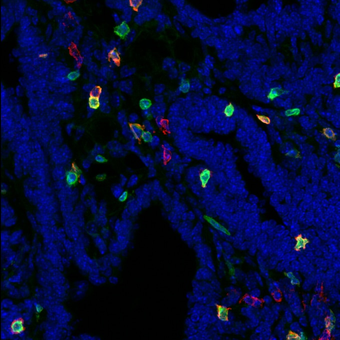
Cryosections from PLP immersion fixed PyMT mammary gland tumors were stained for CD69 (1:100) for 4h @ RT. After washing, tissue was stained with Donkey anti-Goat alexa546 2ndary antibody for 1h at RT. CD3 in green, CD69 in red, nuclear staining (Hoechst) in blue.


