Human STEAP1 APC-conjugated Antibody Summary
Met1-Trp71
Accession # Q9UHE8
Applications
Please Note: Optimal dilutions should be determined by each laboratory for each application. General Protocols are available in the Technical Information section on our website.
Scientific Data
 View Larger
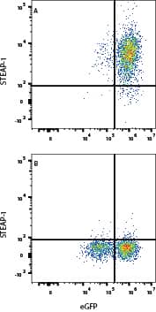
Detection of STEAP1 in HEK293 cells by Flow Cytometry. HEK293 cells transfected with Human STEAP-1 and eGFP (A) vs irrelevant cells (B) were stained with Rabbit Anti-Human STEAP1 APC-conjugated MonoclonalAntibody (Catalog # FAB55871A). View our protocol for Staining Membrane-associated Proteins.
 View Larger
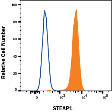
Detection of STEAP1 in LnCaP cells by Flow Cytometry LnCaP cells were stained with Rabbit Anti-Human STEAP1 APC‑conjugated Monoclonal Antibody (Catalog # FAB55871A, filled histogram) or isotype control antibody (Catalog # IC1051A, open histogram). To facilitate intracellular staining, cells were fixed with Flow Cytometry Fixation Buffer (Catalog # FC004) and permeabilized with saponin. View our protocol for Staining Intracellular Molecules.
Reconstitution Calculator
Preparation and Storage
- 12 months from date of receipt, 2 to 8 °C as supplied.
Background: STEAP1
STEAP1 (six-transmembrane epithelial antigen of the prostate-1) is a 40 kDa protein (predicted) of the STEAP family of metalloreductases. It is expressed mainly at cell‑cell junctions between prostate secretory epithelium, and up‑regulated in prostate and some bladder, colon and ovarian cancers and Ewing’s sarcomas. Human STEAP1 is 339 amino acids (aa) in length. It contains a ferric oxidoreductase domain (aa 119‑265) that includes transmembrane sequences (# 2, 3, 4, and part of 5, out of 6 sequences). Two splice forms diverge at aa 255, terminating after aa 58 or 259. Over aa 1‑71, human and mouse STEAP1 share 68% aa identity.
Product Datasheets
FAQs
No product specific FAQs exist for this product, however you may
View all Antibody FAQsReviews for Human STEAP1 APC-conjugated Antibody
Average Rating: 1 (Based on 1 Review)
Have you used Human STEAP1 APC-conjugated Antibody?
Submit a review and receive an Amazon gift card.
$25/€18/£15/$25CAN/¥75 Yuan/¥2500 Yen for a review with an image
$10/€7/£6/$10 CAD/¥70 Yuan/¥1110 Yen for a review without an image
Filter by:

