Human Neprilysin/CD10 Antibody Summary
Tyr52-Trp750
Accession # P08473
*Small pack size (-SP) is supplied either lyophilized or as a 0.2 µm filtered solution in PBS.
Applications
Please Note: Optimal dilutions should be determined by each laboratory for each application. General Protocols are available in the Technical Information section on our website.
Scientific Data
 View Larger
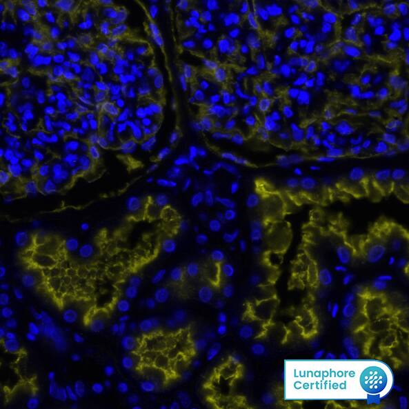
Detection of Neprilysin/CD10 in Human Kidney via seqIF™ staining on COMET™ Neprilysin/CD10 Antibody was detected in immersion fixed paraffin-embedded sections of human Kidney using Mouse Anti-Human Neprilysin/CD10, Monoclonal Antibody (Catalog # MAB11821) at 0.25ug/mL at 37 ° Celsius for 2 minutes. Before incubation with the primary antibody, tissue underwent an all-in-one dewaxing and antigen retrieval preprocessing using PreTreatment Module (PT Module) and Dewax and HIER Buffer H (pH 9; Epredia Catalog # TA-999-DHBH). Tissue was stained using the Alexa Fluor™ 555 Goat anti-Mouse IgG Secondary Antibody at 1:100 at 37 ° Celsius for 2 minutes. (Yellow; Lunaphore Catalog # DR555MS) and counterstained with DAPI (blue; Lunaphore Catalog # DR100). Specific staining was localized to the membrane. Protocol available in COMET™ Panel Builder.
 View Larger
Detection of Human Neprilysin/CD10 by Western Blot. Western blot shows lysates of Daudi human Burkitt's lymphoma cell line and Ramos human Burkitt's lymphoma cell line. PVDF membrane was probed with 0.1 µg/mL of Mouse Anti-Human Neprilysin/CD10 Monoclonal Antibody (Catalog # MAB11821) followed by HRP-conjugated Anti-Mouse IgG Secondary Antibody (Catalog # HAF007). A specific band was detected for Neprilysin/CD10 at approximately 100 kDa (as indicated). This experiment was conducted under reducing conditions and using Immunoblot Buffer Group 1.
 View Larger
Neprilysin/CD10 in Human Kidney. Neprilysin/CD10 was detected in immersion fixed paraffin-embedded sections of human kidney using Mouse Anti-Human Neprilysin/CD10 Monoclonal Antibody (Catalog # MAB11821) at 0.5 µg/mL for 1 hour at room temperature followed by incubation with the Anti-Mouse IgG VisUCyte™ HRP Polymer Antibody (Catalog # VC001). Tissue was stained using DAB (brown) and counterstained with hematoxylin (blue). Specific staining was localized to convoluted tubules and glomeruli. View our protocol for IHC Staining with VisUCyte HRP Polymer Detection Reagents.
 View Larger
Detection of Human Neprilysin/CD10 by Western Blot Western blot analysis and quantitative densitometry for NEP levels in RA-differentiated SH-SY5Y cells.The cells were incubated with 0.5% DMSO (control) or 5 μM curcumin (no. 1) or compound 7 or 8 for 24 h, then NEP protein in the cell membrane fraction was measured by western blotting. (a) Western blotting results of three independent experiments. GAPDH was used as the loading control. (b) The quantification of NEP protein levels by Image J. The NEP intensities were normalized to the GAPDH intensities for three independent experiments. Data are presented as the mean ± SD; ns, not significant; **p < 0.01 compared to the DMSO-treated control group by Student’s t-test. Image collected and cropped by CiteAb from the following open publication (https://pubmed.ncbi.nlm.nih.gov/27407064), licensed under a CC-BY license. Not internally tested by R&D Systems.
Reconstitution Calculator
Preparation and Storage
- 12 months from date of receipt, -20 to -70 °C as supplied.
- 1 month, 2 to 8 °C under sterile conditions after reconstitution.
- 6 months, -20 to -70 °C under sterile conditions after reconstitution.
Background: Neprilysin/CD10
Neprilysin/CD10, also known as NEP and neutral endopeptidase 24.11, is a zinc metallopeptidase expressed at the cell surface of a variety of cells. The enzyme functions both as an endopeptidase with a thermolysin-like specificity and as a dipeptidylcarboxypeptidase. NEP has been shown to be involved in the degradation of enkephalins in the mammalian brain and the inactivation of circulating atrial natriuretic peptide (1, 2). NEP has also been identified as the common acute lymphocytic leukemia antigen (CALLA), and is expressed on the surface of lymphocytes in some disease states (3, 4). These and other observations have resulted in considerable interest in NEP as a target for analgesics and antihypertensive drugs. NEP is also a major degrading enzyme of amyloid beta peptide (A beta ) in the brain, indicating that down-regulation of NEP activity, which could be caused by aging, can contribute to the development of Alzheimer’s disease by promoting A beta accumulation (5).
- Malfroy, B. et al. (1978) Nature 276:523.
- Kenny, A.J. and Stephenson, S.L. (1988) FEBS Lett. 232:1.
- LeTarte, M. et al. (1988) J. Exp. Med. 168:1247.
- Shipp, M.A. et al. (1988) Proc. Natl. Acad. Sci. USA 85:4819.
- Itwata, N. et al. (2001) Science 292:1550.
Product Datasheets
Citation for Human Neprilysin/CD10 Antibody
R&D Systems personnel manually curate a database that contains references using R&D Systems products. The data collected includes not only links to publications in PubMed, but also provides information about sample types, species, and experimental conditions.
1 Citation: Showing 1 - 1
-
Polyhydroxycurcuminoids but not curcumin upregulate neprilysin and can be applied to the prevention of Alzheimer's disease
Sci Rep, 2016-07-13;6(0):29760.
Species: Human
Sample Types: Cell Lysates
Applications: Western Blot
FAQs
No product specific FAQs exist for this product, however you may
View all Antibody FAQsReviews for Human Neprilysin/CD10 Antibody
There are currently no reviews for this product. Be the first to review Human Neprilysin/CD10 Antibody and earn rewards!
Have you used Human Neprilysin/CD10 Antibody?
Submit a review and receive an Amazon gift card.
$25/€18/£15/$25CAN/¥75 Yuan/¥2500 Yen for a review with an image
$10/€7/£6/$10 CAD/¥70 Yuan/¥1110 Yen for a review without an image


