Human/Mouse TLR9 Antibody Summary
Accession # Q9NR96
*Small pack size (-SP) is supplied either lyophilized or as a 0.2 µm filtered solution in PBS.
Applications
Please Note: Optimal dilutions should be determined by each laboratory for each application. General Protocols are available in the Technical Information section on our website.
Scientific Data
.") View Larger
View Larger
TLR9 in Mouse Splenocytes. TLR9 was detected in immersion fixed mouse splenocytes using Mouse Anti-Human TLR9 Monoclonal Antibody (Catalog # MAB36581) at 3 µg/mL for 3 hours at room temperature. Cells were stained using the NorthernLights™ 557-conjugated Anti-Mouse IgG Secondary Antibody (red; NL007) and counterstained with DAPI (blue). Specific staining was localized to plasma membrane. View our protocol for Fluorescent ICC Staining of Non-adherent Cells.
.") View Larger
View Larger
TLR9 in Mouse Spleen. TLR9 was detected in perfusion fixed frozen sections of mouse spleen using Mouse Anti-Human TLR9 Monoclonal Antibody (Catalog # MAB36581) at 1.7 µg/mL for 1 hour at room temperature followed by incubation with the Anti-Mouse IgG VisUCyte™ HRP Polymer Antibody (VC001). Tissue was stained using DAB (brown) and counterstained with hematoxylin (blue). Specific staining was localized to plasma membrane. View our protocol for IHC Staining with VisUCyte HRP Polymer Detection Reagents.
 View Larger
Detection of TLR9 in Daudi Human Burkitt's Lymphoma Cell Line (positive) and HDLM‑2 Human Hodgkin’s Lymphoma Cell Line (negative). TLR9 was detected in immersion fixed Daudi Human Burkitt's Lymphoma Cell Line (positive) and absent in HDLM‑2 Human Hodgkin’s Lymphoma Cell Line (negative) Cells using Mouse Anti-Human/Mouse TLR9 Monoclonal Antibody (Catalog # MAB36581) at 5 µg/mL for 3 hours at room temperature. Cells were stained using the NorthernLights™ 557-conjugated Anti-Mouse IgG Secondary Antibody (red; Catalog # NL007) and counterstained with DAPI (blue). Specific staining was localized to cytoplasm. View our protocol for Fluorescent ICC Staining of Non-adherent Cells.
 View Larger
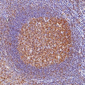
Detection of TLR9 in Human Tonsil. TLR9 was detected in immersion fixed paraffin-embedded sections of human tonsil using Mouse Anti-Human/Mouse TLR9 Monoclonal Antibody (Catalog # MAB36581) at 5 µg/ml for 1 hour at room temperature followed by incubation with the Anti-Mouse IgG VisUCyte™ HRP Polymer Antibody (Catalog # VC001). Before incubation with the primary antibody, tissue was subjected to heat-induced epitope retrieval using VisUCyte Antigen Retrieval Reagent-Basic (Catalog # VCTS021). Tissue was stained using DAB (brown) and counterstained with hematoxylin (blue). Specific staining was localized to the cytoplasm. View our protocol for IHC Staining with VisUCyte HRP Polymer Detection Reagents.
Reconstitution Calculator
Preparation and Storage
- 12 months from date of receipt, -20 to -70 °C as supplied.
- 1 month, 2 to 8 °C under sterile conditions after reconstitution.
- 6 months, -20 to -70 °C under sterile conditions after reconstitution.
Background: TLR9
TLR9 (Toll receptor 9; also CD289) is a 145-150 kDa member of the Toll-like receptor family of molecules. It is expressed by colonic epithelium, CD123+ plasmacytoid dendritic cells, and transitional B cells, and responds to unmethylated DNA CpG motifs that contain either a GTCGTT sequence (in human), or a GACGTT sequence (in mouse). TLR9 is found in the ER and translocates to either the cell membrane or to lysosomes where it binds bacterial DNA. Precursor human TLR9 is a type I transmembrane protein 1032 amino acids (aa) in length. It possesses a 793 aa extracellular region that contains 26 LRRs (aa 26-818) plus a 193 aa cytoplasmic domain. The full-length 150 kDa form is suggested to be ligand-binding but nonsignaling. The active form is believed to be an 80 kDa cleavage product found in the endosome compartment. There are multiple splice forms. One contains a deletion of aa 2-16, a second possesses an alternate start site at Met58, while a third and fourth show alternative start sites aa 23 and 24 upstream of the standard site. Over aa 64-189, human TLR9 shares 76% aa identity with mouse TLR9.
Product Datasheets
FAQs
No product specific FAQs exist for this product, however you may
View all Antibody FAQsReviews for Human/Mouse TLR9 Antibody
Average Rating: 5 (Based on 1 Review)
Have you used Human/Mouse TLR9 Antibody?
Submit a review and receive an Amazon gift card.
$25/€18/£15/$25CAN/¥75 Yuan/¥2500 Yen for a review with an image
$10/€7/£6/$10 CAD/¥70 Yuan/¥1110 Yen for a review without an image
Filter by:


