Human/Mouse Tenascin C Antibody Summary
Applications
Please Note: Optimal dilutions should be determined by each laboratory for each application. General Protocols are available in the Technical Information section on our website.
Scientific Data
 View Larger
Tenascin C in U‑118 MG Human Cell Line. Tenascin C was detected in immersion fixed U-118 MG human glioblastoma/astrocytoma cell line using Rat Anti-Human/Mouse Tenascin C Monoclonal Antibody (Catalog # MAB2138) at 10 µg/mL for 3 hours at room temperature. Cells were stained using the NorthernLights™ 557-conjugated Anti-Rat IgG Secondary Antibody (yellow; Catalog # NL013) and counterstained with DAPI (blue). View our protocol for Fluorescent ICC Staining of Cells on Coverslips.
Reconstitution Calculator
Preparation and Storage
- 12 months from date of receipt, -20 to -70 °C as supplied.
- 1 month, 2 to 8 °C under sterile conditions after reconstitution.
- 6 months, -20 to -70 °C under sterile conditions after reconstitution.
Background: Tenascin C
Tenascin C, also known as hexabrachion, cytotactin, neuronectin, GMEM, JI, myotendinous antigen, glioma-associated-extracellular matrix antigen, and GP 150‑225, is a member of the Tenascin family of extracellular matrix proteins. It is secreted as a disulfide-linked homohexamer whose subunits can vary in size from approximately 200 kDa to over 300 kDa due to differences in glycosylation (1). Rotary-shadowed electron micrographs of the purified molecule show six strands joined to one another at one end in a globular domain with each arm terminating in a knob-like structure (2, 3). The human Tenascin C monomer is synthesized as a precursor with a 22 amino acid (aa) signal sequence and a 2179 aa mature chain. The mature chain consists of a coiled-coil region (aa 118‑145), followed by
15 EGF‑like domains, 15 fibronectin type-III domains, and a fibrinogen C-terminal domain. In addition, there are 23 potential sites of N‑linked glycosylation. Alternative splicing within the fibronectin type-III repeats produces six isoforms for human Tenascin C. Mature human Tenascin C (isoform 1) shares 84% aa sequence identity with mature mouse Tenascin C. In the developing embryo, Tenascin C is expressed during neural, skeletal, and vascular morphogenesis (1, 2). In the adult, it virtually disappears with continued basal expression detectable only in tendon-associated tissues (1, 2). However, great up-regulation in expression occurs in tissues undergoing remodeling processes seen during wound repair and neovascularization or in pathological states such as inflammation or tumorigenesis (1, 4, 5). Biologically, Tenascin C functions as an adhesion-modulatory extracellular matrix protein (1, 4‑8). Specifically, it antagonizes the adhesive effects of fibronectin, and impacts the ability of fibroblasts to deposit and contract the matrix by affecting the morphology and signaling pathways of adherent cells (5‑7). Tenascin C acts by blocking syndecan-4 binding at the edges of the wound and by suppressing fibronectin-mediated activation of RhoA and focal adhesion kinase (FAK) (4‑8). Tenascin C thus promotes epidermal cell migration and proliferation during wound repair.
- Hsia, H.C. and J.E. Schwarzbauer (2005) J. Biol. Chem. 280:26641.
- Nies, D.E. et al. (1991) J. Biol. Chem. 266:2818.
- Erickson, H.P and J.L. Iglesias (1984) Nature 311:267.
- Orend, G. et al. (2003) Oncogene 22:3917.
- Wenk, M.B. et al. (2000) J. Cell Biol. 150:913.
- Midwood, K.S. et al. (2004) Mol. Biol. Cell 15:5670.
- Midwood, K.S. and J. E. Schwarzbauer (2002) Mol. Biol. Cell 13:3601.
- Hsia, H.C. and J.E. Schwarzbauer (2006) J. Surg. Res. 136:92.
Product Datasheets
Citations for Human/Mouse Tenascin C Antibody
R&D Systems personnel manually curate a database that contains references using R&D Systems products. The data collected includes not only links to publications in PubMed, but also provides information about sample types, species, and experimental conditions.
29
Citations: Showing 1 - 10
Filter your results:
Filter by:
-
Fibroblast activation and abnormal extracellular matrix remodelling as common hallmarks in three cancer‐prone genodermatoses
Authors: E. Chacón‐Solano, C. León, F. Díaz, F. García‐García, M. García, M.J. Escámez et al.
British Journal of Dermatology
-
The Matrikine Tenascin-C Protects Multipotential Stromal Cells/Mesenchymal Stem Cells from Death Cytokines Such as FasL
Authors: Melanie Rodrigues, Cecelia C. Yates, Austin Nuschke, Linda Griffith, Alan Wells
Tissue Engineering Part A
-
Regulation of IL-6 Secretion by Astrocytes via TLR4 in the Fragile X Mouse Model
Authors: Victoria Krasovska, Laurie C. Doering
Frontiers in Molecular Neuroscience
-
CAQK, a peptide associating with extracellular matrix components targets sites of demyelinating injuries
Authors: Charly Abi-Ghanem, Deepa Jonnalagadda, Jerold Chun, Yasuyuki Kihara, Barbara Ranscht
Frontiers in Cellular Neuroscience
-
Scaffold-free 3D cell culture of primary skin fibroblasts induces profound changes of the matrisome
Authors: Bich Vu, Glauco R. Souza, Jörn Dengjel
Matrix Biology Plus
-
Proteomic Analysis of Laser Microdissected Melanoma Cells from Skin Organ Cultures
Authors: Brian L. Hood, Jelena Grahovac, Melanie S. Flint, Mai Sun, Nuno Charro, Dorothea Becker et al.
Journal of Proteome Research
-
Tenascin-C-mediated suppression of extracellular matrix adhesion force promotes entheseal new bone formation through activation of Hippo signalling in ankylosing spondylitis
Authors: Zihao Li, Siwen Chen, Haowen Cui, Xiang Li, Dongying Chen, Wenjun Hao et al.
Annals of the Rheumatic Diseases
-
Tenascin-C promotes angiogenesis in fibrovascular membranes in eyes with proliferative diabetic retinopathy
Authors: Yoshiyuki Kobayashi, Shigeo Yoshida, Yedi Zhou, Takahito Nakama, Keijiro Ishikawa, Mitsuru Arima et al.
Mol. Vis
-
Global remodelling of cellular microenvironment due to loss of collagen VII
Authors: Victoria Küttner, Claudia Mack, Kristoffer T G Rigbolt, Johannes S Kern, Oliver Schilling, Hauke Busch et al.
Molecular Systems Biology
-
Mesothelial cells with mesenchymal features enhance peritoneal dissemination by forming a protumorigenic microenvironment
Authors: Yonemura, A;Semba, T;Zhang, J;Fan, Y;Yasuda-Yoshihara, N;Wang, H;Uchihara, T;Yasuda, T;Nishimura, A;Fu, L;Hu, X;Wei, F;Kitamura, F;Akiyama, T;Yamashita, K;Eto, K;Iwagami, S;Iwatsuki, M;Miyamoto, Y;Matsusaki, K;Yamasaki, J;Nagano, O;Saya, H;Song, S;Tan, P;Baba, H;Ajani, JA;Ishimoto, T;
Cell reports
Species: Mouse
Sample Types: Whole Cells
Applications: Immunocytochemistry -
Mechanical tension mobilizes Lgr6+ epidermal stem cells to drive skin growth
Authors: Y Xue, C Lyu, A Taylor, A Van Ee, A Kiemen, Y Choi, N Khavanian, D Henn, C Lee, L Hwang, E Wier, S Wang, S Lee, A Li, C Kirby, G Wang, PH Wu, D Wirtz, LA Garza, SK Reddy
Science Advances, 2022-04-27;8(17):eabl8698.
Species: Mouse
Sample Types: Whole Tissue
Applications: IHC/IF -
Intestinal fibroblastic reticular cell niches control innate lymphoid cell homeostasis and function
Authors: HW Cheng, U Mörbe, M Lütge, C Engetschwi, L Onder, M Novkovic, C Gil-Cruz, C Perez-Shib, T Hehlgans, E Scandella, B Ludewig
Nature Communications, 2022-04-19;13(1):2027.
Species: Mouse
Sample Types: Whole Tissue
Applications: IHC -
Pro-inflammatory immunity supports fibrosis advancement in epidermolysis bullosa: intervention with Ang-(1-7)
Authors: R Bernasconi, K Thriene, E Romero-Fer, C Gretzmeier, T Kühl, M Maler, P Nauroy, S Kleiser, AC Rühl-Muth, M Stumpe, D Kiritsi, SF Martin, B Hinz, L Bruckner-T, J Dengjel, A Nyström
Embo Molecular Medicine, 2021-08-30;0(0):e14392.
Species: Human, Mouse
Sample Types: Tissue Homogenates, Whole Tissue
Applications: IHC, Western Blot -
Capturing human trophoblast development with naive pluripotent stem cells in�vitro
Authors: S Io, M Kabata, Y Iemura, K Semi, N Morone, A Minagawa, B Wang, I Okamoto, T Nakamura, Y Kojima, C Iwatani, H Tsuchiya, B Kaswandy, E Kondoh, S Kaneko, K Woltjen, M Saitou, T Yamamoto, M Mandai, Y Takashima
Cell Stem Cell, 2021-04-07;28(6):1023-1039.e13.
Species: Human
Sample Types: Whole Cells, Whole Tissue
Applications: Flow Cytometry, IHC -
Expansion and characterization of epithelial stem cells with potential for cyclical hair regeneration
Authors: M Takeo, K Asakawa, KE Toyoshima, M Ogawa, J Tong, T Irié, M Yanagisawa, A Sato, T Tsuji
Scientific Reports, 2021-02-10;11(1):1173.
Species: Mouse
Sample Types: Whole Cells
Applications: Neutralization -
Autophagy deficiency promotes triple-negative breast cancer resistance to T cell-mediated cytotoxicity by blocking tenascin-C degradation
Authors: ZL Li, HL Zhang, Y Huang, JH Huang, P Sun, NN Zhou, YH Chen, J Mai, Y Wang, Y Yu, LH Zhou, X Li, D Yang, XD Peng, GK Feng, J Tang, XF Zhu, R Deng
Nat Commun, 2020-07-30;11(1):3806.
Species: Human, Mouse
Sample Types: Whole Cells
Applications: Functional Assay, Neutralization -
Transforming Growth Factor-Beta and Sonic Hedgehog Signaling in Palatal Epithelium Regulate Tenascin-C Expression in Palatal Mesenchyme During Soft Palate Development
Authors: S Ohki, K Oka, K Ogata, S Okuhara, M Rikitake, M Toda-Nakam, S Tamura, M Ozaki, S Iseki, T Sakai
Front Physiol, 2020-06-04;11(0):532.
Species: Mouse
Sample Types: Whole Tissue
Applications: IHC -
Single-cell analysis of progenitor cell dynamics and lineage specification in the human fetal kidney.
Authors: Menon R, Otto E, Kokoruda A, Zhou J, Zhang Z, Yoon E, Chen Y, Troyanskaya O, Spence J, Kretzler M, Cebrian C
Development, 2018-08-30;145(16):.
Species: Human
Sample Types: Whole Tissue
Applications: IHC -
Combined CSL and p53 downregulation promotes cancer-associated fibroblast activation.
Authors: Procopio M, Laszlo C, Al Labban D, Kim D, Bordignon P, Jo S, Goruppi S, Menietti E, Ostano P, Ala U, Provero P, Hoetzenecker W, Neel V, Kilarski W, Swartz M, Brisken C, Lefort K, Dotto G
Nat Cell Biol, 2015-08-24;17(9):1193-204.
Species: Mouse
Sample Types: Whole Tissue
Applications: IHC -
Temporal expression of growth factors triggered by epiregulin regulates inflammation development.
Authors: Harada M, Kamimura D, Arima Y, Kohsaka H, Nakatsuji Y, Nishida M, Atsumi T, Meng J, Bando H, Singh R, Sabharwal L, Jiang J, Kumai N, Miyasaka N, Sakoda S, Yamauchi-Takihara K, Ogura H, Hirano T, Murakami M
J Immunol, 2015-01-02;194(3):1039-46.
Species: Mouse
Sample Types: In Vivo
Applications: Neutralization -
The missense mutation p.R1303Q in type XVII collagen underlies junctional epidermolysis bullosa resembling Kindler syndrome.
Authors: Has, Cristina, Kiritsi, Dimitra, Mellerio, Jemima E, Franzke, Claus-We, Wedgeworth, Emma, Tantcheva-Poor, Iliana, Kernland-Lang, Kristin, Itin, Peter, Simpson, Michael, Dopping-Hepenstal, Patricia, Fujimoto, Wataru, McGrath, John A, Bruckner-Tuderman, Leena
J Invest Dermatol, 2013-09-04;134(3):845-9.
Species: Human
Sample Types: Whole Tissue
Applications: IHC -
Melanoma cell invasiveness is promoted at least in part by the epidermal growth factor-like repeats of tenascin-C.
Authors: Grahovac, Jelena, Becker, Dorothea, Wells, Alan
J Invest Dermatol, 2012-09-06;133(1):210-20.
Species: Human
Sample Types: Cell Lysates, Whole Cells
Applications: IHC, Western Blot -
Mechanisms of fibroblast cell therapy for dystrophic epidermolysis bullosa: high stability of collagen VII favors long-term skin integrity.
Authors: Kern JS, Loeckermann S, Fritsch A, Hausser I, Roth W, Magin TM, Mack C, Muller ML, Paul O, Ruther P, Bruckner-Tuderman L
Mol. Ther., 2009-06-30;17(9):1605-15.
Species: Mouse
Sample Types: Whole Tissue
Applications: IHC -
ELR-negative CXC chemokine CXCL11 (IP-9/I-TAC) facilitates dermal and epidermal maturation during wound repair.
Authors: Yates CC, Whaley D, Y-Chen A, Kulesekaran P, Hebda PA, Wells A
Am. J. Pathol., 2008-07-31;173(3):643-52.
Species: Mouse
Sample Types: Whole Tissue
Applications: IHC-P -
A hypomorphic mouse model of dystrophic epidermolysis bullosa reveals mechanisms of disease and response to fibroblast therapy.
Authors: Fritsch A, Loeckermann S, Kern JS, Braun A, Bosl MR, Bley TA, Schumann H, von Elverfeldt D, Paul D, Erlacher M, Berens von Rautenfeld D, Hausser I, Fassler R, Bruckner-Tuderman L
J. Clin. Invest., 2008-05-01;118(5):1669-79.
Species: Mouse
Sample Types: Whole Tissue
Applications: IHC-Fr -
Essential role of Smad3 in infarct healing and in the pathogenesis of cardiac remodeling.
Authors: Bujak M, Ren G, Kweon HJ, Dobaczewski M, Reddy A, Taffet G, Wang XF, Frangogiannis NG
Circulation, 2007-10-22;116(19):2127-38.
Species: Mouse
Sample Types: Whole Tissue
Applications: IHC-P -
Adipose tissue derived stem cells differentiate into carcinoma-associated fibroblast-like cells under the influence of tumor derived factors
Authors: Constantin Jotzu, Eckhard Alt, Gabriel Welte, Jie Li, Bryan T. Hennessy, Eswaran Devarajan et al.
Cell Oncol (Dordr)
-
Human Subacromial Bursal Cells Display Superior Engraftment Versus Bone Marrow Stromal Cells in Murine Tendon Repair
Authors: Felix Dyrna, Philip Zakko, Leo Pauzenberger, Mary Beth McCarthy, Augustus D. Mazzocca, Nathaniel A. Dyment et al.
The American Journal of Sports Medicine
-
Genetic Background is a Key Determinant of Glomerular Extracellular Matrix Composition and Organization
Authors: Michael J. Randles, Adrian S. Woolf, Jennifer L. Huang, Adam Byron, Jonathan D. Humphries, Karen L. Price et al.
Journal of the American Society of Nephrology
FAQs
No product specific FAQs exist for this product, however you may
View all Antibody FAQsReviews for Human/Mouse Tenascin C Antibody
Average Rating: 4.5 (Based on 2 Reviews)
Have you used Human/Mouse Tenascin C Antibody?
Submit a review and receive an Amazon gift card.
$25/€18/£15/$25CAN/¥75 Yuan/¥2500 Yen for a review with an image
$10/€7/£6/$10 CAD/¥70 Yuan/¥1110 Yen for a review without an image
Filter by:
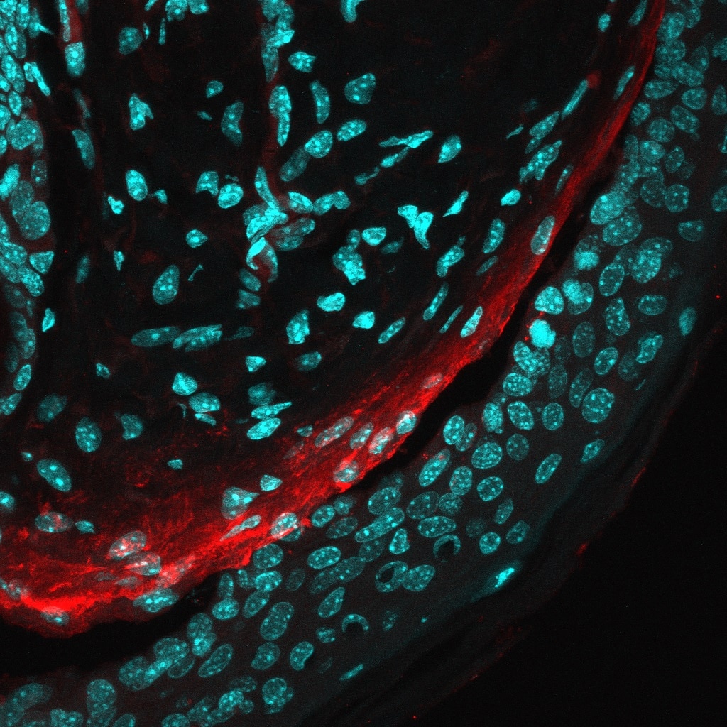
Skin tissue derived from a mouse model of epidermolysis bullosa. The skin displays a splitting in the dermal-epidermal junction, and an exacerbated tenascin C deposition in the dermis.


