Human/Mouse/Rat JNK1/JNK2 Antibody Summary
Applications
Please Note: Optimal dilutions should be determined by each laboratory for each application. General Protocols are available in the Technical Information section on our website.
Scientific Data
 View Larger
Detection of Human/Mouse/Rat JNK1/JNK2 by Western Blot. Western blot shows lysates of HeLa human cervical epithelial carcinoma cell line and C2C12 mouse myoblast cell line. PVDF membrane was probed with 0.2 µg/mL Mouse Anti-Human/Mouse/Rat JNK1/JNK2 Monoclonal Antibody (Catalog # MAB2076) followed by HRP-conjugated Anti-Mouse IgG Secondary Antibody (Catalog # HAF007). For additional reference, recombinant human JNK1, JNK2, and JNK3 (1 ng/lane) were included. Specific bands for JNK1 and JNK2 were detected at approximately 46 kDa and 54 kDa (as indicated). This experiment was conducted under reducing conditions and using Immunoblot Buffer Group 1.
.") View Larger
View Larger
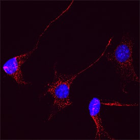
JNK1/JNK2 in HeLa Human Cervical Epithelial Carcinoma Cell Line. JNK1/JNK2 was detected in immersion fixed HeLa human cervical epithelial carcinoma cell line using Mouse Anti-Human/Mouse/Rat JNK1/JNK2 Monoclonal Antibody (Catalog # MAB2076) at 10 µg/mL for 3 hours at room temperature. Cells were stained using the NorthernLights™ 557-conjugated Anti-Mouse IgG Secondary Antibody (red; Catalog # NL007) and counterstained with DAPI (blue). Specific staining was localized to cytoplasm. View our protocol for Fluorescent ICC Staining of Cells on Coverslips.
 View Larger
JNK1/JNK2 in C2C12 Mouse Cell Line. JNK1/JNK2 was detected in immersion fixed C2C12 mouse myoblast cell line using Mouse Anti-Human/Mouse/Rat JNK1/JNK2 Monoclonal Antibody (Catalog # MAB2076) at 25 µg/mL for 3 hours at room temperature. Cells were stained using the NorthernLights™ 557-conjugated Anti-Mouse IgG Secondary Antibody (red; NL007) and counterstained with DAPI (blue). Specific staining was localized to cytoplasm. Staining was performed using our protocol for Fluorescent ICC Staining of Non-adherent Cells.
 View Larger
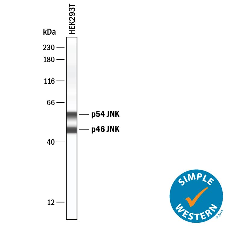
Detection of Human JNK1/JNK2 by Simple WesternTM. Simple Western lane view shows lysates of HEK293T human embryonic kidney cell line, loaded at 0.2 mg/mL. Specific bands were detected for JNK1/JNK2 at approximately 48 and 58 kDa (as indicated) using 10 µg/mL of Mouse Anti-Human/Mouse/Rat JNK1/JNK2 Monoclonal Antibody (Catalog # MAB2076). This experiment was conducted under reducing conditions and using the 12-230 kDa separation system.
Reconstitution Calculator
Preparation and Storage
- 12 months from date of receipt, -20 to -70 °C as supplied.
- 1 month, 2 to 8 °C under sterile conditions after reconstitution.
- 6 months, -20 to -70 °C under sterile conditions after reconstitution.
Background: JNK1/JNK2
The c-Jun N-terminal kinases (JNKs) are encoded by three genes: JNK1, JNK2, and JNK3. Members of the MAPK superfamily, JNKs are activated by environmental stresses and inflammatory cytokines. JNK1, also known as SAPK1 gamma and MAPK8, is expressed as four isoforms generated by alternative splicing. JNK1 is activated by dual phosphorylation at T183 and Y185 by the MAPK kinases MKK4 and/or MKK7.
Product Datasheets
Citations for Human/Mouse/Rat JNK1/JNK2 Antibody
R&D Systems personnel manually curate a database that contains references using R&D Systems products. The data collected includes not only links to publications in PubMed, but also provides information about sample types, species, and experimental conditions.
6
Citations: Showing 1 - 6
Filter your results:
Filter by:
-
Activity of fibroblast-like synoviocytes in rheumatoid arthritis was impaired by dickkopf-1 targeting siRNA
Authors: YY Liu, SY Wang, YN Li, WJ Bian, LQ Zhang, YH Li, L Long, X Liu, XW Zhang, ZG Li
Chin. Med. J., 2020-03-20;0(6):679-686.
Species: Human
Sample Types: Cell Lysate
Applications: Western Blot -
Ulinastatin Ameliorates Pulmonary Capillary Endothelial Permeability Induced by Sepsis Through Protection of Tight Junctions via Inhibition of TNF-? and Related Pathways
Authors: M Fang, WH Zhong, WL Song, YY Deng, DM Yang, B Xiong, HK Zeng, HD Wang
Front Pharmacol, 2018-08-13;9(0):823.
Species: Rat
Sample Types: Tissue Homogenates
Applications: Western Blot -
WWOX sensitises ovarian cancer cells to paclitaxel via modulation of the ER stress response
Authors: S Janczar, J Nautiyal, Y Xiao, E Curry, M Sun, E Zanini, AJ Paige, H Gabra
Cell Death Dis, 2017-07-27;8(7):e2955.
Species: Human
Sample Types: Cell Lysates
Applications: Western Blot -
High glucose alters retinal astrocytes phenotype through increased production of inflammatory cytokines and oxidative stress.
Authors: Shin E, Huang Q, Gurel Z, Sorenson C, Sheibani N
PLoS ONE, 2014-07-28;9(7):e103148.
Species: Mouse
Sample Types: Cell Lysates
Applications: Western Blot -
Pre-exposure of Mycobacterium tuberculosis-infected macrophages to crystalline silica impairs control of bacterial growth by deregulating the balance between apoptosis and necrosis.
Authors: Chavez-Galan L, Ramon-Luing L, Torre-Bouscoulet L, Perez-Padilla R, Sada-Ovalle I
PLoS ONE, 2013-11-22;8(11):e80971.
Species: Human
Sample Types: Cell Lysates
Applications: Western Blot -
Signaling by Fyn-ADAP via the Carma1-Bcl-10-MAP3K7 signalosome exclusively regulates inflammatory cytokine production in NK cells.
Authors: Rajasekaran K, Kumar P, Schuldt KM et al.
Nat Immunol.
FAQs
No product specific FAQs exist for this product, however you may
View all Antibody FAQsReviews for Human/Mouse/Rat JNK1/JNK2 Antibody
There are currently no reviews for this product. Be the first to review Human/Mouse/Rat JNK1/JNK2 Antibody and earn rewards!
Have you used Human/Mouse/Rat JNK1/JNK2 Antibody?
Submit a review and receive an Amazon gift card.
$25/€18/£15/$25CAN/¥75 Yuan/¥2500 Yen for a review with an image
$10/€7/£6/$10 CAD/¥70 Yuan/¥1110 Yen for a review without an image


