Human LIN-28A Antibody Summary
Met1-Asn209
Accession # Q9H9Z2
Applications
Please Note: Optimal dilutions should be determined by each laboratory for each application. General Protocols are available in the Technical Information section on our website.
Scientific Data
 View Larger
Detection of Human LIN‑28A by Western Blot. Western blot shows lysates of JAR human choriocarcinoma cell line and NTera-2 human testicular embryonic carcinoma cell line. PVDF membrane was probed with 0.1 µg/mL of Goat Anti-Human LIN-28A Antigen Affinity-purified Polyclonal Antibody (Catalog # AF3757) followed by HRP-conjugated Anti-Goat IgG Secondary Antibody (Catalog # HAF109). A specific band was detected for LIN-28A at approximately 30 kDa (as indicated). This experiment was conducted under reducing conditions and using Immunoblot Buffer Group 1.
 View Larger
LIN‑28A in BG01V Human Stem Cells. LIN-28A was detected in immersion fixed BG01V human embryonic stem cells using 10 µg/mL Goat Anti-Human LIN-28A Antigen Affinity-purified Polyclonal Antibody (Catalog # AF3757) for 3 hours at room temperature. Cells were stained with the NorthernLights™ 557-conjugated Anti-Goat IgG Secondary Antibody (red; Catalog # NL001) and counter-stained with DAPI (blue). View our protocol for Fluorescent ICC Staining of Cells on Coverslips.
 View Larger
LIN‑28A in D3 Mouse Stem Cells. LIN-28A was detected in immersion fixed D3 mouse embryonic stem cell line using Goat Anti-Human LIN-28A Antigen Affinity-purified Polyclonal Antibody (Catalog # AF3757) at 10 µg/mL for 3 hours at room temperature. Cells were stained using the Northern-Lights™ 557-conjugated Anti-Goat IgG Secondary Antibody (red; Catalog # NL001) and counterstained with DAPI (blue). Specific staining was localized to cytoplasm. View our protocol for Fluorescent ICC Staining of Cells on Coverslips.
 View Larger
Detection of Human LIN-28A by Simple WesternTM. Simple Western lane view shows lysates of NTera‑2 human testicular embryonic carcinoma cell line and JAR human choriocarcinoma cell line, loaded at 0.2 mg/mL. A specific band was detected for LIN-28A at approximately 45 kDa (as indicated) using 10 µg/mL of Goat Anti-Human LIN-28A Antigen Affinity-purified Polyclonal Antibody (Catalog # AF3757). This experiment was conducted under reducing conditions and using the 12-230 kDa separation system.
 View Larger
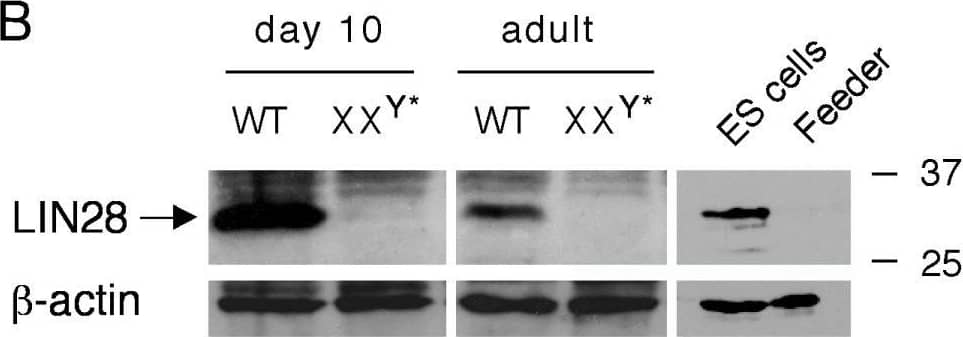
Detection of LIN-28A by Western Blot Expression of LIN28 in mouse testis. Western blot analysis was performed on 20 μg of protein extracts for each sample. beta -actin served as a control. Molecular weight standards were marked in kDa. (A) Western blot analysis of LIN28 in adult mouse tissues. (B) Absence of LIN28 in germ cell-deficient XXY* testes. Testes were collected from adult and post-natal day 10-old mice. V6.5 mouse embryonic stem (ES) cells served as a positive control. LIN28 was absent in fibroblast feeder cells. (C) Developmental expression of LIN28 in postnatal testes. Testes were collected from mice of postnatal day 1 through adulthood. Image collected and cropped by CiteAb from the following open publication (https://pubmed.ncbi.nlm.nih.gov/19563657), licensed under a CC-BY license. Not internally tested by R&D Systems.
 View Larger
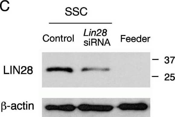
Detection of LIN-28A by Western Blot Expression and siRNA knockdown of LIN28 in cultured spermatogonia highly enriched for spermatogonial stem cells (SSCs). (A) Immunostaining of SSCs with anti-LIN28 and anti-PLZF or anti-GFRA1 antibodies. Scale bar, 50 μm. (B) Quantitative PCR measurement of Lin28 mRNA levels (n = 3, mean ± SE) in SSCs after siRNA treatment for 30 hours. (C) Decreased LIN28 protein abundance (43% compared to the control) in SSCs after 30 hours of siRNA treatment. The control SSCs were not treated with Lin28 siRNA. Feeder cells served as a negative control. beta -actin served as a loading control. (D) The number of SSCs (n = 3, mean ± SE) with and without Lin28 siRNA treatment. (E) Quantitative measurement of mature let-7g miRNA levels (n = 3, mean ± SE) in SSCs after siRNA treatment for 30 hours. Image collected and cropped by CiteAb from the following open publication (https://pubmed.ncbi.nlm.nih.gov/19563657), licensed under a CC-BY license. Not internally tested by R&D Systems.
Reconstitution Calculator
Preparation and Storage
- 12 months from date of receipt, -20 to -70 °C as supplied.
- 1 month, 2 to 8 °C under sterile conditions after reconstitution.
- 6 months, -20 to -70 °C under sterile conditions after reconstitution.
Background: LIN-28A
Human LIN-28 (Protein lin-28 homolog A; CSDD1, LIN28 and ZCCHC1) is a 30 kDa (209 amino acids) cytoplasmic RNA-binding protein with an N-terminal cold shock domain and two C-terminal CCHC zinc finger domains. It is expressed by various undifferentiated embryonic cell types and is also present in adult cardiac and skeletal muscle. Expression of LIN-28 has been shown to be regulated by micro-RNA. Human LIN-28 shares 98% and 97% amino acid sequence homology with rat and mouse LIN-28, respectively.
Product Datasheets
Citations for Human LIN-28A Antibody
R&D Systems personnel manually curate a database that contains references using R&D Systems products. The data collected includes not only links to publications in PubMed, but also provides information about sample types, species, and experimental conditions.
22
Citations: Showing 1 - 10
Filter your results:
Filter by:
-
Identification of dynamic undifferentiated cell states within the male germline
Authors: HM La, JA Mäkelä, AL Chan, FJ Rossello, CM Nefzger, JMD Legrand, M De Seram, JM Polo, RM Hobbs
Nat Commun, 2018-07-19;9(1):2819.
-
Transgene-Free Cynomolgus Monkey iPSCs Generated under Chemically Defined Conditions
Authors: Tereshchenko, Y;Esiyok, N;Garea-Rodríguez, E;Repetto, D;Behr, R;Rodríguez-Polo, I;
Cells
Species: Human
Sample Types: Whole Cells
Applications: Immunocytochemistry -
Inactivation of Tumor Suppressor CYLD Inhibits Fibroblast Reprogramming to Pluripotency
Authors: Bekas, N;Samiotaki, M;Papathanasiou, M;Mokos, P;Pseftogas, A;Xanthopoulos, K;Thanos, D;Mosialos, G;Dafou, D;
Cancers
Species: Mouse
Sample Types: Whole Cells
Applications: ICC -
Semi-automated optimized method to isolate CRISPR/Cas9 edited human pluripotent stem cell clones
Authors: Elie Frank, Michel Cailleret, Constantin Nelep, Pascal Fragner, Jérome Polentes, Elise Herardot et al.
Stem Cell Research & Therapy
-
Modeling of early neural development in vitro by direct neurosphere formation culture of chimpanzee induced pluripotent stem cells
Authors: R Kitajima, R Nakai, T Imamura, T Kameda, D Kozuka, H Hirai, H Ito, H Imai, M Imamura
Stem Cell Res, 2020-02-28;44(0):101749.
Species: Pan troglodytes (Chimpanzee)
Sample Types: Whole Cells
Applications: ICC -
Exonuclease Domain-Containing 1 Enhances MIWI2 piRNA Biogenesis via Its Interaction with TDRD12
Authors: RR Pandey, D Homolka, O Olotu, R Sachidanan, N Kotaja, RS Pillai
Cell Rep, 2018-09-25;24(13):3423-3432.e4.
Species: Mouse
Sample Types: Whole Tissue
Applications: IHC -
Derivation of induced pluripotent stem cells in Japanese macaque (Macaca fuscata)
Authors: R Nakai, M Ohnuki, K Kuroki, H Ito, H Hirai, R Kitajima, T Fujimoto, M Nakagawa, W Enard, M Imamura
Sci Rep, 2018-08-15;8(1):12187.
Species: Primate - Macaca mulatta (Rhesus Macaque)
Sample Types: Whole Cells
Applications: ICC -
Biallelic loss of human CTNNA2, encoding ?N-catenin, leads to ARP2/3 complex overactivity and disordered cortical neuronal migration
Authors: AE Schaffer, MW Breuss, AO Caglayan, N Al-Sanaa, HY Al-Abdulwa, H Kaymakçala, C Y?lmaz, MS Zaki, RO Rosti, B Copeland, ST Baek, D Musaev, EC Scott, T Ben-Omran, A Karimineja, H Kayserili, F Mojahedi, M Kara, N Cai, JL Silhavy, S Elsharif, E Fenerciogl, BA Barshop, B Kara, R Wang, V Stanley, KN James, R Nachnani, A Kalur, H Megahed, F Incecik, S Danda, Y Alanay, E Faqeih, G Melikishvi, L Mansour, I Miller, B Sukhudyan, J Chelly, WB Dobyns, K Bilguvar, RA Jamra, M Gunel, JG Gleeson
Nat. Genet., 2018-07-16;0(0):.
Species: Human
Sample Types: Whole Cells
Applications: ICC -
Autologous and Heterologous Cell Therapy for Hemophilia B toward Functional Restoration of Factor IX
Authors: S Ramaswamy, N Tonnu, T Menon, BM Lewis, KT Green, D Wampler, PE Monahan, IM Verma
Cell Rep, 2018-05-01;23(5):1565-1580.
Species: Human
Sample Types: Whole Cells
Applications: ICC -
Modeling Short QT Syndrome Using Human‐Induced Pluripotent Stem Cell–Derived Cardiomyocytes
Authors: Ibrahim El‐Battrawy, Huan Lan, Lukas Cyganek, Zhihan Zhao, Xin Li, Fanis Buljubasic et al.
Journal of the American Heart Association
-
Embryonic lethality and defective male germ cell development in mice lacking UTF1
Authors: SD Kasowitz, M Luo, J Ma, NA Leu, PJ Wang
Sci Rep, 2017-12-08;7(1):17259.
Species: Mouse
Sample Types: Cell Lysates
Applications: Western Blot -
Protein-driven RNA nanostructured devices that function in vitro and control mammalian cell fate
Authors: T Shibata, Y Fujita, H Ohno, Y Suzuki, K Hayashi, KR Komatsu, S Kawasaki, K Hidaka, S Yonehara, H Sugiyama, M Endo, H Saito
Nat Commun, 2017-09-14;8(1):540.
Species: Human
Sample Types: Cell Lysates
Applications: Simple Western -
Synthetic mRNA devices that detect endogenous proteins and distinguish mammalian cells
Authors: Shunsuke Kawasaki, Yoshihiko Fujita, Takashi Nagaike, Kozo Tomita, Hirohide Saito
Nucleic Acids Research
-
Glycolysis-Optimized Conditions Enhance Maintenance of Regenerative Integrity in Mouse Spermatogonial Stem Cells during Long-Term Culture
Authors: AR Helsel, MJ Oatley, JM Oatley
Stem Cell Reports, 2017-04-06;0(0):.
Species: Mouse
Sample Types: Whole Tissue
Applications: IHC-P -
Characterizing the Spermatogonial Response to Retinoic Acid During the Onset of Spermatogenesis and Following Synchronization in the Neonatal Mouse Testis1
Authors: Kellie S. Agrimson, Jennifer Onken, Debra Mitchell, Traci B. Topping, Hélio Chiarini-Garcia, Cathryn A. Hogarth et al.
Biology of Reproduction
-
The cancer/testis-antigen PRAME supports the pluripotency network and represses somatic and germ cell differentiation programs in seminomas
Authors: Daniel Nettersheim, Isabell Arndt, Rakesh Sharma, Stefanie Riesenberg, Sina Jostes, Simon Schneider et al.
British Journal of Cancer
-
Polyglutamine-expanded androgen receptor interferes with TFEB to elicit pathological autophagy defects in SBMA
Authors: Constanza J. Cortes, Helen C. Miranda, Harald Frankowski, Yakup Batlevi, Jessica E. Young, Amy Le et al.
Nature Neuroscience
-
Reconstruction of mouse testicular cellular microenvironments in long-term seminiferous tubule culture.
Authors: Makela J, Toppari J, Rivero-Muller A, Ventela S
PLoS ONE, 2014-03-11;9(3):e90088.
Species: Mouse
Sample Types: Whole Cells
Applications: ICC -
SALL4 Expression in Gonocytes and Spermatogonial Clones of Postnatal Mouse Testes
Authors: Kathrin Gassei, Kyle E. Orwig
PLoS ONE
-
Activation of pluripotency genes in human fibroblast cells by a novel mRNA based approach.
Authors: Plews JR, Li J, Jones M, Moore HD, Mason C, Andrews PW, Na J
PLoS ONE, 2010-12-30;5(12):e14397.
Species: Human
Sample Types: Cell Lysates
Applications: Western Blot -
Labeling human embryonic stem cell-derived cardiomyocytes with indocyanine green for noninvasive tracking with optical imaging: an FDA-compatible alternative to firefly luciferase
Authors: Sophie E. Boddington, Tobias D. Henning, Priyanka Jha, Christopher R. Schlieve, Lydia Mandrussow, David DeNardo et al.
Cell Transplant
-
Induction of pluripotent stem cells from adult human fibroblasts by defined factors.
Authors: Takahashi K, Tanabe K, Ohnuki M, Narita M, Ichisaka T, Tomoda K, Yamanaka S
Cell, 2007-11-30;131(5):861-72.
Species: Human
Sample Types: Cell Lysates
Applications: Western Blot
FAQs
No product specific FAQs exist for this product, however you may
View all Antibody FAQsReviews for Human LIN-28A Antibody
There are currently no reviews for this product. Be the first to review Human LIN-28A Antibody and earn rewards!
Have you used Human LIN-28A Antibody?
Submit a review and receive an Amazon gift card.
$25/€18/£15/$25CAN/¥75 Yuan/¥2500 Yen for a review with an image
$10/€7/£6/$10 CAD/¥70 Yuan/¥1110 Yen for a review without an image

