Mouse TREM2 Antibody Summary
Leu19-Pro168
Accession # Q99NH8
Applications
This antibody functions as an ELISA capture antibody when paired with Sheep Anti-Mouse TREM2 Biotinylated Antigen Affinity-purified Polyclonal Antibody (Catalog # BAF1729).
This product is intended for assay development on various assay platforms requiring antibody pairs.
Please Note: Optimal dilutions should be determined by each laboratory for each application. General Protocols are available in the Technical Information section on our website.
Scientific Data
 View Larger
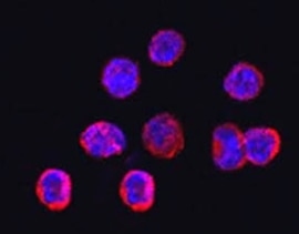
TREM2 in RAW 264.7 Mouse Cell Line. TREM2 was detected in immersion fixed RAW 264.7 mouse monocyte/macrophage cell line using Sheep Anti-Mouse TREM2 Antigen Affinity-purified Polyclonal Antibody (Catalog # AF1729) at 1.7 µg/mL for 3 hours at room temperature. Cells were stained using the NorthernLights™ 557-conjugated Anti-Sheep IgG Secondary Antibody (red; NL010) and counterstained with DAPI (blue). Specific staining was localized to cytoplasm. View our protocol for Fluorescent ICC Staining of Non-adherent Cells.
 View Larger
Mouse TREM2 ELISA Standard Curve. Recombinant Mouse TREM2 protein was serially diluted 2-fold and captured by Sheep Anti-Mouse TREM2 Antigen Affinity-purified Polyclonal Antibody (Catalog # AF1729) coated on a Clear Polystyrene Microplate (DY990). Sheep Anti-Mouse TREM2 Biotinylated Antigen Affinity-purified Polyclonal Antibody (BAF1729) was incubated with the protein captured on the plate. Detection of the standard curve was achieved by incubating Streptavidin-HRP (DY998) followed by Substrate Solution (DY999) and stopping the enzymatic reaction with Stop Solution (DY994).
 View Larger
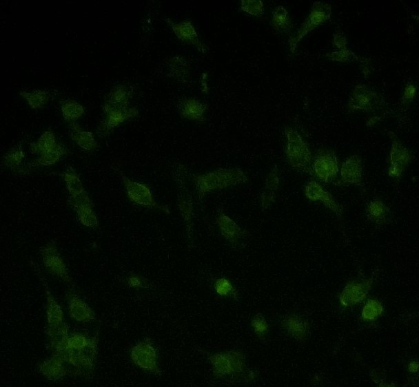
TREM2 Specificity is Shown by Immunocytochemistry in Knockout Cell Line. TREM2 was detected in immersion fixed RAW 264.7 mouse monocyte/macrophage cell line (left panel) but is not detected in TREM2 knockout (KO) RAW 264.7 Mouse Cell Line cell line (right panel) using Sheep Anti-Mouse TREM2 Antigen Affinity-purified Polyclonal Antibody (Catalog # AF1729) at 1.7 µg/mL for 3 hours at room temperature. Cells were stained using the NorthernLights™ 557-conjugated Anti-Sheep IgG Secondary Antibody (red; NL010) and counterstained with DAPI (blue). Specific staining was localized to cytoplasm. View our protocol for Fluorescent ICC Staining of Non-adherent Cells.
 View Larger
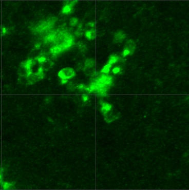
Detection of Mouse TREM2 by Immunocytochemistry/Immunofluorescence SCF+G-CSF treatment increases TREM2 expression in the Iba1+ microglia/macrophages surrounding the 6E10+ senile plaques. (A) Representative confocal images of TREM2 (red), 6E10 (purple) and Iba1 (green) triple immunofluorescence staining in the brains of aged APP/PS1 mice. Blue: Nuclear counterstaining by DAPI. (B) Representative orthographic view of z-stack images (12 z-stacks with 1μm intervals) illustrates the location and interaction of TREM2 + cells (red) and 6E10+ A beta plaques (white) in the brains of aged APP/PS1 mice. (C) Quantification data show the percentage of TREM2+ area surrounding the 6E10+ A beta plaques (within 10μm from the border of the A beta plaques) in the brains of aged APP/PS1 mice with or without SCF+G-CSF treatment. (D) Representative orthographic view of z-stack images (12 z-stacks with 1μm intervals) displays the location and interaction of TREM2+/Iba1+ co-expressing cells (yellow) and 6E10+ A beta plaques (white) in the brains of APP/PS1 mice. (E) Quantification data show the percentage of TREM2+/Iba1+ co-expression area in the total of Iba1+ area in the vicinity of 6E10+ A beta plaques in the brains of aged APP/PS1 mice with or without SCF+G-CSF treatment. N=4-5. Mean ± SEM. * p<0.05 by Student’s t-test. Image collected and cropped by CiteAb from the following publication (https://pubmed.ncbi.nlm.nih.gov/33269098), licensed under a CC-BY license. Not internally tested by R&D Systems.
 View Larger
Detection of Mouse Mouse TREM2 Antibody by Western Blot A beta oligomers induce TREM2 proteolysis and sTREM2 release, which then binds A beta oligomers, but R47H sTREM2 binds less. A, western blot of cell lysate and unprocessed supernatant (sTREM2) of HEK293 cells coexpressing human DAP12 and full-length N-terminally-tagged wild-type (WT) human TREM2 (FL-TREM2) 16 h after adding A beta oligomers. This blot and those for A beta monomers and fibrils are reproduced in Figure S5 for comparison. B, quantification of sTREM2 release from transfected HEK293 cells expressing wild-type (green line) and R47H TREM2 (red line). C, quantification of sTREM2 release from wild-type TREM2 expressing HEK293 cells induced by doses of A beta oligomers (red line), monomers (green line), or fibrils (blue line). For both (B and C) error bars = SEM; ∗p < 0.05 ∗∗p < 0.01 ∗∗∗p < 0.001, n = 3 independent experiments; one-way ANOVA with Tukey's post-hoc multiple comparisons test. D, example field of single-molecule TIRF imaging of mixture of A beta oligomers (green) and wild-type TREM2 ectodomain (red), where colocalized spots appear yellow. Scale bar: 1 micron. Magnified image of three sections of field at right. E, proportion of monomeric or oligomeric A beta colocalized with wild-type sTREM2. F, proportion of A beta oligomers colocalized with wild-type or R47H TREM2 ectodomain. For (E and F), error bars = SEM; ∗∗∗∗p < 0.0001, n = 3 independent preparations, each analyzed in nine fields each; two-tailed t-test of significance. Image collected and cropped by CiteAb from the following publication (https://pubmed.ncbi.nlm.nih.gov/33823153), licensed under a CC-BY license. Not internally tested by R&D Systems.
Reconstitution Calculator
Preparation and Storage
- 12 months from date of receipt, -20 to -70 °C as supplied.
- 1 month, 2 to 8 °C under sterile conditions after reconstitution.
- 6 months, -20 to -70 °C under sterile conditions after reconstitution.
Background: TREM2
TREM2 (Triggering Receptor Expressed by Myeloid cells) is an Ig superfamily cell surface receptor that activates a number of myeloid cell types (1). It is a member of a small gene family located on human chromosome 6p21 and mouse chromosome 17 in a region linked to the MHC (2). A single human TREM2 gene has been described, however, two closely related orthologs were reported in mouse (3). The proteins differ by only three amino acids and were designated TREM2a and TREM2b. TREM2 is type I transmembrane protein consisting of a single extracellular immunoglobulin (V-like) domain, a transmembrane domain with a positively charged lysine residue, and a short cytoplasmic tail (1). It associates with the signal adapter protein, DAP12, for signaling and function. DAP12 has a cytoplasmic ITAM that is phosphorylated upon ligand binding leading to the subsequent activation of cytoplasmic tyrosine kinases. TREM2 is expressed by immature monocyte-derived dendritic cells (DC), and expression is down-regulated upon activation of DC by microbial products and costimulatory signals (4). Ligation of TREM2 on immature DC with anti-TREM2 antibodies results in partial DC activation and the up-regulation of CCR7 and some co-stimulatory molecules. A role for TREM2 in the functioning of osteoclasts and microglia is suggested by the discovery that homozygous loss-of-function mutations in either TREM2 or DAP12 result in Nasu-Hakola disease characterized by a combination of presenile demetia and bone cysts (5). In vitro studies indicate that the differentiation of myeloid precursors into osteoclasts is dramatically impaired in TREM2 deficient individuals (6).
- Colonna, M. (2003) Nature Rev. Immunol. 3:445.
- Allcock, R. et al. (2003) Eur. J. Immunol. 33:567.
- Daws, M. et al. (2001) Eur. J. Immunol. 31:783.
- Bouchon, A. et al. (2001) J. Exp. Med. 194:1111.
- Paloneva, J. et al. (2002) Am. J. Hum. Genet. 71:656.
- Cella, M. et al. (2003) J. Exp. Med. 198:645.
Product Datasheets
Citations for Mouse TREM2 Antibody
R&D Systems personnel manually curate a database that contains references using R&D Systems products. The data collected includes not only links to publications in PubMed, but also provides information about sample types, species, and experimental conditions.
57
Citations: Showing 1 - 10
Filter your results:
Filter by:
-
A Distinct Microglial Cell Population Expressing Both CD86 and CD206 Constitutes a Dominant Type and Executes Phagocytosis in Two Mouse Models of Retinal Degeneration
Authors: Zhang, Y;Park, YS;Kim, IB;
International journal of molecular sciences
-
Rescue of a lysosomal storage disorder caused by Grn loss of function with a brain penetrant progranulin biologic
Authors: Logan T, Simon MJ, Rana A Et al.
Cell
-
A multifaceted role of progranulin in regulating amyloid-beta dynamics and responses
Authors: Du H, Wong MY, Zhang T et al.
Life science alliance
-
Enhancing protective microglial activities with a dual function TREM2 antibody to the stalk region
Authors: K Schlepckow, KM Monroe, G Kleinberge, L Cantuti-Ca, S Parhizkar, D Xia, M Willem, G Werner, N Pettkus, B Brunner, A Sülzen, B Nuscher, H Hampel, X Xiang, R Feederle, S Tahirovic, JI Park, R Prorok, C Mahon, CC Liang, J Shi, DJ Kim, H Sabelström, F Huang, G Di Paolo, M Simons, JW Lewcock, C Haass
EMBO Mol Med, 2020-03-10;0(0):e11227.
-
Dark Microglia Are Abundant in Normal Postnatal Development, where they Remodel Synapses via Phagocytosis and Trogocytosis, and Are Dependent on TREM2
Authors: Vecchiarelli, HA;Bisht, K;Sharma, K;Weiser Novak, S;Traetta, ME;Garcia-Segura, ME;St-Pierre, MK;Savage, JC;Willis, C;Picard, K;Bordeleau, M;Vernoux, N;Khakpour, M;Garg, R;Loewen, SM;Murray, CJ;Grinberg, YY;Faustino, J;Halvorson, T;Lau, V;Pluchino, S;Vexler, ZS;Carson, MJ;Manor, U;Peruzzotti-Jametti, L;Tremblay, MÈ;
bioRxiv : the preprint server for biology
Species: Transgenic Mouse
Sample Types: Whole Tissue
Applications: Immunohistochemistry -
Intermittent hypoxia exacerbates anxiety in high-fat diet-induced diabetic mice by inhibiting TREM2-regulated IFNAR1 signaling
Authors: Ni, W;Niu, Y;Cao, S;Fan, C;Fan, J;Zhu, L;Wang, X;
Journal of neuroinflammation
Species: Mouse
Sample Types: Tissue Homogenates, Whole Cells
Applications: Western Blot, Immunocytochemistry -
A novel phenotype of B cells associated with enhanced phagocytic capability and chemotactic function after ischemic stroke
Authors: Rui Wang, Huaming Li, Chenhan Ling, Xiaotao Zhang, Jianan Lu, Weimin Luan et al.
Neural Regeneration Research
-
TREM2 is down-regulated by HSV1 in microglia and involved in antiviral defense in the brain
Authors: Fruhwürth, S;Reinert, LS;Öberg, C;Sakr, M;Henricsson, M;Zetterberg, H;Paludan, SR;
Science advances
Species: Mouse
Sample Types: Whole Tissue
Applications: IHC -
Progranulin deficiency results in sex-dependent alterations in microglia in response to demyelination
Authors: Zhang T, Feng T, Wu K et al.
Acta Neuropathologica
-
Ldlr-/-.Leiden mice develop neurodegeneration, age-dependent astrogliosis and obesity-induced changes in microglia immunophenotype which are partly reversed by complement component 5 neutralizing antibody
Authors: Florine Seidel, Kees Fluiter, Robert Kleemann, Nicole Worms, Anita van Nieuwkoop, Martien P. M. Caspers et al.
Frontiers in Cellular Neuroscience
-
Galectin-3 activates spinal microglia to induce inflammatory nociception in wild type but not in mice modelling Alzheimer's disease
Authors: Sideris-Lampretsas, G;Oggero, S;Zeboudj, L;Silva, R;Bajpai, A;Dharmalingam, G;Collier, DA;Malcangio, M;
Nature communications
Species: Transgenic Mouse
Sample Types: Whole Tissue
Applications: IHC -
Complement C3aR depletion reverses HIF-1 alpha –induced metabolic impairment and enhances microglial response to A beta pathology
Authors: Manasee Gedam, Michele M. Comerota, Nicholas E. Propson, Tao Chen, Feng Jin, Meng C. Wang et al.
Journal of Clinical Investigation
-
BRI2-mediated regulation of TREM2 processing in microglia and its potential implications for Alzheimer's disease and related dementias
Authors: Yin, T;D'Adamio, L;
bioRxiv : the preprint server for biology
Species: Human, Mouse
Sample Types: Cell Lysates
Applications: Immunoprecipitation, Western Blot -
The microglial innate immune receptors TREM-1 and TREM-2 in the anterior cingulate cortex (ACC) drive visceral hypersensitivity and depressive-like behaviors following DSS-induced colitis
Authors: Wu, K;Liu, YY;Shao, S;Song, W;Chen, XH;Dong, YT;Zhang, YM;
Brain, behavior, and immunity
Species: Mouse
Sample Types: Tissue Homogenates
Applications: Western Blot -
TMEM106B regulates microglial proliferation and survival in response to demyelination
Authors: Zhang, T;Pang, W;Feng, T;Guo, J;Wu, K;Nunez Santos, M;Arthanarisami, A;Nana, AL;Nguyen, Q;Kim, PJ;Jankowsky, JL;Seeley, WW;Hu, F;
Science advances
Species: Mouse
Sample Types: Whole Tissue
Applications: IHC -
Identification of fecal microbiome signatures associated with familial longevity and candidate metabolites for healthy aging
Authors: Gong, J;Liu, S;Wang, S;Ruan, H;Mou, Q;Fan, P;Chen, T;Cai, W;Lu, Y;Lu, Z;
Aging cell
Species: Mouse
Sample Types: Whole Cells
Applications: ICC -
Negative regulation of TREM2-mediated C9orf72 poly-GA clearance by the NLRP3 inflammasome
Authors: X Shu, C Wei, WY Tu, K Zhong, S Qi, A Wang, L Bai, SX Zhang, B Luo, ZZ Xu, K Zhang, C Shen
Cell Reports, 2023-02-16;42(2):112133.
Species: Mouse
Sample Types: Cell Lysates
Applications: Western Blot -
Genetic models of cleavage-reduced and soluble TREM2 reveal distinct effects on myelination and microglia function in the cuprizone model
Authors: N Beckmann, A Neuhaus, S Zurbruegg, P Volkmer, C Patino, S Joller, D Feuerbach, A Doelemeyer, T Schweizer, S Rudin, U Neumann, R Berth, W Frieauff, F Gasparini, DR Shimshek
Journal of Neuroinflammation, 2023-02-08;20(1):29.
Species: Mouse
Sample Types: Tissue Homogenates
Applications: Western Blot -
Experimental evidence for temporal uncoupling of brain Abeta deposition and neurodegenerative sequelae
Authors: C Rother, RE Uhlmann, SA Müller, J Schelle, A Skodras, U Obermüller, LM Häsler, M Lambert, F Baumann, Y Xu, C Bergmann, G Salvadori, M Loos, I Brzak, D Shimshek, U Neumann, Dominantly, LC Walker, SA Schultz, JP Chhatwal, SA Kaeser, SF Lichtentha, M Staufenbie, M Jucker
Nature Communications, 2022-11-28;13(1):7333.
Species: Mouse
Sample Types: Tissue Homogenates
Applications: ELISA Capture -
Plaque contact and unimpaired Trem2 is required for the microglial response to amyloid pathology
Authors: JI Wood, E Wong, R Joghee, A Balbaa, KS Vitanova, KM Stringer, A Vanshoiack, SJ Phelan, F Launchbury, S Desai, T Tripathi, J Hanrieder, DM Cummings, J Hardy, FA Edwards
Cell Reports, 2022-11-22;41(8):111686.
Species: Transgenic Mouse
Sample Types: Whole Tissue
Applications: IHC -
Reduction of alphaSYN Pathology in a Mouse Model of PD Using a Brain-Penetrating Bispecific Antibody
Authors: S Roshanbin, U Julku, M Xiong, J Eriksson, E Masliah, G Hultqvist, J Bergström, M Ingelsson, S Syvänen, D Sehlin
Pharmaceutics, 2022-07-05;14(7):.
Species: Mouse
Sample Types: Tissue Homogenates, Whole Tissue
Applications: ELISA Capture, IHC -
Intranasal delivery of pro-resolving lipid mediators rescues memory and gamma oscillation impairment in AppNL-G-F/NL-G-F mice
Authors: Ceren Emre, Luis E. Arroyo-García, Khanh V. Do, Bokkyoo Jun, Makiko Ohshima, Silvia Gómez Alcalde et al.
Communications Biology
-
Novelty‐like activation of locus coeruleus protects against deleterious human pretangle tau effects while stress‐inducing activation worsens its effects
Authors: Tamunotonye Omoluabi, Sarah E. Torraville, Aida Maziar, Abhinaba Ghosh, Kyron D. Power, Camila Reinhardt et al.
Alzheimer's & Dementia: Translational Research & Clinical Interventions
-
TREM2-dependent lipid droplet biogenesis in phagocytes is required for remyelination
Authors: Garyfallia Gouna, Christian Klose, Mar Bosch-Queralt, Lu Liu, Ozgun Gokce, Martina Schifferer et al.
Journal of Experimental Medicine
-
Absence of Apolipoprotein E is associated with exacerbation of prion pathology and promotes microglial neurodegenerative phenotype
Authors: JE Pankiewicz, AM Lizi?czyk, LA Franco, JR Diaz, M Martá-Ariz, MJ Sadowski
Acta neuropathologica communications, 2021-09-26;9(1):157.
Species: Mouse, Transgenic Mouse
Sample Types: Whole Cells
Applications: IHC -
Age-at-Injury Determines the Extent of Long-Term Neuropathology and Microgliosis After a Diffuse Brain Injury in Male Rats
Authors: Yasmine V. Doust, Rachel K. Rowe, P. David Adelson, Jonathan Lifshitz, Jenna M. Ziebell
Frontiers in Neurology
-
Microglial Calhm2 regulates neuroinflammation and contributes to Alzheimer's disease pathology
Authors: J Cheng, Y Dong, J Ma, R Pan, Y Liao, X Kong, X Li, S Li, P Chen, L Wang, Y Yu, Z Yuan
Science Advances, 2021-08-25;7(35):.
Species: Mouse
Sample Types: Whole Tissue
Applications: IHC -
Age-related changes in brain phospholipids and bioactive lipids in the APP knock-in mouse model of Alzheimer's disease
Authors: C Emre, KV Do, B Jun, E Hjorth, SG Alcalde, MI Kautzmann, WC Gordon, P Nilsson, NG Bazan, M Schultzber
Acta neuropathologica communications, 2021-06-29;9(1):116.
Species: Mouse
Sample Types: Tissue Homogenates
Applications: Western Blot -
Microglia use TAM receptors to detect and engulf amyloid &beta plaques
Authors: Y Huang, KE Happonen, PG Burrola, C O'Connor, N Hah, L Huang, A Nimmerjahn, G Lemke
Nature Immunology, 2021-04-15;0(0):.
Species: Mouse
Sample Types: Whole Tissue
Applications: IHC -
Wild-type sTREM2 blocks A&beta aggregation and neurotoxicity, but the Alzheimer's R47H mutant increases A&beta aggregation
Authors: A Vilalta, Y Zhou, J Sevalle, JK Griffin, K Satoh, DH Allendorf, S De, M Puigdellív, A Bruzas, MA Burguillos, RB Dodd, F Chen, Y Zhang, P Flagmeier, LM Needham, M Enomoto, S Qamar, J Henderson, J Walter, PE Fraser, D Klenerman, SF Lee, P St George-, GC Brown
The Journal of Biological Chemistry, 2021-04-03;0(0):100631.
Species: Rat
Sample Types: Cell Lysates
Applications: Co-Immunoprecipitation -
Diet-dependent regulation of TGF beta impairs reparative innate immune responses after demyelination
Authors: Mar Bosch-Queralt, Ludovico Cantuti-Castelvetri, Alkmini Damkou, Martina Schifferer, Kai Schlepckow, Ioannis Alexopoulos et al.
Nature Metabolism
-
Conditional genetic deletion of CSF1 receptor in microglia ameliorates the physiopathology of Alzheimer's disease.
Authors: Pons V, Levesque P, Plante M, Rivest S
Alzheimers Res Ther, 2021-01-05;13(1):8.
Species: Mouse
Sample Types: Tissue Homogenates
Applications: Western Blot -
Microglial Activation in the Right Amygdala-Entorhinal-Hippocampal Complex is Associated with Preserved Spatial Learning in AppNL-G-F mice
Authors: G Biechele, K Wind, T Blume, C Sacher, L Beyer, F Eckenweber, N Franzmeier, M Ewers, B Zott, S Lindner, FJ Gildehaus, B von Ungern, S Tahirovic, M Willem, P Bartenstei, P Cumming, A Rominger, J Herms, M Brendel
Neuroimage, 2020-12-29;0(0):117707.
Species: Mouse
Sample Types: Cell Lysates
Applications: ELISA Capture -
Reparative Effects of Stem Cell Factor and Granulocyte Colony-Stimulating Factor in Aged APP/PS1 Mice
Authors: Guo X, Liu Y, Morgan D, Zhao LR.
Aging and disease
-
Differential Roles of TREM2+ Microglia in Anterograde and Retrograde Axonal Injury Models
Authors: Gemma Manich, Ariadna Regina Gómez-López, Beatriz Almolda, Nàdia Villacampa, Mireia Recasens, Kalpana Shrivastava et al.
Frontiers in Cellular Neuroscience
-
Trem2 Y38C mutation and loss of Trem2 impairs neuronal synapses in adult mice
Authors: VS Jadhav, PBC Lin, T Pennington, GV Di Prisco, AJ Jannu, G Xu, M Moutinho, J Zhang, BK Atwood, SS Puntambeka, SJ Bissel, AL Oblak, GE Landreth, BT Lamb
Mol Neurodegener, 2020-10-28;15(1):62.
Species: Mouse
Sample Types: Whole Tissue
Applications: IHC -
TREM2 ameliorates neuroinflammatory response and cognitive impairment via PI3K/AKT/FoxO3a signaling pathway in Alzheimer's disease mice
Authors: Y Wang, Y Lin, L Wang, H Zhan, X Luo, Y Zeng, W Wu, X Zhang, F Wang
Aging (Albany NY), 2020-10-16;12(0):.
Species: Mouse
Sample Types: Cell Culture Supernates, Whole Cells, Whole Tissue
Applications: ICC, IHC, Western Blot -
Microglia Demonstrate Local Mixed Inflammation and a Defined Morphological Shift in an APP/PS1 Mouse Model
Authors: Olivia G. Holloway, Anna E. King, Jenna M. Ziebell
Journal of Alzheimer's Disease
-
Loss of TMEM 106B potentiates lysosomal and FTLD ‐like pathology in progranulin‐deficient mice
Authors: Georg Werner, Markus Damme, Martin Schludi, Johannes Gnörich, Karin Wind, Katrin Fellerer et al.
EMBO reports
-
Peroxiredoxin 6 mediates protective function of astrocytes in A&beta proteostasis
Authors: JE Pankiewicz, JR Diaz, M Martá-Ariz, AM Lizi?czyk, LA Franco, MJ Sadowski
Mol Neurodegener, 2020-09-09;15(1):50.
Species: Mouse
Sample Types: Whole Tissue
Applications: IHC -
Muramyl dipeptide-mediated immunomodulation on monocyte subsets exerts therapeutic effects in a mouse model of Alzheimer's disease
Authors: A Fani Malek, G Cisbani, MM Plante, P Préfontain, N Laflamme, J Gosselin, S Rivest
J Neuroinflammation, 2020-07-22;17(1):218.
Species: Mouse
Sample Types: Cell Lysates
Applications: Western Blot -
Fibrillar A beta triggers microglial proteome alterations and dysfunction in Alzheimer mouse models
Authors: Laura Sebastian Monasor, Stephan A Müller, Alessio Vittorio Colombo, Gaye Tanrioever, Jasmin König, Stefan Roth et al.
eLife
-
Longitudinal PET Monitoring of Amyloidosis and Microglial Activation in a Second-Generation Amyloid-beta Mouse Model
Authors: Christian Sacher, Tanja Blume, Leonie Beyer, Finn Peters, Florian Eckenweber, Carmelo Sgobio et al.
Journal of Nuclear Medicine
-
APOE genotype and sex affect microglial interactions with plaques in Alzheimer's disease mice
Authors: TL Stephen, M Cacciottol, D Balu, TE Morgan, MJ LaDu, CE Finch, CJ Pike
Acta Neuropathol Commun, 2019-05-21;7(1):82.
Species: Mouse
Sample Types: Whole Tissue
Applications: IHC -
Sodium rutin ameliorates Alzheimer’s disease–like pathology by enhancing microglial amyloid-beta clearance
Authors: Rui-Yuan Pan, Jun Ma, Xiang-Xi Kong, Xiao-Feng Wang, Shuo-Shuo Li, Xiao-Long Qi et al.
Science Advances
-
The Trem2 R47H variant confers loss-of-function-like phenotypes in Alzheimer's disease
Authors: PJ Cheng-Hath, EG Reed-Geagh, TR Jay, BT Casali, SM Bemiller, SS Puntambeka, VE von Saucke, RY Williams, JC Karlo, M Moutinho, G Xu, RM Ransohoff, BT Lamb, GE Landreth
Mol Neurodegener, 2018-06-01;13(1):29.
Species: Mouse
Sample Types: Whole Tissue
Applications: IHC -
Elevated TREM2 Gene Dosage Reprograms Microglia Responsivity and Ameliorates Pathological Phenotypes in Alzheimer's Disease Models
Authors: CYD Lee, A Daggett, X Gu, LL Jiang, P Langfelder, X Li, N Wang, Y Zhao, CS Park, Y Cooper, I Ferando, I Mody, G Coppola, H Xu, XW Yang
Neuron, 2018-03-07;97(5):1032-1048.e5.
Species: Transgenic Mouse
Sample Types: Tissue Homogenates
Applications: Western Blot -
TREM2 Is a Receptor for ?-Amyloid that Mediates Microglial Function
Authors: Y Zhao, X Wu, X Li, LL Jiang, X Gui, Y Liu, Y Sun, B Zhu, JC Piña-Cresp, M Zhang, N Zhang, X Chen, G Bu, Z An, TY Huang, H Xu
Neuron, 2018-03-07;97(5):1023-1031.e7.
Species: Mouse
Sample Types: Cell Lysates
Applications: Western Blot -
Intracellular trafficking of TREM2 is regulated by presenilin 1
Authors: Y Zhao, X Li, T Huang, LL Jiang, Z Tan, M Zhang, IH Cheng, X Wang, G Bu, YW Zhang, Q Wang, H Xu
Exp. Mol. Med., 2017-12-01;49(12):e405.
Species: Mouse
Sample Types: Protein
Applications: Western Blot -
Effect of high fat diet on phenotype, brain transcriptome and lipidome in Alzheimer's model mice
Authors: KN Nam, A Mounier, CM Wolfe, NF Fitz, AY Carter, EL Castranio, HI Kamboh, VL Reeves, J Wang, X Han, J Schug, I Lefterov, R Koldamova
Sci Rep, 2017-06-27;7(1):4307.
Species: Mouse
Sample Types: Whole Tissue
Applications: IHC -
MicroRNA-101a regulates microglial morphology and inflammation
Authors: R Saika, H Sakuma, D Noto, S Yamaguchi, T Yamamura, S Miyake
J Neuroinflammation, 2017-05-30;14(1):109.
Species: Mouse
Sample Types: Whole Cells
Applications: ICC -
Gene co-expression networks identify Trem2 and Tyrobp as major hubs in human APOE expressing mice following traumatic brain injury
Authors: EL Castranio, A Mounier, CM Wolfe, KN Nam, NF Fitz, F Letronne, J Schug, R Koldamova, I Lefterov
Neurobiol. Dis., 2017-05-11;0(0):.
Species: Mouse
Sample Types: Whole Tissue
Applications: IHC -
Deficiency of a sulfotransferase for sialic acid-modified glycans mitigates Alzheimer's pathology
Authors: Z Zhang, Y Takeda-Uch, T Foyez, S Ohtake-Nii, Narentuya, H Akatsu, K Nishitsuji, M Michikawa, T Wyss-Coray, K Kadomatsu, K Uchimura
Proc. Natl. Acad. Sci. U.S.A, 2017-03-20;0(0):.
Species: Mouse
Sample Types: Whole Tissue
Applications: IHC -
Differential modulation of TREM2 protein during postnatal brain development in mice.
Authors: Chertoff M, Shrivastava K, Gonzalez B, Acarin L, Gimenez-Llort L
PLoS ONE, 2013-08-19;8(8):e72083.
Species: Mouse
Sample Types: Whole Tissue
Applications: IHC -
In Situ Dividing and Phagocytosing Retinal Microglia Express Nestin, Vimentin, and NG2 In Vivo.
Authors: Wohl SG, Schmeer CW, Friese T, Witte OW, Isenmann S
PLoS ONE, 2011-08-05;6(8):e22408.
Species: Rat
Sample Types: Whole Tissue
Applications: IHC-Fr -
Adaptable toolbox to characterize Alzheimer’s disease pathology in mouse models
Authors: Youtong H, Greg L
STAR Protocols
FAQs
No product specific FAQs exist for this product, however you may
View all Antibody FAQsReviews for Mouse TREM2 Antibody
Average Rating: 3.3 (Based on 4 Reviews)
Have you used Mouse TREM2 Antibody?
Submit a review and receive an Amazon gift card.
$25/€18/£15/$25CAN/¥75 Yuan/¥2500 Yen for a review with an image
$10/€7/£6/$10 CAD/¥70 Yuan/¥1110 Yen for a review without an image
Filter by:
Only works for IF staining in cells
30 and 60 micrograms loaded for each experiment. We tried AB concentratrations from 1:10.000 to 1:1000, none of them working
Technical Support is following up


