Mouse Serum Amyloid A1/A2 Antibody Summary
Gly20-Tyr122
Accession # P05366
Applications
This antibody functions as an ELISA detection antibody when paired with Rat Anti-Mouse Serum Amyloid A1/A2 Monoclonal Antibody (Catalog # MAB2948).
This product is intended for assay development on various assay platforms requiring antibody pairs. We recommend the Mouse Serum Amyloid A DuoSet ELISA Kit (Catalog # DY2948-05) for convenient development of a sandwich ELISA or the Mouse Serum Amyloid A Quantikine ELISA Kit (Catalog # MSAA00) for a complete optimized ELISA.
Please Note: Optimal dilutions should be determined by each laboratory for each application. General Protocols are available in the Technical Information section on our website.
Scientific Data
 View Larger
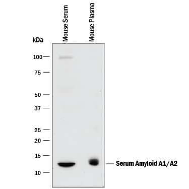
Detection of Mouse Serum Amyloid A1/A2 by Western Blot. Western blot shows mouse serum and mouse plasma. PVDF membrane was probed with 1 µg/mL of Goat Anti-Mouse Serum Amyloid A1/A2 Antigen Affinity-purified Polyclonal Antibody (Catalog # AF2948) followed by HRP-conjugated Anti-Goat IgG Secondary Antibody (Catalog # HAF017). A specific band was detected for Serum Amyloid A1/A2 at approximately 12 kDa (as indicated). This experiment was conducted under reducing conditions and using Immunoblot Buffer Group 1.
 View Larger
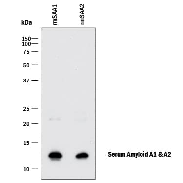
Detection of Mouse Serum Amyloid A1/A2 by Western Blot. Western blot shows recombinant mouse Serum Amyloid A1 and recombinant mouse Serum Amyloid A2. PVDF membrane was probed with 1 µg/mL of Goat Anti-Mouse Serum Amyloid A1/A2 Antigen Affinity-purified Polyclonal Antibody (Catalog # AF2948) followed by HRP-conjugated Anti-Goat IgG Secondary Antibody (Catalog # HAF017). A specific band was detected for Serum Amyloid A1/A2 at approximately 12 kDa (as indicated). This experiment was conducted under reducing conditions and using Immunoblot Buffer Group 1.
 View Larger
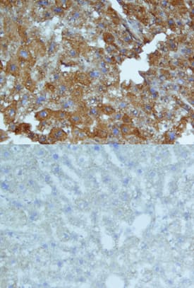
Serum Amyloid A1/A2 in Mouse Liver. Serum Amyloid A1/A2 was detected in perfusion fixed frozen sections of mouse liver using Goat Anti-Mouse Serum Amyloid A1/A2 Antigen Affinity-purified Polyclonal Antibody (Catalog # AF2948) at 15 µg/mL overnight at 4 °C. Tissue was stained using the Anti-Goat HRP-DAB Cell & Tissue Staining Kit (brown; Catalog # CTS008) and counterstained with hematoxylin (blue). Lower panel shows a lack of labeling when primary antibodies are omitted and tissue is stained only with secondary antibody followed by incubation with detection reagents. Specific staining was localized to cytoplasm. View our protocol for Chromogenic IHC Staining of Frozen Tissue Sections.
 View Larger
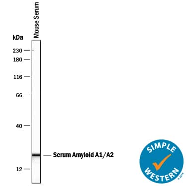
Detection of Mouse Serum Amyloid A1/A2 by Simple WesternTM. Simple Western lane view shows mouse serum, loaded at a 1:100 dilution. A specific band was detected for Serum Amyloid A1/A2 at approximately 14 kDa (as indicated) using 50 µg/mL of (Catalog # AF2948) followed by 1:50 dilution of HRP-conjugated Anti-Goat IgG Secondary Antibody (Catalog # HAF109). This experiment was conducted under reducing conditions and using the 12-230 kDa separation system.
 View Larger
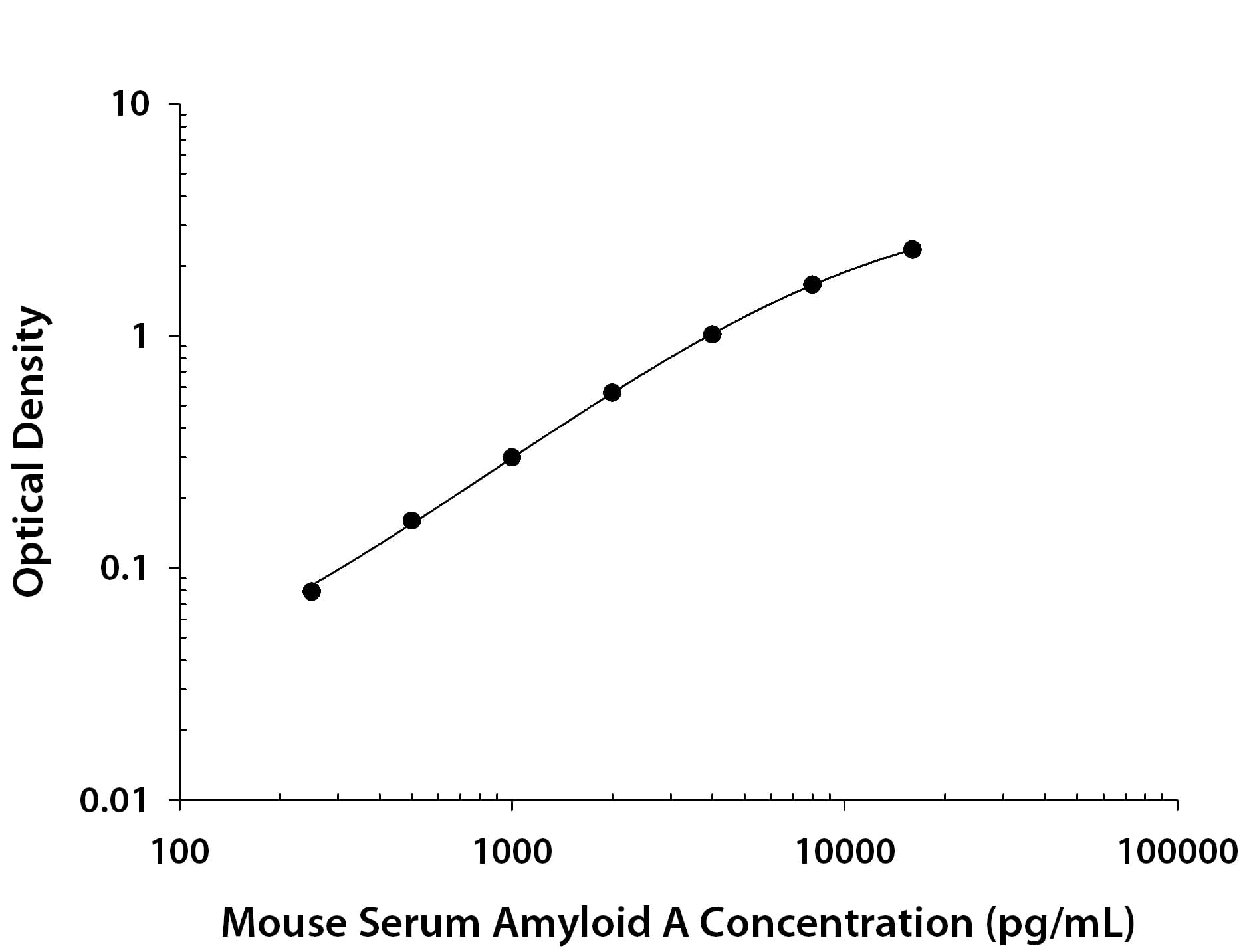
Mouse Serum Amyloid A1/A2 ELISA Standard Curve. Recombinant Mouse Serum Amyloid A1/A2 protein was serially diluted 2-fold and captured by Rat Anti-Mouse Serum Amyloid A1/A2 Monoclonal Antibody (Catalog # MAB2948) coated on a Clear Polystyrene Microplate (Catalog # DY990). Goat Anti-Mouse Serum Amyloid A1/A2 Antigen Affinity-purified Polyclonal Antibody (Catalog # AF2948) was biotinylated and incubated with the protein captured on the plate. Detection of the standard curve was achieved by incubating Streptavidin-HRP (Catalog # DY998) followed by Substrate Solution (Catalog # DY999) and stopping the enzymatic reaction with Stop Solution (Catalog # DY994).
 View Larger
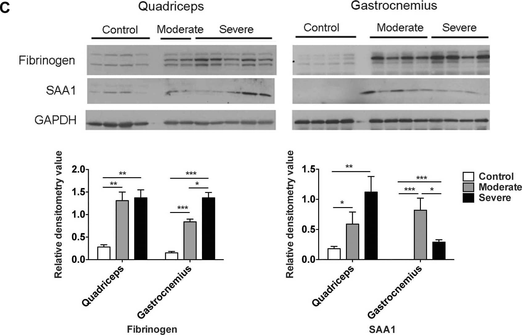
Detection of Serum Amyloid A1/A2 by Western Blot Robust expression of acute phase response proteins in skeletal muscle versus liver in C26 cachexia.A. Western blotting and quantitation of fibrinogen levels in control and C26 quadriceps and liver. Data (mean ± SEM) are expressed as relative densitometry value. **P<0.01, ***P<0.001. B, Western blotting analysis of fibrinogen standard proteins and quadriceps and liver extracts for control, CHO-IL6 injected nude mice and C26 injected CD2F1 mice. Quantitation was performed on the band indicated by the arrow. Data (means ± SEM) are expressed as ng fibrinogen / µg protein. *P<0.05, **P<0.01, ***P<0.001. C, Western blotting analysis demonstrates significantly increased fibrinogen and SAA1 protein levels in quadriceps and gastrocnemius in moderate and severe C26 cachexia. *P<0.05, **P<0.01, ***P<0.001. Image collected and cropped by CiteAb from the following open publication (https://pubmed.ncbi.nlm.nih.gov/21799891), licensed under a CC-BY license. Not internally tested by R&D Systems.
Reconstitution Calculator
Preparation and Storage
- 12 months from date of receipt, -20 to -70 °C as supplied.
- 1 month, 2 to 8 °C under sterile conditions after reconstitution.
- 6 months, -20 to -70 °C under sterile conditions after reconstitution.
Background: Serum Amyloid A1/A2
Mouse serum amyloid A protein-1 (SAA1) is a multifunctional apolipoprotein produced by hepatocytes in response to pro-inflammatory cytokines. It is secreted as a 12 kDa, 103 amino acid (aa), nonglycosylated polypeptide and circulates as part of the HDL complex. The SAA1 gene is one of three SAA genes in mouse, and, based on human, is likely to be allelic. The SAA1 gene product differs from the SAA2 gene product by only nine amino acids. In human, circulating SAA1 shows multiple proteolytically-generated isoforms, with anywhere from one to three amino acids being cleaved from either the N- or C-terminus. The same situation may exist in mouse. The amino acid sequence of mature mouse SAA1 is 72%, 72%, 67%, and 74% identical to mature human, rabbit, equine, and hamster SAA1, respectively.
Product Datasheets
Citations for Mouse Serum Amyloid A1/A2 Antibody
R&D Systems personnel manually curate a database that contains references using R&D Systems products. The data collected includes not only links to publications in PubMed, but also provides information about sample types, species, and experimental conditions.
37
Citations: Showing 1 - 10
Filter your results:
Filter by:
-
Hepatic Forkhead Box Protein A3 Regulates ApoA-I (Apolipoprotein A-I) Expression, Cholesterol Efflux, and Atherogenesis
Authors: Li Y, Xu Y, Jadhav K et al.
Arterioscler. Thromb. Vasc. Biol.
-
STAT3 Activation in Skeletal Muscle Links Muscle Wasting and the Acute Phase Response in Cancer Cachexia
Authors: Andrea Bonetto, Tufan Aydogdu, Noelia Kunzevitzky, Denis C. Guttridge, Sawsan Khuri, Leonidas G. Koniaris et al.
PLoS ONE
-
Nanosensor dosimetry of mouse blood proteins after exposure to ionizing radiation
Authors: Dokyoon Kim, Francesco Marchetti, Zuxiong Chen, Sasa Zaric, Robert J. Wilson, Drew A. Hall et al.
Scientific Reports
-
De novo Synthesis of SAA1 in the Placenta Participates in Parturition
Authors: Xiao-Wen Gan, Wang-Sheng Wang, Jiang-Wen Lu, Li-Jun Ling, Qiong Zhou, Hui-Juan Zhang et al.
Frontiers in Immunology
-
10,12-conjugated linoleic acid supplementation improves HDL composition and function in mice
Authors: Vaisar T, Wang S, Omer M et al.
Journal of lipid research
-
Serum Amyloid A Facilitates the Binding of High-Density Lipoprotein From Mice Injected With Lipopolysaccharide to Vascular Proteoglycans
Authors: Tsuyoshi Chiba, Mary Y. Chang, Shari Wang, Thomas N. Wight, Timothy S. McMillen, John F. Oram et al.
Arteriosclerosis, Thrombosis, and Vascular Biology
-
Probiotic treatment causes sex-specific neuroprotection after traumatic brain injury in mice
Authors: Holcomb, M;Marshall, A;Flinn, H;Lozano, M;Soriano, S;Gomez-Pinilla, F;Treangen, TJ;Villapol, S;
Research square
Species: Mouse
Sample Types: Serum
Applications: Western Blot -
Serum amyloid A promotes glycolysis of neutrophils during PD-1 blockade resistance in hepatocellular carcinoma
Authors: He, M;Liu, Y;Chen, S;Deng, H;Feng, C;Qiao, S;Chen, Q;Hu, Y;Chen, H;Wang, X;Jiang, X;Xia, X;Zhao, M;Lyu, N;
Nature communications
Species: Mouse
Sample Types: In Vivo
Applications: In vivo assay -
Serum amyloid A-dependent inflammasome activation and acute injury in a mouse model of experimental stroke
Authors: Yu, J;Zhu, H;Taheri, S;Lee, JY;Diamond, DM;Kirstein, C;Kindy, MS;
Research square
Species: Mouse
Sample Types: Cell Lysates
Applications: Western Blot -
Fecal Microbiota Transplantation Derived from Alzheimer's Disease Mice Worsens Brain Trauma Outcomes in Wild-Type Controls
Authors: S Soriano, K Curry, Q Wang, E Chow, TJ Treangen, S Villapol
International Journal of Molecular Sciences, 2022-04-19;23(9):.
Species: Mouse
Sample Types: Serum
Applications: Western Blot -
Promoting mechanism of serum amyloid a family expression in mouse intestinal epithelial cells
Authors: M Wakai, R Hayashi, Y Ueno, K Onishi, T Takasago, T Uchida, H Takigawa, R Yuge, Y Urabe, S Oka, Y Kitadai, S Tanaka
PLoS ONE, 2022-03-18;17(3):e0264836.
Species: Mouse
Sample Types: Whole Tissue
Applications: IHC -
Fractionated Irradiation of Right Thorax Induces Abscopal Damage on Bone Marrow Cells via TNF-alpha and SAA
Authors: Y Song, S Hu, J Zhang, L Zhu, X Zhao, Q Chen, J Zhang, Y Bai, Y Pan, C Shao
International Journal of Molecular Sciences, 2021-09-15;22(18):.
Species: Mouse
Sample Types: Cell Lysates
Applications: Western Blot -
Serum Amyloid A1/Toll-Like Receptor-4 Axis, an Important Link between Inflammation and Outcome of TBI Patients
Authors: V Farré-Alin, A Palomino-A, P Narros-Fer, AB Lopez-Rodr, C Decouty-Pe, A Muñoz-Mont, J Zamorano-F, B Mansilla-F, J Giner-Garc, P García-Fei, M Sáez-Alegr, AJ Palpán-Flo, JM Roda-Frade, CS Carabias, JM Rosa, B Civantos-M, S Yus-Teruel, L Gandía, A Lagares, BJ Hernández-, J Egea
Biomedicines, 2021-05-25;9(6):.
Species: Mouse
Sample Types: Cell Lysate, Whole Tissue
Applications: IHC, Western Blot -
Marked Increased Production of Acute Phase Reactants by Skeletal Muscle during Cancer Cachexia
Authors: IS Massart, G Paulissen, A Loumaye, P Lause, SA Pötgens, MM Thibaut, E Balan, L Deldicque, A Atfi, E Louis, D Gruson, LB Bindels, MA Meuwis, JP Thissen
Cancers (Basel), 2020-10-31;12(11):.
Species: Mouse
Sample Types: Cell Lysates
Applications: Western Blot -
Comparative plasma proteomics in muscle atrophy during cancer-cachexia and disuse: The search for atrokines
Authors: S Lim, KR Dunlap, ME Rosa-Caldw, WS Haynie, LT Jansen, TA Washington, NP Greene
Physiol Rep, 2020-10-01;8(19):e14608.
Species: Mouse
Sample Types: Plasma
Applications: Western Blot -
Serum amyloid A-containing HDL binds adipocyte-derived versican and macrophage-derived biglycan, reducing its anti-inflammatory properties
Authors: CY Han, I Kang, M Omer, S Wang, T Wietecha, TN Wight, A Chait
JCI Insight, 2020-09-24;0(0):.
Species: Human, Mouse
Sample Types: Whole Cells, Whole Tissue
Applications: ICC, IHC -
Presence of serum amyloid A3 in mouse plasma is dependent on the nature and extent of the inflammatory stimulus
Authors: A Chait, LJ den Hartig, S Wang, L Goodspeed, I Babenko, WA Altemeier, T Vaisar
Sci Rep, 2020-06-25;10(1):10397.
Species: Mouse
Sample Types: Tissue Homogenates
Applications: Western Blot -
Serum amyloid A is a soluble pattern recognition receptor that drives type 2 immunity
Authors: U Smole, N Gour, J Phelan, G Hofer, C Köhler, B Kratzer, PA Tauber, X Xiao, N Yao, J Dvorak, L Caraballo, L Puerta, S Rosskopf, J Chakir, E Malle, AP Lane, WF Pickl, S Lajoie, M Wills-Karp
Nat. Immunol., 2020-06-22;21(7):756-765.
Species: Mouse
Sample Types: In Vivo
Applications: Neutralization -
Serum amyloid A exhibits pH dependent antibacterial action and contributes to host defense against Staphylococcus aureus cutaneous infection
Authors: H Zheng, H Li, J Zhang, H Fan, L Jia, W Ma, S Ma, S Wang, H You, Z Yin, X Li
J. Biol. Chem., 2019-12-09;0(0):.
Species: Mouse
Sample Types: Protein, Whole Tissue
Applications: IHC-P, Western Blot -
Serum Amyloid A Protein as a Potential Biomarker for Severity and Acute Outcome in Traumatic Brain Injury
Authors: E Wicker, L Benton, K George, W Furlow, S Villapol
Biomed Res Int, 2019-04-16;2019(0):5967816.
Species: Mouse
Sample Types: Serum
Applications: Western Blot -
Increased hypothalamic microglial activation after viral-induced pneumococcal lung infection is associated with excess serum amyloid A production
Authors: H Wang, M Blackall, L Sominsky, SJ Spencer, R Vlahos, M Churchill, S Bozinovski
J Neuroinflammation, 2018-07-06;15(1):200.
Species: Mouse
Sample Types: Whole Tissue
Applications: IHC -
Serum Amyloid A1 is an epithelial pro-restitutive factor
Authors: BH Hinrichs, JD Matthews, D Siuda, MN O'Leary, AA Wolfarth, BJ Saeedi, A Nusrat, AS Neish
Am. J. Pathol., 2018-01-31;0(0):.
Species: Human
Sample Types: Whole Cells
Applications: Cell Culture -
Amyloid deposition in a mouse model humanized at the transthyretin and retinol-binding protein 4 loci
Authors: X Li, Y Lyu, J Shen, Y Mu, L Qiang, L Liu, K Araki, BP Imbimbo, KI Yamamura, S Jin, Z Li
Lab. Invest., 2018-01-12;0(0):.
Species: Mouse
Sample Types: Whole Tissue
Applications: IHC -
Scavenger receptor A1 prevents metastasis of non-small cell lung cancer via suppression of macrophage serum amyloid A1
Authors: Y Zhang, Y Wei, B Jiang, L Chen, H Bai, X Zhu, X Li, H Zhang, Q Yang, J Ma, Y Xu, J Ben, DC Christiani, Q Chen
Cancer Res, 2017-02-15;0(0):.
Species: Human
Sample Types: Whole Tissue
Applications: IHC -
Zymosan-mediated inflammation impairs in vivo reverse cholesterol transport.
Authors: Malik P, Berisha SZ, Santore J
J. Lipid Res., 2011-02-19;52(5):951-7.
Species: Mouse
Sample Types: Plasma
Applications: Western Blot -
Transmission of circulating cell-free AA amyloid oligomers in exosomes vectors via a prion-like mechanism.
Authors: Tasaki M, Ueda M, Ochiai S, Tanabe Y, Murata S, Misumi Y, Su Y, Sun X, Shinriki S, Jono H, Shono M, Obayashi K, Ando Y
Biochem. Biophys. Res. Commun., 2010-08-31;400(4):559-62.
Species: Mouse
Sample Types: Cell Culture Supernates
Applications: Western Blot -
Role of APAF-1, E-cadherin and peritumoral lymphocytic infiltration in tumour budding in colorectal cancer.
Authors: Zlobec I, Lugli A, Baker K, Roth S, Minoo P, Hayashi S, Terracciano L, Jass JR
J. Pathol., 2007-07-01;212(3):260-8.
Species: Mouse
Sample Types: Serum
Applications: ELISA Development -
Fish oil increases cholesterol storage in white adipose tissue with concomitant decreases in inflammation, hepatic steatosis, and atherosclerosis in mice.
Authors: Saraswathi V, Gao L, Morrow JD, Chait A, Niswender KD, Hasty AH
J. Nutr., 2007-07-01;137(7):1776-82.
Species: Mouse
Sample Types: Serum
Applications: ELISA Development -
Serum amyloid alpha 1-2 are not required for liver inflammation in the 4T1 murine breast cancer model
Authors: Chenfeng He, Riyo Konishi, Ayano Harata, Yuki Nakamura, Rin Mizuno, Mayuko Yoda et al.
Frontiers in Immunology
-
Serum amyloid A impairs the antiinflammatory properties of HDL
Authors: Chang Yeop Han, Chongren Tang, Myriam E. Guevara, Hao Wei, Tomasz Wietecha, Baohai Shao et al.
Journal of Clinical Investigation
-
CETP Inhibition Improves HDL Function but Leads to Fatty Liver and Insulin Resistance in CETP-Expressing Transgenic Mice on a High-Fat Diet
Authors: Lin Zhu, Thao Luu, Christopher H. Emfinger, Bryan A. Parks, Jeanne Shi, Elijah Trefts et al.
Diabetes
-
Role of myeloperoxidase in abdominal aortic aneurysm formation: mitigation by taurine
Authors: Ha Won Kim, Andra L. Blomkalns, Mourad Ogbi, Manesh Thomas, Daniel Gavrila, Bonnie S. Neltner et al.
American Journal of Physiology-Heart and Circulatory Physiology
-
Experimental transmission of systemic AA amyloidosis in autoimmune disease and type 2 diabetes mellitus model mice
Authors: Mayuko Maeda, Tomoaki Murakami, Naeem Muhammad, Yasuo Inoshima, Naotaka Ishiguro
Experimental Animals
-
Serum Amyloid A is Expressed in the Brain After Traumatic Brain Injury in a Sex-Dependent Manner
Authors: Sirena Soriano, Bridget Moffet, Evan Wicker, Sonia Villapol
Cellular and Molecular Neurobiology
-
Hepatic Expression of Serum Amyloid A1 Is Induced by Traumatic Brain Injury and Modulated by Telmisartan
Authors: Sonia Villapol, Dmitry Kryndushkin, Maria G. Balarezo, Ashley M. Campbell, Juan M. Saavedra, Frank P. Shewmaker et al.
The American Journal of Pathology
-
Amyloid persistence in decellularized liver: biochemical and histopathological characterization
Authors: Giuseppe Mazza, J. Paul Simons, Raya Al-Shawi, Stephan Ellmerich, Luca Urbani, Sofia Giorgetti et al.
Amyloid
-
Serum Amyloid A Proteins Induce Pathogenic Th17 Cells and Promote Inflammatory Disease
Authors: June-Yong Lee, Jason A. Hall, Lina Kroehling, Lin Wu, Tariq Najar, Henry H. Nguyen et al.
Cell
FAQs
No product specific FAQs exist for this product, however you may
View all Antibody FAQsReviews for Mouse Serum Amyloid A1/A2 Antibody
Average Rating: 3.7 (Based on 3 Reviews)
Have you used Mouse Serum Amyloid A1/A2 Antibody?
Submit a review and receive an Amazon gift card.
$25/€18/£15/$25CAN/¥75 Yuan/¥2500 Yen for a review with an image
$10/€7/£6/$10 CAD/¥70 Yuan/¥1110 Yen for a review without an image
Filter by:
Protein loaded:
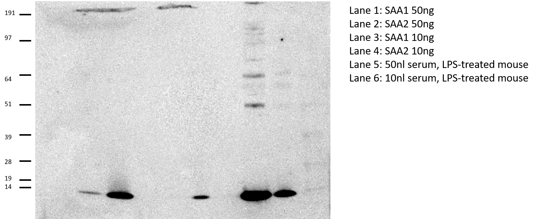
Lane 1: SAA1 50ng
Lane 2: SAA2 50ng
Lane 3: SAA1 10ng
Lane 4: SAA2 10ng
Lane 5: 50nl serum, LPS-treated mouse
Lane 6: 10nl serum, LPS-treated mouse
Recombinant proteins were from R and D systems
Prepared in Invitrogen 1x LDS sample buffer plus 1x reducing agent.
Run on 4-12% Bis-tris gel under reducing conditions in MOPS buffer. Transferred using iBlot 20V 7 min
Blocked 1 hour 5% BSA/TBS-T
Incubated overnight 4 C, antibody prepared 1/1000 in 5% BSA/TBS-T
Secondary antibody was anti-goat from Jackson Immuno 1:5000 for 30 minutes.
West Pico Chemiluminescent Reagent
ImageQuant LAS4000 for imaging
*This antibody gives great signal, but predominantly detects SAA2 over SAA1.
