Mouse/Rat FABP4/A-FABP Antibody Summary
Met1-Ala132
Accession # P04117
Applications
Please Note: Optimal dilutions should be determined by each laboratory for each application. General Protocols are available in the Technical Information section on our website.
Scientific Data
 View Larger
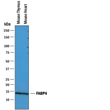
Detection of Mouse FABP4/A‑FABP by Western Blot. Western blot shows lysates of mouse thymus tissue and mouse heart tissue. PVDF membrane was probed with 0.25 µg/mL of Goat Anti-Mouse/Rat FABP4/A-FABP Antigen Affinity-purified Polyclonal Antibody (Catalog # AF1443) followed by HRP-conjugated Anti-Goat IgG Secondary Antibody (Catalog # HAF019). A specific band was detected for FABP4/A-FABP at approximately 14 kDa (as indicated). This experiment was conducted under reducing conditions and using Immunoblot Buffer Group 1.
 View Larger
FABP4 in Mouse Adipocytes. FABP4 was detected in immersion fixed mouse mesenchymal stem cell-derived adipocytes using 10 µg/mL Goat Anti-Mouse/Rat FABP4 Antigen Affinity-purified Polyclonal Antibody (Catalog # AF1443) for 3 hours at room temperature. Cells were stained with the NorthernLights™ 557-conjugated Anti-Goat IgG Secondary Antibody (red; Catalog # NL001) and counterstained with DAPI (blue). View our protocol for Fluorescent ICC Staining of Cells on Coverslips.
 View Larger
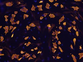
FABP4 in Rat Mesenchymal Stem Cells. FABP4 was detected in immersion fixed rat mesenchymal stem cells differentiated to adipocytes using Goat Anti-Mouse/Rat FABP4 Antigen Affinity-purified Polyclonal Antibody (Catalog # AF1443) at 10 µg/mL for 3 hours at room temperature. Cells were stained using the NorthernLights™ 557-conjugated Anti-Goat IgG Secondary Antibody (yellow; Catalog # NL001) and counterstained with DAPI (blue). View our protocol for Fluorescent ICC Staining of Cells on Coverslips.
 View Larger
FABP4 in Mouse Intestine. FABP4 was detected in perfusion fixed frozen sections of mouse intestine using Goat Anti-Mouse/Rat FABP4 Antigen Affinity-purified Polyclonal Antibody (Catalog # AF1443) at 5 µg/mL overnight at 4 °C. Tissue was stained using the Anti-Goat HRP-DAB Cell & Tissue Staining Kit (brown; Catalog # CTS008) and counterstained with hematoxylin (blue). Specific labeling was localized to the plasma membrane of epithelial cells in intestinal glands. View our protocol for Chromogenic IHC Staining of Frozen Tissue Sections.
 View Larger
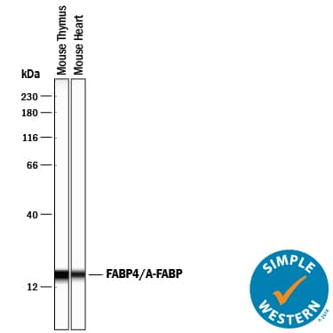
Detection of Mouse FABP4/A‑FABP by Simple WesternTM. Simple Western lane view shows lysates of mouse thymus tissue and mouse heart tissue, loaded at 0.2 mg/mL. A specific band was detected for FABP4/A-FABP at approximately 17 kDa (as indicated) using 2.5 µg/mL of Goat Anti-Mouse/Rat FABP4/A-FABP Antigen Affinity-purified Polyclonal Antibody (Catalog # AF1443) followed by 1:50 dilution of HRP-conjugated Anti-Goat IgG Secondary Antibody (Catalog # HAF109). This experiment was conducted under reducing conditions and using the 12-230 kDa separation system.
 View Larger
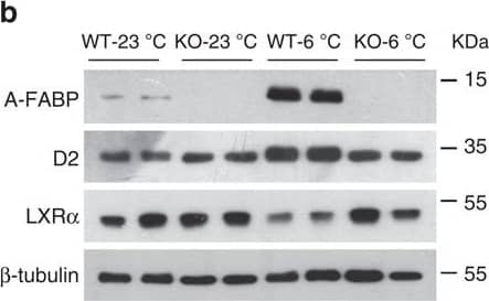
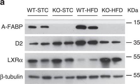
Detection of Mouse FABP4/A-FABP by Western Blot A-FABP mediates expression of Dio2 via inhibition of LXR alpha.(a,b) BAT isolated from WT and A-FABP KO mice (a) fed with STC or HFD for 24 weeks or (b) subjected to room temperature (23 °C) or cold exposure (6 °C) for 24 h as indicated in Fig. 2 were subjected to immunoblotting using an antibody against A-FABP, type II iodothyronine deiodinase (D2), liver X receptor alpha (LXR alpha ) and beta -tubulin. The bar charts below are the band intensity of each protein relative to beta -tubulin and expressed as arbitrary units, N.D., not detected (n=6). (c,d) The mRNA abundance of A-FABP, Dio2 and LXR alpha in BAT of above WT and A-FABP KO mice (c) fed with STC or HFD for 24 weeks or (d) subjected to cold exposure (6 °C) for 24 h (n=6). (e) The mRNA abundance of A-FABP, LXR alpha and Dio2 in WT or A-FABP-deficient primary brown adipocytes incubated with PBS or recombinant A-FABP (rA-FABP, 2 μg ml−1) for 24 h (n=6). (f) The mRNA abundance of LXR alpha downstream target genes (SCD-1, SREBP-1c) and Dio2 in WT and A-FABP-deficient primary brown adipocytes treated with or without LXR alpha agonist TO901317 (TO;1 μM) and/or rA-FABP (2 μg ml−1) for 24 h (n=6). Uncropped western blot images are shown in Supplementary Fig. 13. Data are represented as mean±s.e.m. *P<0.05, **P<0.01 (One-way analysis of variance with Bonferroni correction for multiple comparisons). Image collected and cropped by CiteAb from the following open publication (https://pubmed.ncbi.nlm.nih.gov/28128199), licensed under a CC-BY license. Not internally tested by R&D Systems.
 View Larger
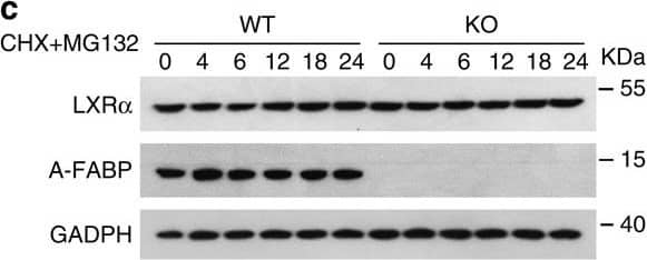
Detection of Mouse FABP4/A-FABP by Western Blot A-FABP accelerates proteasomal degradation of LXR alpha.(a) Primary adipocytes derived from male 6-week-old A-FABP KO mice or WT littermates were treated with actinomycin D (actD, 1 μg ml−1) or vehicle (PBS). The mRNA level of LXR alpha was determined by real-time PCR at time points as indicated (n=4). (b,c) Primary brown adipocytes derived from male 6-week-old A-FABP KO mice or WT littermates were treated with (b) cycloheximide (CHX, 50 μg ml−1) or (c) CHX (50 μg ml−1) together with MG132 (10 μM) for different periods were subjected to immunoblotting using an antibody against LXR alpha, A-FABP and GADPH as indicated (n=4). (d) Primary brown adipocytes derived male 6-week-old A-FABP KO mice or WT littermates were infected with adenovirus overexpressing A-FABP (Ad-AFABP) or luciferase (Ad-Luci) for 48 h, followed by treatment with CHX (50 μg ml−1) for 0,6,12 and 24 h, and then subjected to immunoblotting using an antibody against LXR alpha, A-FABP and GADPH as indicated (n=4). The right panel is the band intensity of LXR alpha normalized with GAPDH, and expressed as percentage relative to baseline (0 h). Uncropped western blot images are shown in Supplementary Fig. 14. Data are represented as mean±s.e.m. *P<0.05, **P<0.01 (Students' t-test). Image collected and cropped by CiteAb from the following open publication (https://pubmed.ncbi.nlm.nih.gov/28128199), licensed under a CC-BY license. Not internally tested by R&D Systems.
 View Larger
Detection of Mouse FABP4/A-FABP by Western Blot NPGPx is a suppressor of adipogenesis via ROS regulationRelative NPGPx expression assayed by qRT-PCR (left panel) and Western blots (right panel) in the stromal vascular fraction (SVF) and adipocyte fraction isolated from the gonadal fat of C57BL/6 mice (n = 3 per group).Relative NPGPx expression in preadipocytes (Lin−CD34+CD29+Sca1+), endothelial cells (CD31+), macrophages (CD11b+) and adipocytes of adipose tissue measured by in C57BL/6 mice (n = 3 per group).NPGPx, CCAAT/enhancer-binding protein beta (C/EBP beta ) and peroxisome proliferator-activated receptor gamma (PPAR gamma ) mRNA level during induced 3T3-L1 adipogenesis (n = 5 per group).NPGPx expression assayed by Western blots during 3T3L1 adipogenesis.NBT reduction staining of wild-type SVF cells (WT), NPGPx-knockout SVF cells (KO) and NPGPx-knockout SVF cells expressing human NPGPx (KO + hNPGPx) (left panels). Quantification of NBT reduction stains 8 days after induction (n = 3 per group) (right panels). *p = 0.0002 for WT vs. KO by independent Student's t-test.Oil Red O stain of WT, KO and KO + hNPGPx SVF (left panels). Quantification of Oil Red O stain 8 days after induction (n = 3 per group) (right panels). *p = 0.0002 for WT vs. KO by independent Student's t-test.Western blots showing ap2 and adiponectin expression in WT, KO and KO + hNPGPx SVF cells 8 days after induction.NBT reduction stain of NPGPx knockdown (KD) and control (V) 3T3-L1 preadipocytes after adipogenic induction (left panels). Quantification of NBT reduction stain at day 8 (n = 3 per group) (right panels). *p = 0.0004 by independent Student's t-test.Oil Red O staining of KD and V 3T3-L1 preadipocytes after adipogenic induction (left panels). Quantification of Oil Red O stain at day 8 (n = 3 per group) (right panels). *p = 0.0005 by independent Student's t-test.Western blots showing ap2 and adiponectin expression in NPGPx KD and V 3T3-L1 preadipocytes after adipogenic stimulation.NBT reduction stain and Oil Red O stain of NPGPx KD and V 3T3-L1 cells 8 days after induction. Cells were treated at with NAC at various doses the first 2 days after induction.Quantification of NBT reduction stain and Oil Red O stain in (K) (n = 3 per group).Correlation (R = 0.96) between ROS levels (Rel. NBT staining) and triglyceride contents (Rel. Oil-Red-O staining) in NPGPx KD and V 3T3-L1 cells 8 days after induction.Quantification of Oil Red O staining of NPGPx KD and V 3T3-L1 cells 8 days after induction. Cells were treated with H2O2 at different concentrations the first 2 days after induction (n = 3 per group). *p = 0.006, **p = 0.006, #p < 0.0001, ##p < 0.0001 by independent Student's t-test. All values are presented as means ± S.E.M. Image collected and cropped by CiteAb from the following open publication (https://pubmed.ncbi.nlm.nih.gov/23828861), licensed under a CC-BY license. Not internally tested by R&D Systems.
 View Larger
Detection of Mouse FABP4/A-FABP by Western Blot A-FABP mediates expression of Dio2 via inhibition of LXR alpha.(a,b) BAT isolated from WT and A-FABP KO mice (a) fed with STC or HFD for 24 weeks or (b) subjected to room temperature (23 °C) or cold exposure (6 °C) for 24 h as indicated in Fig. 2 were subjected to immunoblotting using an antibody against A-FABP, type II iodothyronine deiodinase (D2), liver X receptor alpha (LXR alpha ) and beta -tubulin. The bar charts below are the band intensity of each protein relative to beta -tubulin and expressed as arbitrary units, N.D., not detected (n=6). (c,d) The mRNA abundance of A-FABP, Dio2 and LXR alpha in BAT of above WT and A-FABP KO mice (c) fed with STC or HFD for 24 weeks or (d) subjected to cold exposure (6 °C) for 24 h (n=6). (e) The mRNA abundance of A-FABP, LXR alpha and Dio2 in WT or A-FABP-deficient primary brown adipocytes incubated with PBS or recombinant A-FABP (rA-FABP, 2 μg ml−1) for 24 h (n=6). (f) The mRNA abundance of LXR alpha downstream target genes (SCD-1, SREBP-1c) and Dio2 in WT and A-FABP-deficient primary brown adipocytes treated with or without LXR alpha agonist TO901317 (TO;1 μM) and/or rA-FABP (2 μg ml−1) for 24 h (n=6). Uncropped western blot images are shown in Supplementary Fig. 13. Data are represented as mean±s.e.m. *P<0.05, **P<0.01 (One-way analysis of variance with Bonferroni correction for multiple comparisons). Image collected and cropped by CiteAb from the following open publication (https://pubmed.ncbi.nlm.nih.gov/28128199), licensed under a CC-BY license. Not internally tested by R&D Systems.
 View Larger
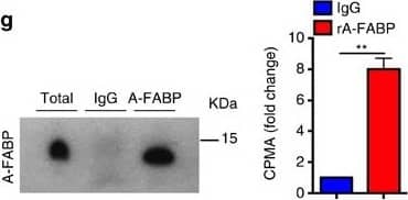
Detection of Mouse FABP4/A-FABP by Western Blot Circulating A-FABP facilitates the uptake of free fatty acid into adipocytes.(a) Circulating A-FABP and (b) FFA profile of male 4-week-old C57BL/6N mice fed with HFD for 24 weeks (n=8). (c) Circulating A-FABP and (d) FFA level of male 8-week-old C57BL/6N mice during cold exposure (6 °C) for 4 h (n=8). (e) Circulating A-FABP and (f) FFA level of male 8-week-old C57BL/6N mice intraperitoneally injected with norepinephrine (NE; 1 mg kg−1) or PBS (vehicle) for 4 h under fasting condition (n=6). (g) Co-immunoprecipitation (Co-IP) of A-FABP and 3H-palmitate in serum of male 8-week-old C57BL/6N mice after administration of 3H-palmitate (2 μCi) for 4 h. Right panel is the 3H-palmitate radioactivity of the co-immunoprecipitated A-FABP protein (n=6). (h,i) 3H-palmitate uptake in BAT and WAT of 8-week-old A-FABP KO mice and their WT littermates infused with PBS or (h) recombinant A-FABP (rA-FABP; 1 μg h−1) or (i) mutant R126Q (1 μg h−1) (n=6). (j) BODIPY-FA uptake in WT or A-FABP-deficient brown adipocytes treated with PBS or rA-FABP (2 μg ml−1) for 10 min (min) (n=6). (k) 3H-palmitate uptake in A-FABP-deficient adipocytes incubated with PBS, bovine serum albumin (BSA; 3 μg ml−1) or rA-FABP (2 μg ml−1) (n=6). (l) In vitro fluorescent imaging analysis of brown adipocytes treated with BODIPY-FA (2 μM) with or without pre-incubation with fluorescent-labelled rA-FABP (2 μg ml−1). Images were taken at 5, 10 and 30 min after treatment. Control image was taken at 30 min in which A-FABP-deficient brown adipocytes were incubated with BODIPY-FA without pre-incubation with rA-FABP. Scale bar, 20 μm, with magnification of 400 ×. Representative images from three independent experiments are shown (n=6). (m) Oxygen consumption rate (OCR) and its mean value (lower panel) of A-FABP-deficient brown adipocytes treated with palmitate (PA: 200 nM) with or without pre-incubation with rA-FABP (2 μg ml−1) (n=6). CPMA, count per minutes for beta particles; RFU, relative fluorescence units; OCR, oxygen consumption rate; FCCP, carbonyl cyanide-4-(trifluoromethoxy) phenylhydrazone; R/A, rotenone/antimycin A. Uncropped image for co-immunoprecipitation is shown in Supplementary Fig. 13. Data are represented as mean±s.e.m. *P<0.05, **P<0.01, ***P<0.001 (Student's t-test (a–g), one-way analysis of variance with Bonferroni correction for multiple comparisons (h–k,m)). Image collected and cropped by CiteAb from the following open publication (https://pubmed.ncbi.nlm.nih.gov/28128199), licensed under a CC-BY license. Not internally tested by R&D Systems.
 View Larger
Detection of Mouse FABP4/A-FABP by Western Blot NPGPx is a suppressor of adipogenesis via ROS regulationRelative NPGPx expression assayed by qRT-PCR (left panel) and Western blots (right panel) in the stromal vascular fraction (SVF) and adipocyte fraction isolated from the gonadal fat of C57BL/6 mice (n = 3 per group).Relative NPGPx expression in preadipocytes (Lin−CD34+CD29+Sca1+), endothelial cells (CD31+), macrophages (CD11b+) and adipocytes of adipose tissue measured by in C57BL/6 mice (n = 3 per group).NPGPx, CCAAT/enhancer-binding protein beta (C/EBP beta ) and peroxisome proliferator-activated receptor gamma (PPAR gamma ) mRNA level during induced 3T3-L1 adipogenesis (n = 5 per group).NPGPx expression assayed by Western blots during 3T3L1 adipogenesis.NBT reduction staining of wild-type SVF cells (WT), NPGPx-knockout SVF cells (KO) and NPGPx-knockout SVF cells expressing human NPGPx (KO + hNPGPx) (left panels). Quantification of NBT reduction stains 8 days after induction (n = 3 per group) (right panels). *p = 0.0002 for WT vs. KO by independent Student's t-test.Oil Red O stain of WT, KO and KO + hNPGPx SVF (left panels). Quantification of Oil Red O stain 8 days after induction (n = 3 per group) (right panels). *p = 0.0002 for WT vs. KO by independent Student's t-test.Western blots showing ap2 and adiponectin expression in WT, KO and KO + hNPGPx SVF cells 8 days after induction.NBT reduction stain of NPGPx knockdown (KD) and control (V) 3T3-L1 preadipocytes after adipogenic induction (left panels). Quantification of NBT reduction stain at day 8 (n = 3 per group) (right panels). *p = 0.0004 by independent Student's t-test.Oil Red O staining of KD and V 3T3-L1 preadipocytes after adipogenic induction (left panels). Quantification of Oil Red O stain at day 8 (n = 3 per group) (right panels). *p = 0.0005 by independent Student's t-test.Western blots showing ap2 and adiponectin expression in NPGPx KD and V 3T3-L1 preadipocytes after adipogenic stimulation.NBT reduction stain and Oil Red O stain of NPGPx KD and V 3T3-L1 cells 8 days after induction. Cells were treated at with NAC at various doses the first 2 days after induction.Quantification of NBT reduction stain and Oil Red O stain in (K) (n = 3 per group).Correlation (R = 0.96) between ROS levels (Rel. NBT staining) and triglyceride contents (Rel. Oil-Red-O staining) in NPGPx KD and V 3T3-L1 cells 8 days after induction.Quantification of Oil Red O staining of NPGPx KD and V 3T3-L1 cells 8 days after induction. Cells were treated with H2O2 at different concentrations the first 2 days after induction (n = 3 per group). *p = 0.006, **p = 0.006, #p < 0.0001, ##p < 0.0001 by independent Student's t-test. All values are presented as means ± S.E.M. Image collected and cropped by CiteAb from the following open publication (https://pubmed.ncbi.nlm.nih.gov/23828861), licensed under a CC-BY license. Not internally tested by R&D Systems.
 View Larger
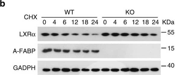
Detection of Mouse FABP4/A-FABP by Western Blot A-FABP accelerates proteasomal degradation of LXR alpha.(a) Primary adipocytes derived from male 6-week-old A-FABP KO mice or WT littermates were treated with actinomycin D (actD, 1 μg ml−1) or vehicle (PBS). The mRNA level of LXR alpha was determined by real-time PCR at time points as indicated (n=4). (b,c) Primary brown adipocytes derived from male 6-week-old A-FABP KO mice or WT littermates were treated with (b) cycloheximide (CHX, 50 μg ml−1) or (c) CHX (50 μg ml−1) together with MG132 (10 μM) for different periods were subjected to immunoblotting using an antibody against LXR alpha, A-FABP and GADPH as indicated (n=4). (d) Primary brown adipocytes derived male 6-week-old A-FABP KO mice or WT littermates were infected with adenovirus overexpressing A-FABP (Ad-AFABP) or luciferase (Ad-Luci) for 48 h, followed by treatment with CHX (50 μg ml−1) for 0,6,12 and 24 h, and then subjected to immunoblotting using an antibody against LXR alpha, A-FABP and GADPH as indicated (n=4). The right panel is the band intensity of LXR alpha normalized with GAPDH, and expressed as percentage relative to baseline (0 h). Uncropped western blot images are shown in Supplementary Fig. 14. Data are represented as mean±s.e.m. *P<0.05, **P<0.01 (Students' t-test). Image collected and cropped by CiteAb from the following open publication (https://pubmed.ncbi.nlm.nih.gov/28128199), licensed under a CC-BY license. Not internally tested by R&D Systems.
 View Larger
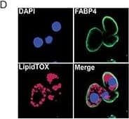
Detection of Mouse FABP4/A-FABP by Immunocytochemistry/ Immunofluorescence Chemotherapy leads to fat loss in diet induced obese miceA. Weight of epididymal white adipose tissue (eWAT) from LFD, HFD, CY200 and MTX50 treated mice 1 week after last chemotherapy treatment. Each treatment group consisted of 6 mice, showing mean ± SD, data representative of 2 biological replicates. Comparisons by Student's t-test: ns - not significant, * p<0.02, ** p<0.004, *** p<0.002, **** p<0.0007 B. Representative illustration of abdominal cavities measured in (A), showing that large visceral fat deposits (arrows) were reduced post chemotherapy in HFD mice. C. Relative percent frequency of eWAT-associated adipocyte stem-like cells (ASC) in HFD mice treated with MTX12.5 or CY100. Each treatment group consisted of 3 mice, showing mean ± SD, data representative of 2 biological replicates. Statistics as in (A). D. Representative confocal imagery of sorted ASC cultured in adipogenic medium displaying lipid droplets. DAPI (blue)- DNA nuclear stain, FABP4 (green)- lipid transport protein in adipocytes, LipidTOX (red)- neutral lipid stain. Image collected and cropped by CiteAb from the following open publication (https://www.oncotarget.com/lookup/doi/10.18632/oncotarget.14576), licensed under a CC-BY license. Not internally tested by R&D Systems.
 View Larger
Detection of Porcine FABP4/A-FABP by Immunocytochemistry/ Immunofluorescence In vitro differentiation capacity of young and old BM-MSCs: (A) Representative micrographs from each experimental group showing morphological change after 21 days of differentiation. (B) Results of adipogenic, osteogenic and chondrogenic differentiation. Representative micrographs showing anti-FABP4, Oil Red O, anti-osteocalcin, Alizarin Red-S and anti-aggrecan staining in cultured BM-MSCs from each experimental group. Image collected and cropped by CiteAb from the following open publication (https://pubmed.ncbi.nlm.nih.gov/21992089), licensed under a CC-BY license. Not internally tested by R&D Systems.
 View Larger
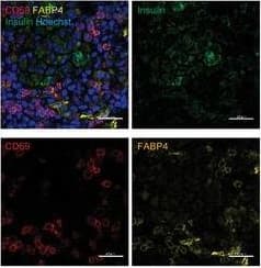
Detection of Mouse FABP4/A-FABP by Immunohistochemistry-Frozen Pancreatic TRM cell recruitment during T1D progression is FABP4‐dependent. G) Representative images of immunofluorescent co‐staining of FABP4, CD69,&insulin in the pancreas of 10‐week‐old NOD mice (Scale bar 20 µm). Image collected & cropped by CiteAb from the following open publication (https://pubmed.ncbi.nlm.nih.gov/38884133), licensed under a CC-BY license. Not internally tested by R&D Systems.
Reconstitution Calculator
Preparation and Storage
- 12 months from date of receipt, -20 to -70 °C as supplied.
- 1 month, 2 to 8 °C under sterile conditions after reconstitution.
- 6 months, -20 to -70 °C under sterile conditions after reconstitution.
Background: FABP4/A-FABP
FABP-4 (fatty acid binding protein-4; also Adipocyte-type FABP, ALBP and aP2) is a 14-15 kDa member of the fatty acid binding protein family, calycin superfamily of molecules. It is expressed by adipocytes, macrophages, monocyte-derived DC and myoepithelial cells, and is known to serve as a carrier for select PPAR gamma fatty acid ligands as they enter the nucleus. These ligands, or fatty acids, facilitate PPAR gamma -mediated gene transcription. Mouse FABP-4 is 132 amino acids (aa) in length. It is a two beta -sheet molecule that contains an NLS (aa 22-32) and a fatty acid binding segment (aa 127-129). Mouse FABP-4 is 94% and 92% aa identical to rat and human FABP-4, respectively.
Product Datasheets
Citations for Mouse/Rat FABP4/A-FABP Antibody
R&D Systems personnel manually curate a database that contains references using R&D Systems products. The data collected includes not only links to publications in PubMed, but also provides information about sample types, species, and experimental conditions.
41
Citations: Showing 1 - 10
Filter your results:
Filter by:
-
Endothelial APLNR regulates tissue fatty acid uptake and is essential for apelin’s glucose-lowering effects
Authors: Cheol Hwangbo, Jingxia Wu, Irinna Papangeli, Takaomi Adachi, Bikram Sharma, Saejeong Park et al.
Science Translational Medicine
-
Fast and high-fidelity in situ 3D imaging protocol for stem cells and niche components for mouse organs and tissues
Authors: Mehmet Saçma, Francesca Matteini, Medhanie A. Mulaw, Ali Hageb, Ruzhica Bogeska, Vadim Sakk et al.
STAR Protocols
-
Vav1 Regulates Mesenchymal Stem Cell Differentiation Decision Between Adipocyte and Chondrocyte via Sirt1
Authors: Peng Qu, Lizhen Wang, Yongfen Min, Lois McKennett, Jonathan R. Keller, P. Charles Lin
Stem Cells
-
Lipolysis of bone marrow adipocytes is required to fuel bone and the marrow niche during energy deficits
Authors: Ziru Li, Emily Bowers, Junxiong Zhu, Hui Yu, Julie Hardij, Devika P Bagchi et al.
eLife
-
Novel Populations of Lung Capillary Endothelial Cells and Their Functional Significance
Authors: James J, Dekan A, Niihori M et al.
Research square
-
Secreted frizzled‑related protein 4 promotes brown adipocyte differentiation
Authors: Hua Guan, Huiyuan Zheng, Jin Zhang, Aoqi Xiang, Yongxin Li, Huadong Zheng et al.
Experimental and Therapeutic Medicine
-
Expression and enhancement of FABP4 in septoclasts of the growth plate in FABP5-deficient mouse tibiae
Authors: Yasuhiko Bando, Nobuko Tokuda, Yudai Ogasawara, Go Onozawa, Arata Nagasaka, Koji Sakiyama et al.
Histochemistry and Cell Biology
-
A vicious loop of fatty acid-binding protein 4 and DNA methyltransferase 1 promotes acute myeloid leukemia and acts as a therapeutic target
Authors: F Yan, N Shen, J X Pang, N Zhao, Y W Zhang, A M Bode et al.
Leukemia
-
Chemical Landscape of Adipocytes Derived from 3T3-L1 Cells Investigated by Fourier Transform Infrared and Raman Spectroscopies
Authors: Augustyniak, K;Lesniak, M;Golan, MP;Latka, H;Wojtan, K;Zdanowski, R;Kubiak, JZ;Malek, K;
International journal of molecular sciences
Species: Mouse
Sample Types: Whole Cells
Applications: Immunocytochemistry -
FABP4-mediated lipid accumulation and lipolysis in tumor-associated macrophages promote breast cancer metastasis
Authors: Yorek, M;Jiang, X;Liu, S;Hao, J;Yu, J;Avellino, A;Liu, Z;Curry, M;Keen, H;Shao, J;Kanagasabapathy, A;Kong, M;Xiong, Y;Sauter, ER;Sugg, SL;Li, B;
eLife
Species: Mouse, Transgenic Mouse
Sample Types: Whole Cells
Applications: Flow Cytometry -
Rosiglitazone and trametinib exhibit potent anti-tumor activity in a mouse model of muscle invasive bladder cancer
Authors: Plumber, SA;Tate, T;Al-Ahmadie, H;Chen, X;Choi, W;Basar, M;Lu, C;Viny, A;Batourina, E;Li, J;Gretarsson, K;Alija, B;Molotkov, A;Wiessner, G;Lee, BHL;McKiernan, J;McConkey, DJ;Dinney, C;Czerniak, B;Mendelsohn, CL;
Nature communications
Species: Mouse
Sample Types: Whole Tissue
Applications: Immunohistochemistry -
Intraoperative bioprinting of human adipose-derived stem cells and extra-cellular matrix induces hair follicle-like downgrowths and adipose tissue formation during full-thickness craniomaxillofacial skin reconstruction
Authors: Kang, Y;Yeo, M;Derman, ID;Ravnic, DJ;Singh, YP;Alioglu, MA;Wu, Y;Makkar, J;Driskell, RR;Ozbolat, IT;
Bioactive materials
Species: Xenograft
Sample Types: Whole Tissue
Applications: Immunohistochemistry -
Intraoperative Bioprinting of Human Adipose-derived Stem cells and Extra-cellular Matrix Induces Hair Follicle-Like Downgrowths and Adipose Tissue Formation during Full-thickness Craniomaxillofacial Skin Reconstruction
Authors: Kang, Y;Yeo, M;Derman, ID;Ravnic, DJ;Singh, YP;Alioglu, MA;Wu, Y;Makkar, J;Driskell, RR;Ozbolat, IT;
bioRxiv : the preprint server for biology
Species: Xenograft
Sample Types: Whole Tissue
Applications: Immunohistochemistry -
Combined Mek inhibition and Pparg activation Eradicates Muscle Invasive Bladder cancer in a Mouse Model of BBN-induced Carcinogenesis
Authors: Tate, T;Plumber, SA;Al-Ahmadie, H;Chen, X;Choi, W;Lu, C;Viny, A;Batourina, E;Gartensson, K;Alija, B;Molotkov, A;Wiessner, G;McKiernan, J;McConkey, D;Dinney, C;Czerniak, B;Mendelsohn, CL;
bioRxiv : the preprint server for biology
Species: Mouse
Sample Types: Whole Tissue
Applications: IHC -
Inflammatory signals from fatty bone marrow support DNMT3A driven clonal hematopoiesis
Authors: N Zioni, AA Bercovich, N Chapal-Ila, T Bacharach, N Rappoport, A Solomon, R Avraham, E Kopitman, Z Porat, M Sacma, G Hartmut, M Scheller, C Muller-Tid, D Lipka, E Shlush, M Minden, N Kaushansky, LI Shlush
Nature Communications, 2023-04-12;14(1):2070.
Species: Mouse
Sample Types: Whole Tissue
Applications: IHC -
Early enforcement of cell identity by a functional component of the terminally differentiated state
Authors: Z Bahrami-Ne, ZB Zhang, S Tholen, S Sharma, A Rabiee, ML Zhao, FB Kraemer, MN Teruel
PloS Biology, 2022-12-05;20(12):e3001900.
Species: Mouse
Sample Types: Recombinant Protein
Applications: Western Blot -
The circadian clock mediates daily bursts of cell differentiation by periodically restricting cell-differentiation commitment
Authors: ZB Zhang, J Sinha, Z Bahrami-Ne, MN Teruel
Proceedings of the National Academy of Sciences of the United States of America, 2022-08-08;119(33):e2204470119.
Species: Mouse
Sample Types: Whole Cell
Applications: ICC -
1,2,3,4,6-Penta-O-galloyl-d-glucose Interrupts the Early Adipocyte Lifecycle and Attenuates Adiposity and Hepatic Steatosis in Mice with Diet-Induced Obesity
Authors: AR Sathyanara, CK Lu, CC Liaw, CC Chang, HY Han, BD Green, WJ Huang, C Huang, WD He, LC Lee, HK Liu
International Journal of Molecular Sciences, 2022-04-06;23(7):.
Species: Rat
Sample Types: Cell Lysates
Applications: Western Blot -
Adipocytes disrupt the translational programme of acute lymphoblastic leukaemia to favour tumour survival and persistence
Authors: Q Heydt, C Xintaropou, A Clear, M Austin, I Pislariu, F Miraki-Mou, P Cutillas, K Korfi, M Calaminici, W Cawthorn, K Suchacki, A Nagano, JG Gribben, M Smith, JD Cavenagh, H Oakervee, A Castleton, D Taussig, B Peck, A Wilczynska, L McNaughton, D Bonnet, F Mardakheh, B Patel
Nature Communications, 2021-09-17;12(1):5507.
Species: Human
Sample Types: Whole Cells
Applications: ICC/IF -
Fatty acid binding protein-4 promotes autoimmune diabetes by recruitment and activation of pancreatic islet macrophages
Authors: Y Xiao, L Shu, X Wu, Y Liu, LY Cheong, B Liao, X Xiao, RL Hoo, Z Zhou, A Xu
JCI Insight, 2021-04-08;0(0):.
Species: Mouse
Sample Types: Cell Lysates
Applications: Western Blot -
Distinct Regulatory Programs Control the Latent Regenerative Potential of Dermal Fibroblasts during Wound Healing
Authors: S Abbasi, S Sinha, E Labit, NL Rosin, G Yoon, W Rahmani, A Jaffer, N Sharma, A Hagner, P Shah, R Arora, J Yoon, A Islam, A Uchida, CK Chang, JA Stratton, RW Scott, FMV Rossi, TM Underhill, J Biernaskie
Cell Stem Cell, 2020-08-04;27(3):396-412.e6.
Species: Mouse, Transgenic Mouse
Sample Types: Whole Cells, Whole Tissue
Applications: ICC, IHC -
Adipocyte Fatty Acid Transfer Supports Megakaryocyte Maturation
Authors: C Valet, A Batut, A Vauclard, A Dortignac, M Bellio, B Payrastre, P Valet, S Severin
Cell Rep, 2020-07-07;32(1):107875.
Species: Mouse
Sample Types: Whole Tissue
Applications: IHC -
Molecular Competition in G1 Controls When Cells Simultaneously Commit to Terminally Differentiate and Exit the Cell Cycle
Authors: ML Zhao, A Rabiee, KM Kovary, Z Bahrami-Ne, B Taylor, MN Teruel
Cell Rep, 2020-06-16;31(11):107769.
Species: Mouse
Sample Types: Whole Cells
Applications: ICC -
Haematopoietic stem cells in perisinusoidal niches are protected from ageing
Authors: M Saçma, J Pospiech, R Bogeska, W de Back, JP Mallm, V Sakk, K Soller, G Marka, A Vollmer, R Karns, N Cabezas-Wa, A Trumpp, S Méndez-Fer, MD Milsom, MA Mulaw, H Geiger, MC Florian
Nat. Cell Biol., 2019-11-04;21(11):1309-1320.
Species: Mouse
Sample Types: Whole Cells
Applications: ICC -
Pparg promotes differentiation and regulates mitochondrial gene expression in bladder epithelial cells
Authors: C Liu, T Tate, E Batourina, ST Truschel, S Potter, M Adam, T Xiang, M Picard, M Reiley, K Schneider, M Tamargo, C Lu, X Chen, J He, H Kim, CL Mendelsohn
Nat Commun, 2019-10-09;10(1):4589.
Species: Mouse
Sample Types: Whole Tissue
Applications: IHC-P -
Comparison of Immunosuppressive and Angiogenic Properties of Human Amnion-Derived Mesenchymal Stem Cells between 2D and 3D Culture Systems
Authors: V Miceli, M Pampalone, S Vella, AP Carreca, G Amico, PG Conaldi
Stem Cells Int, 2019-02-18;2019(0):7486279.
Species: Human
Sample Types: Whole Cells
Applications: ICC -
Single-cell analysis reveals fibroblast heterogeneity and myeloid-derived adipocyte progenitors in murine skin wounds
Authors: CF Guerrero-J, PH Dedhia, S Jin, R Ruiz-Vega, D Ma, Y Liu, K Yamaga, O Shestova, DL Gay, Z Yang, K Kessenbroc, Q Nie, WS Pear, G Cotsarelis, MV Plikus
Nat Commun, 2019-02-08;10(1):650.
Species: Mouse
Sample Types: Whole Tissue
Applications: IHC-P -
Maintenance of the bladder cancer precursor urothelial hyperplasia requires FOXA1 and persistent expression of oncogenic HRAS
Authors: CH Yee, Z Zheng, L Shuman, H Yamashita, JI Warrick, XR Wu, JD Raman, DJ DeGraff
Sci Rep, 2019-01-22;9(1):270.
Species: Mouse
Sample Types: Whole Tissue
Applications: IHC-P -
A-FABP mediates adaptive thermogenesis by promoting intracellular activation of thyroid hormones in brown adipocytes
Authors: L Shu, RL Hoo, X Wu, Y Pan, IP Lee, LY Cheong, SR Bornstein, X Rong, J Guo, A Xu
Nat Commun, 2017-01-27;8(0):14147.
Species: Mouse
Sample Types: Serum, Tissue Homogenates
Applications: Immunoprecipitation, Western Blot -
Chemotherapy can induce weight normalization of morbidly obese mice despite undiminished ingestion of high fat diet
Authors: CE Myers, DB Hoelzinger, TN Truong, LA Chew, A Myles, L Chaudhuri, JB Egan, J Liu, SJ Gendler, PA Cohen
Oncotarget, 2017-01-17;8(3):5426-5438.
Species: Mouse
Sample Types: Whole Cells
Applications: ICC -
Epigenetic programming of Dnmt3a mediated by AP2? is required for granting preadipocyte the ability to differentiate
Cell Death Dis, 2016-12-01;7(12):e2496.
Species: Mouse
Sample Types: Cell Lysates
Applications: Western Blot -
Epidermal Wnt/beta-catenin signaling regulates adipocyte differentiation via secretion of adipogenic factors.
Authors: Donati G, Proserpio V, Lichtenberger B, Natsuga K, Sinclair R, Fujiwara H, Watt F
Proc Natl Acad Sci U S A, 2014-03-31;111(15):E1501-9.
Species: Mouse
Sample Types: Whole Tissue
Applications: IHC -
Distinct fibroblast lineages determine dermal architecture in skin development and repair.
Authors: Driskell R, Lichtenberger B, Hoste E, Kretzschmar K, Simons B, Charalambous M, Ferron S, Herault Y, Pavlovic G, Ferguson-Smith A, Watt F
Nature, 2013-12-12;504(7479):277-81.
Species: Mouse
Sample Types: Whole Tissue
Applications: IHC-Fr -
Artificial sweeteners stimulate adipogenesis and suppress lipolysis independently of sweet taste receptors.
Authors: Simon B, Parlee S, Learman B, Mori H, Scheller E, Cawthorn W, Ning X, Gallagher K, Tyrberg B, Assadi-Porter F, Evans C, MacDougald O
J Biol Chem, 2013-09-24;288(45):32475-89.
Species: Mouse
Sample Types: Cell Lysates
Applications: Western Blot -
Age-related changes in rat bone-marrow mesenchymal stem cell plasticity.
Authors: Asumda FZ, Chase PB
BMC Cell Biol., 2011-10-12;12(0):44.
Species: Rat
Sample Types: Whole Cells
Applications: ICC -
Cyclooxygenase-2 deficiency attenuates adipose tissue differentiation and inflammation in mice.
Authors: Ghoshal S, Trivedi DB, Graf GA, Loftin CD
J. Biol. Chem., 2010-10-20;286(1):889-98.
Species: Mouse
Sample Types: Cell Lysates
Applications: Western Blot -
Chordin-like 1, a Novel Adipokine, Markedly Promotes Adipogenesis and Lipid Accumulation
Authors: J Ahn, Y Suh, K Lee
Cells, 2023-02-15;12(4):.
-
Krüppel-like factor KLF9 regulates PPAR gamma transactivation at the middle stage of adipogenesis
Authors: H Pei, Y Yao, Y Yang, K Liao, J-R Wu
Cell Death & Differentiation
-
The Antidiabetic Drug Ciglitazone Induces High Grade Bladder Cancer Cells Apoptosis through the Up-Regulation of TRAIL
Authors: Marie-Laure Plissonnier, Sylvie Fauconnet, Hugues Bittard, Isabelle Lascombe
PLoS ONE
-
Adipose PD-L1 Modulates PD-1/PD-L1 Checkpoint Blockade Immunotherapy Efficacy in Breast Cancer
Authors: Bogang Wu, Xiujie Sun, Harshita B. Gupta, Bin Yuan, Jingwei Li, Fei Ge et al.
OncoImmunology
-
Lentiviral Delivery of PPAR gamma shRNA Alters the Balance of Osteogenesis and Adipogenesis, Improving Bone Microarchitecture
Authors: Aaron W. James, Jia Shen, Kevork Khadarian, Shen Pang, Greg Chung, Raghav Goyal et al.
Tissue Engineering Part A
FAQs
No product specific FAQs exist for this product, however you may
View all Antibody FAQsReviews for Mouse/Rat FABP4/A-FABP Antibody
There are currently no reviews for this product. Be the first to review Mouse/Rat FABP4/A-FABP Antibody and earn rewards!
Have you used Mouse/Rat FABP4/A-FABP Antibody?
Submit a review and receive an Amazon gift card.
$25/€18/£15/$25CAN/¥75 Yuan/¥2500 Yen for a review with an image
$10/€7/£6/$10 CAD/¥70 Yuan/¥1110 Yen for a review without an image

