Mouse PD-L1/B7-H1 Antibody Summary
Phe19-Thr238
Accession # Q9EP73
*Small pack size (-SP) is supplied either lyophilized or as a 0.2 µm filtered solution in PBS.
Applications
Please Note: Optimal dilutions should be determined by each laboratory for each application. General Protocols are available in the Technical Information section on our website.
Scientific Data
 View Larger
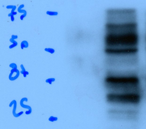
Detection of Mouse PD-L1/B7-H1 by Western Blot. Western blot shows lysates of RAW 264.7 mouse monocyte/macrophage cell line untreated (-) or treated (+) with 10 µg/mL LPS for 4 hours. PVDF membrane was probed with 0.5 µg/mL of Goat Anti-Mouse PD-L1/B7-H1 Antigen Affinity-purified Polyclonal Antibody (Catalog # AF1019) followed by HRP-conjugated Anti-Goat IgG Secondary Antibody (Catalog # HAF017). A specific band was detected for PD-L1/B7-H1 at approximately 50-55 kDa (as indicated). This experiment was conducted under reducing conditions and using Immunoblot Buffer Group 1.
 View Larger
Detection of PD-L1/B7-H1 in RAW 264.7 Mouse Cell Line by Flow Cytometry. RAW 264.7 mouse monocyte/macrophage cell line either treated with LPS overnight (filled histogram) or untreated (open histogram) was stained with Goat Anti-Mouse PD-L1/B7-H1 Antigen Affinity-purified Polyclonal Antibody (Catalog # AF1019), followed by Allophycocyanin-conjugated Anti-Goat IgG Secondary Antibody (Catalog # F0108). View our protocol for Staining Membrane-associated Proteins.
 View Larger
Detection of PD-L1/B7-H1 in HEK293 Human Cell Line Transfected with Mouse PD-L1/B7-H1 and eGFP by Flow Cytometry. HEK293 human embryonic kidney cell line transfected with either (A) mouse PD-L1/B7-H1 or (B) irrelevant transfectants and eGFP was stained with Goat Anti-Mouse PD-L1/B7-H1 Antigen Affinity-purified Polyclonal Antibody (Catalog # AF1019) followed by Allophycocyanin-conjugated Anti-Goat IgG Secondary Antibody (Catalog # F0108). Quadrant markers were set based on control antibody staining (Catalog # AB-108-C). View our protocol for Staining Membrane-associated Proteins.
 View Larger
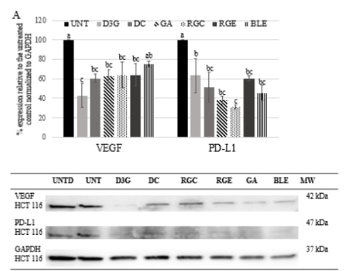
Detection of Mouse PD-L1 by Western Blot Pyruvate kinase isoform M2 (PKM2) is required for LPS-induced expression of PD-L1. RAW cells (0.5 × 10⁶ cells/ml) (A), bone marrow-derived macrophages (BMDMs) (B), or bone marrow-derived dendritic cells (C) were treated with TEPP-46 at 50 µM for 1 h prior to stimulation with LPS (100 ng/ml, 24 h), lysed and assayed for expression of PD-L1 by flow cytometry [(A–C) top panels] and pdl1 mRNA by RT-PCR [(A,B) bottom panels]. Statistical analysis was performed on cells from three separate experiments. Error bars represent mean ± SEM. BMDM cells were transfected with scrambled control (SC) or anti-PKM2 siRNA (D). 24 h post-transfection, cells were stimulated with LPS (100 ng/ml) for 24 h after which expression of PKM2 (top panel) and PDL1 (middle panel) was measured by western blot. Image collected and cropped by CiteAb from the following publication (https://pubmed.ncbi.nlm.nih.gov/29081778), licensed under a CC-BY license. Not internally tested by R&D Systems.
 View Larger
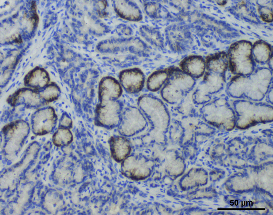
Detection of PD-L1/B7-H1 in Mouse Intestine. Formalin-fixed paraffin-embedded tissue sections of mouse intestines were probed for PD-L1 mRNA (ACD RNAScope Probe, catalog #420508; Fast Red chromogen, ACD catalog # 322750). Adjacent tissue section was processed for immunohistochemistry using goat anti-mouse PD-L1 polyclonal antibody (R&D Systems catalog # AF1019) at 3ug/mL with one-hour incubation at room temperature followed by incubation with anti-goat IgG VisUCyte HRP Polymer Antibody (Catalog # VC004) and DAB chromogen (yellow-brown). Tissue was counterstained with hematoxylin (blue). Specific staining was localized to lamina propria of villi.
 View Larger
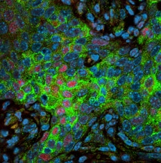
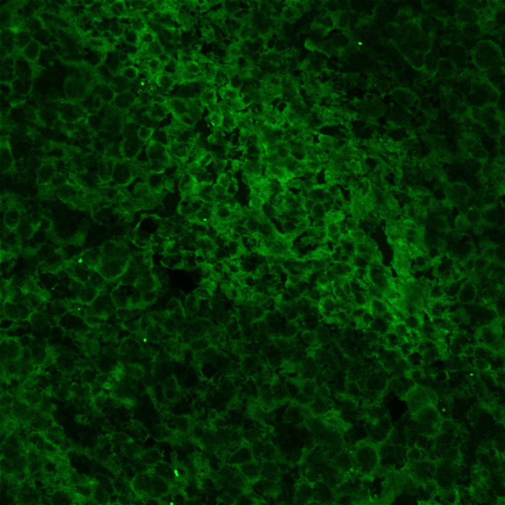
Detection of Mouse Mouse PD-L1/B7-H1 Antibody by Immunohistochemistry Protumor roles for TAMs and granulocytic myeloid-derived suppressor cells are obviated in neoantigen+ PDA.(A) Immunofluorescence staining of CB+ (nAg+) or CB– (nAg–) orthotopic tumors on day 21. Arrows show CD8+ T cell contact with a macrophage. Scale bar: 50 μm. Original magnification of insets, 2.25×. The number of CD8+ T cell and TAM (F4/80+) contacts per field of view. The box plots depict the minimum and maximum values (whiskers), the upper and lower quartiles, and the median. The length of the box represents the interquartile range. Image collected and cropped by CiteAb from the following publication (https://pubmed.ncbi.nlm.nih.gov/35393950), licensed under a CC-BY license. Not internally tested by R&D Systems.
 View Larger
Detection of Mouse Mouse PD-L1/B7-H1 Antibody by Immunohistochemistry Representative images (×20 magnification) of H&E and PD-L1 IHC-stained sections (A-C): (A) Nude mouse lymph node showing normal histology (H&E) and diffuse staining for PD-L1 (mouse) with vessels and cells within the paracortical regions displaying increased staining intensity; (B) Nude mouse spleen showing normal histology (H&E) and intense membranous staining for PD-L1 (mouse) with the most intense staining at the periphery of the white pulp; (C) MDA-MB231 tumor showing a viable region (H&E necrotic regions were observed in all sections) with membranous and cytoplasmic staining for PD-L1 (human) on the tumor cells; (D) IHC quantitative analysis (staining intensity score, H-score) of PD-L1 expression levels in lymph nodes, spleen, and MDA-MB231 tumors from mouse xenografts; each bar represents the mean H-score ± standard deviation (SD; n = 3, spleen and lymph nodes; n= 6, tumors). Image collected and cropped by CiteAb from the following publication (https://pubmed.ncbi.nlm.nih.gov/31044647), licensed under a CC-BY license. Not internally tested by R&D Systems.
Reconstitution Calculator
Preparation and Storage
- 12 months from date of receipt, -20 to -70 °C as supplied.
- 1 month, 2 to 8 °C under sterile conditions after reconstitution.
- 6 months, -20 to -70 °C under sterile conditions after reconstitution.
Background: PD-L1/B7-H1
Mouse B7 homolog 1(B7-H1), also called programmed death ligand 1 (PD-L1) and programmed cell death 1 ligand 1 (PDCD1L1), is a member of the B7 family of proteins that provide signals for regulating T-cell activation and tolerance (1‑4). Other family members include B7-1, B7-2, B7-H2, B7-H3 and PD-L2. B7 proteins are immunoglobulin (Ig) superfamily members with extracellular Ig-V-like and Ig-C-like domains and a short cytoplasmic region. Among the family members, they share from 20‑40% amino acid (aa) sequence identity. The cloned mouse B7-H1/PD-L1 cDNA encodes a 290 aa type I membrane precursor protein with a putative 18 aa signal peptide, a 220 aa extracellular region containing one V-like and one C-like Ig domain, a 22 aa transmembrane region, and a 30 aa cytoplasmic domain. Mouse and human B7-H1/PD-L1 share approximately 70% aa sequence identity. B7-H1/PD-L1 is one of two ligands for programmed death-1 (PD-1), a member of the CD28 family of immunoreceptors. The other identified ligand is PD-L2. Mouse B7-H1/PD-L1 and PD-L2 share approximately 34% aa sequence identity and have similar functions. B7-H1/PD-L1 is constitutively expressed in various lymphoid and non-lymphoid organs including placenta, heart, pancreas, lung, liver, and endothelium (1‑4). The expression of B7-H1/PD-L1 is detected on B cells, T cells, monocytes, dendritic cells and thymic epithelial cells. IFN-gamma treatment induces B7‑H1/PD‑L1 expression in monocytes, dendritic cells, and endothelial cells. B7-H1/PD-L1 expression is also upregulated in a variety of tumor cell lines. On previously activated T cells, B7-H1/PD-L1 interaction with PD-1 inhibits TCR-mediated proliferation and cytokine production, suggesting an inhibitory role in regulating immune responses. In contrast, a costimulatory function for the PD-1 ligands on resting T cells has also been reported (1‑4).
- Tamura, H. et al. (2001) Blood 97:1809.
- Freeman, G. et al. (2000) J. Exp. Med. 192:1027.
- Sharpe, A.H. and G. J. Freeman (2002) Nat. Rev. Immunol. 2:116.
- Coyle, A. and J. Gutierrez-Ramos (2001) Nat. Immunol. 2:203.
Product Datasheets
Citations for Mouse PD-L1/B7-H1 Antibody
R&D Systems personnel manually curate a database that contains references using R&D Systems products. The data collected includes not only links to publications in PubMed, but also provides information about sample types, species, and experimental conditions.
66
Citations: Showing 1 - 10
Filter your results:
Filter by:
-
NF-kappaB Activator 1 downregulation in macrophages activates STAT3 to promote adenoma-adenocarcinoma transition and immunosuppression in colorectal cancer
Authors: S Wang, Y Kuai, S Lin, L Li, Q Gu, X Zhang, X Li, Y He, S Chen, X Xia, Z Ruan, C Lin, Y Ding, Q Zhang, C Qi, J Li, X He, JL Pathak, W Zhou, S Liu, L Wang, L Zheng
Bmc Medicine, 2023-03-29;21(1):115.
-
Epithelial-mesenchymal transition induced by tumor cell-intrinsic PD-L1 signaling predicts a poor response to immune checkpoint inhibitors in PD-L1-high lung cancer
Authors: Jeong, H;Koh, J;Kim, S;Song, SG;Lee, SH;Jeon, Y;Lee, CH;Keam, B;Lee, SH;Chung, DH;Jeon, YK;
British journal of cancer
Species: Human
Sample Types: Cell Lysates
Applications: Western Blot -
A novel retinoic acid receptor-? agonist antagonizes immune checkpoint resistance in lung cancers by altering the tumor immune microenvironment
Authors: Wei CH, Huang L, Kreh B et al.
Sci Rep
-
Design, synthesis, and biological activity of dual monoamine oxidase A and heat shock protein 90 inhibitors, N-Methylpropargylamine-conjugated 4-isopropylresorcinol for glioblastoma
Authors: Tseng, HJ;Banerjee, S;Qian, B;Lai, MJ;Wu, TY;Hsu, TI;Lin, TE;Hsu, KC;Chuang, KH;Liou, JP;Shih, JC;
European journal of medicinal chemistry
Species: Mouse
Sample Types: Whole Cells
Applications: Flow Cytometry -
Tryptophan metabolism promotes immune evasion in human pancreatic beta cells
Authors: Rachdi, L;Zhou, Z;Berthault, C;Lourenço, C;Fouque, A;Domet, T;Armanet, M;You, S;Peakman, M;Mallone, R;Scharfmann, R;
EBioMedicine
Species: Human
Sample Types: Cell Lysates
Applications: Western Blot -
CDK4/6 inhibitors and the pRB-E2F1 axis suppress PVR and PD-L1 expression in triple-negative breast cancer
Authors: Shrestha, M;Wang, DY;Ben-David, Y;Zacksenhaus, E;
Oncogenesis
Species: Mouse
Sample Types: Cell Lysates
Applications: Western Blot -
Macrophages facilitate tumor cell PD‐L1 expression via an IL‐1 beta ‐centered loop to attenuate immune checkpoint blockade
Authors: Cheng Xu, Yu Xia, Bai‐Wei Zhang, Emmanuel Kwateng Drokow, Hua‐Yi Li, Sen Xu et al.
MedComm (2020)
-
Angiogenic inhibitor pre-administration improves the therapeutic effects of immunotherapy
Authors: M Sato, N Maishi, Y Hida, A Yanagawa-M, MT Alam, J Sakakibara, JM Nam, Y Onodera, S Konno, K Hida
Cancer Medicine, 2023-02-19;0(0):.
Species: Mouse
Sample Types: Whole Tissue
Applications: IHC -
Mevalonate improves anti-PD-1/PD-L1 efficacy by stabilizing CD274 mRNA
Authors: Zhang, W;Pan, X;Xu, Y;Guo, H;Zheng, M;Chen, X;Wu, H;Luan, F;He, Q;Ding, L;Yang, B;
Acta Pharmaceutica Sinica B
Species: Human
Sample Types: Cell Lysates
Applications: Western Blot -
Pancreatic Islet Cells Response to IFNgamma Relies on Their Spatial Location within an Islet
Authors: M De Burghgr, C Lourenço, C Berthault, V Aiello, A Villalba, A Fouque, M Diedisheim, S You, M Oshima, R Scharfmann
Cells, 2022-12-28;12(1):.
Species: Mouse
Sample Types: Whole Tissue
Applications: IHC -
Breast cancer cells survive chemotherapy by activating targetable immune modulatory programs characterized by either PD-L1 or CD80
Authors: Ashkan Shahbandi, Fang-Yen Chiu, Nathan A. Ungerleider, Raegan Kvadas, Zeinab Mheidly, Meijuan J. S. Sun et al.
Nature Cancer
-
Multiscale imaging of therapeutic anti-PD-L1 antibody localization using molecularly defined imaging agents
Authors: Iris M. Hagemans, Peter J. Wierstra, Kas Steuten, Janneke D. M. Molkenboer-Kuenen, Duco van Dalen, Martin ter Beest et al.
Journal of Nanobiotechnology
-
The expression of PD-1 ligand 1 on macrophages and its clinical impacts and mechanisms in lung adenocarcinoma
Authors: Yusuke Shinchi, Shiho Ishizuka, Yoshihiro Komohara, Eri Matsubara, Remi Mito, Cheng Pan et al.
Cancer Immunology, Immunotherapy
-
Lentiviral interferon: A novel method for gene therapy in bladder cancer
Authors: Sharada Mokkapati, Vikram M. Narayan, Ganiraju C. Manyam, Amy H. Lim, Jonathan J. Duplisea, Andrea Kokorovic et al.
Molecular Therapy - Oncolytics
-
WEE1 inhibition enhances the antitumor immune response to PD-L1 blockade by the concomitant activation of STING and STAT1 pathways in SCLC
Authors: Hirokazu Taniguchi, Rebecca Caeser, Shweta S. Chavan, Yingqian A. Zhan, Andrew Chow, Parvathy Manoj et al.
Cell Reports
-
Inhibiting MNK kinases promotes macrophage immunosuppressive phenotype to limit CD8+ T cell anti-tumor immunity
Authors: TN Pham, C Spaulding, MA Shields, AE Metropulos, DN Shah, MG Khalafalla, DR Principe, DJ Bentrem, HG Munshi
JCI Insight, 2022-05-09;0(0):.
Species: Mouse
Sample Types: Whole Cell
Applications: Western Blot -
Activation of Host-NLRP3 Inflammasome in Myeloid Cells Dictates Response to Anti-PD-1 Therapy in Metastatic Breast Cancers
Authors: Isak W. Tengesdal, Suzhao Li, Nicholas E. Powers, Makenna May, Charles P. Neff, Leo A. B. Joosten et al.
Pharmaceuticals (Basel)
-
A novel multifunctional anti-PD-L1-CD16a-IL15 induces potent cancer cell killing in PD-L1-positive tumour cells
Authors: Y Li, L Wu, Y Liu, S Ma, B Huang, X Feng, H Wang
Translational Oncology, 2022-04-26;21(0):101424.
Species: Mouse
Sample Types: Recombinant Protein
Applications: ELISA -
Mitogen-Activated Protein Kinase Phosphatase-1 Controls PD-L1 Expression by Regulating Type I Interferon during Systemic Escherichia coli Infection
Authors: TJ Barley, PR Murphy, X Wang, BA Bowman, JM Mormol, CE Mager, SG Kirk, CJ Cash, SC Linn, X Meng, LD Nelin, B Chen, M Hafner, J Zhang, Y Liu
The Journal of Biological Chemistry, 2022-04-13;0(0):101938.
Species: Mouse
Sample Types: Cell Lysates
Applications: Western Blot -
PD-L1 Antibody Pharmacokinetics and Tumor Targeting in Mouse Models for Infectious Diseases
Authors: Gerwin G. W. Sandker, Gosse Adema, Janneke Molkenboer-Kuenen, Peter Wierstra, Johan Bussink, Sandra Heskamp et al.
Frontiers in Immunology
-
PD-L1 near Infrared Photoimmunotherapy of Ovarian Cancer Model
Authors: J Jin, I Sivakumar, Y Mironchik, B Krishnamac, F Wildes, JD Barnett, CF Hung, S Nimmagadda, H Kobayashi, ZM Bhujwalla, MF Penet
Cancers, 2022-01-26;14(3):.
Species: Mouse
Sample Types: Cell Lysates
Applications: Western Blot -
SKI‐G‐801, an AXL kinase inhibitor, blocks metastasis through inducing anti‐tumor immune responses and potentiates anti‐PD‐1 therapy in mouse cancer models
Authors: Chun‐Bong Synn, Sung Eun Kim, Hee Kyu Lee, Min‐Hwan Kim, Jae Hwan Kim, Ji Min Lee et al.
Clinical & Translational Immunology
-
Anti?VEGF antibody triggers the effect of anti?PD?L1 antibody in PD?L1low and immune desert?like mouse tumors
Authors: N Ishikura, M Sugimoto, K Yorozu, M Kurasawa, O Kondoh
Oncology reports, 2021-12-27;47(2):.
Species: Mouse
Sample Types: Whole Tissue
Applications: IHC -
A Novel Kinase Inhibitor AX-0085 Inhibits Interferon-gamma-Mediated Induction of PD-L1 Expression and Promotes Immune Reaction to Lung Adenocarcinoma Cells
Authors: J Kim, H Jang, GJ Lee, Y Hur, J Keum, JK Jo, SE Yun, SJ Park, YJ Park, MJ Choi, KS Kim, J Kim
Cells, 2021-12-22;11(1):.
Species: Human
Sample Types: Cell Lysates
Applications: Western Blot -
Loss of H2R signaling disrupts neutrophil homeostasis and promotes inflammation-associated colonic tumorigenesis in mice
Authors: Z Shi, Y Mori-Akiya, W Du, R Fultz, Y Zhao, W Ruan, S Venable, MA Engevik, J Versalovic
Cellular and Molecular Gastroenterology and Hepatology, 2021-11-13;0(0):.
Species: Mouse
Sample Types: Whole Tissue
Applications: IHC -
Tumor-derived exosomes drive immunosuppressive macrophages in a pre-metastatic niche through glycolytic dominant metabolic reprogramming
Authors: Samantha M. Morrissey, Fan Zhang, Chuanlin Ding, Diego Elias Montoya-Durango, Xiaoling Hu, Chenghui Yang et al.
Cell Metabolism
-
Distinct Biomarker Profiles and TCR Sequence Diversity Characterize the Response to PD-L1 Blockade in a Mouse Melanoma Model
Authors: El Meskini R, Atkinson D, Kulaga A et al.
Molecular Cancer Research
-
Type 2 immunity is maintained during cancer-associated adipose tissue wasting
Authors: Patrick J Lenehan, Assunta Cirella, Amiko M Uchida, Stephanie J Crowley, Tatyana Sharova, Genevieve Boland et al.
Immunotherapy Advances
-
Romidepsin (FK228) regulates the expression of the immune checkpoint ligand PD-L1 and suppresses cellular immune functions in colon cancer
Authors: Yehui Shi, Ying Fu, Xin Zhang, Gang Zhao, Yuan Yao, Yan Guo et al.
Cancer Immunology, Immunotherapy
-
Immune receptor inhibition through enforced phosphatase recruitment
Authors: RA Fernandes, L Su, Y Nishiga, J Ren, AM Bhuiyan, N Cheng, CJ Kuo, LK Picton, S Ohtsuki, RG Majzner, SP Rietberg, CL Mackall, Q Yin, LR Ali, X Yang, CS Savvides, J Sage, M Dougan, KC Garcia
Nature, 2020-10-21;586(7831):779-784.
Species: Mouse
Sample Types: Whole Cells
Applications: Flow Cytometry -
CD47 prevents the elimination of diseased fibroblasts in scleroderma
Authors: T Lerbs, L Cui, ME King, T Chai, C Muscat, L Chung, R Brown, K Rieger, T Shibata, G Wernig
JCI Insight, 2020-08-20;5(16):.
Species: Mouse
Sample Types: Whole Cells, Whole Tissue
Applications: ICC, IHC -
Impacts of combining anti-PD-L1 immunotherapy and radiotherapy on the tumour immune microenvironment in a murine prostate cancer model
Authors: Y Philippou, HT Sjoberg, E Murphy, S Alyacoubi, KI Jones, AN Gordon-Wee, S Phyu, EE Parkes, W Gillies Mc, AD Lamb, U Gileadi, V Cerundolo, DA Scheiblin, SJ Lockett, DA Wink, IG Mills, FC Hamdy, RJ Muschel, RJ Bryant
Br. J. Cancer, 2020-07-09;0(0):.
Species: Mouse
Sample Types: Cell Lysates
Applications: Western Blot -
Selective inhibition of HDAC3 targets synthetic vulnerabilities and activates immune surveillance in lymphoma.
Authors: Patrizia Mondello, Saber Tadros, Matt Teater, Lorena Fontan, Aaron Y. Chang, Neeraj Jain et al.
Cancer Discovery
-
Chimeric tumor modeling reveals role of partial PDL1 expression in resistance to virally induced immunotherapy
Authors: Mee Y. Bartee, Parker C. Dryja, Eric Bartee
Journal for ImmunoTherapy of Cancer
-
Degradation of tumour stromal hyaluronan by small extracellular vesicle-PH20 stimulates CD103+ dendritic cells and in combination with PD-L1 blockade boosts anti-tumour immunity
Authors: Yeonsun Hong, Yoon Kyoung Kim, Gi Beom Kim, Gi-Hoon Nam, Seong A Kim, Yoon Park et al.
Journal of Extracellular Vesicles
-
The lytic activity of VSV-GP treatment dominates the therapeutic effects in a syngeneic model of lung cancer
Authors: LM Schreiber, C Urbiola, K Das, B Spiesschae, J Kimpel, F Heinemann, B Stierstorf, P Müller, M Petersson, P Erlmann, D von Laer, G Wollmann
Br. J. Cancer, 2019-09-18;0(0):.
Species: Mouse
Sample Types: Whole Tissue
Applications: IHC-P -
Tumor Lymphatic Function Regulates Tumor Inflammatory and Immunosuppressive Microenvironments
Authors: Raghu P. Kataru, Catherine L. Ly, Jinyeon Shin, Hyeung Ju Park, Jung Eun Baik, Sonia Rehal et al.
Cancer Immunology Research
-
IL-17C-mediated innate inflammation decreases the response to PD-1 blockade in a model of Kras-driven lung cancer
Authors: F Ritzmann, C Jungnickel, G Vella, A Kamyschnik, C Herr, D Li, MM Menger, A Angenendt, M Hoth, A Lis, R Bals, C Beisswenge
Sci Rep, 2019-07-17;9(1):10353.
Species: Mouse
Sample Types: Whole Tissue
Applications: IHC-P -
Immunopotentiator Aikejia improves the therapeutic efficacy of PD-1/PD-L1 immunosuppressive pathway in CT26.WT cancer cell
Authors: Chaoqun Huang, Xiaoqiong Tang, Suhuan Li, Qingshui Wang, Bifeng Xie, Jing Xu et al.
Journal of Cancer
-
Immuno-PET Imaging of the Programmed Cell Death-1 Ligand (PD-L1) Using a Zirconium-89 Labeled Therapeutic Antibody, Avelumab
Authors: Elaine M. Jagoda, Olga Vasalatiy, Falguni Basuli, Ana Christina L. Opina, Mark R. Williams, Karen Wong et al.
Mol Imaging
-
Programmed cell death ligand 1 d isruption by clustered regularly interspaced short palindromic repeats /Cas9‐genome editing promotes antitumor immunity and suppresses ovarian cancer progression
Authors: Tamaki Yahata, Mika Mizoguchi, Akihiko Kimura, Takashi Orimo, Saori Toujima, Yumi Kuninaka et al.
Cancer Science
-
Non-Invasive Fluorescent Monitoring of Ovarian Cancer in an Immunocompetent Mouse Model
Authors: Amy L. Wilson, Kirsty L. Wilson, Maree Bilandzic, Laura R. Moffitt, Ming Makanji, Mark D. Gorrell et al.
Cancers (Basel)
-
Adipose PD-L1 Modulates PD-1/PD-L1 Checkpoint Blockade Immunotherapy Efficacy in Breast Cancer
Authors: Bogang Wu, Xiujie Sun, Harshita B. Gupta, Bin Yuan, Jingwei Li, Fei Ge et al.
OncoImmunology
-
Interferon-gamma drives programmed death-ligand 1 expression on islet ? cells to limit T cell function during autoimmune diabetes
Authors: KC Osum, AL Burrack, T Martinov, NL Sahli, JS Mitchell, CG Tucker, KE Pauken, K Papas, B Appakalai, JA Spanier, BT Fife
Sci Rep, 2018-05-29;8(1):8295.
Species: Mouse
Sample Types: Whole Tissue
Applications: IHC -
PD-L1 checkpoint inhibition and anti-CTLA-4 whole tumor cell vaccination counter adaptive immune resistance: A mouse neuroblastoma model that mimics human disease
Authors: P Srinivasan, X Wu, M Basu, C Rossi, AD Sandler
PLoS Med., 2018-01-29;15(1):e1002497.
Species: Mouse
Sample Types: Whole Tissue
Applications: IHC-P -
Programmed Cell Death Protein Ligand-1 (PD-L1) Silencing with Polyethylenimine-Dermatan Sulfate Complex for Dual Inhibition of Melanoma Growth
Authors: Gijung Kwak, Dongkyu Kim, Gi-hoon Nam, Sun Young Wang, In-San Kim, Sun Hwa Kim et al.
ACS Nano
-
Hormonal vitamin D upregulates tissue-specific PD-L1 and PD-L2 surface glycoprotein expression in human but not mouse
Authors: V Dimitrov, M Bouttier, G Boukhaled, R Salehi-Tab, R Avramescu, B Memari, B Hasaj, GL Lukacs, CM Krawczyk, JH White
J. Biol. Chem., 2017-10-23;0(0):.
Species: Human
Sample Types: Cell Lysates
Applications: Western Blot -
Imaging of Programmed Cell Death Ligand 1: Impact of Protein Concentration on Distribution of Anti-PD-L1 SPECT Agents in an Immunocompetent Murine Model of Melanoma
Authors: Jessie R. Nedrow, Anders Josefsson, Sunju Park, Sagar Ranka, Sanchita Roy, George Sgouros
Journal of Nuclear Medicine
-
Tumor localized secretion of soluble PD1 enhances oncolytic virotherapy
Authors: MY Bartee, KM Dunlap, E Bartee
Cancer Res, 2017-03-17;0(0):.
Species: Mouse
Sample Types: Cell Lysates
Applications: Western Blot -
Dependence of Glomerulonephritis Induction on Novel Intraglomerular Alternatively Activated Bone Marrow-Derived Macrophages and Mac-1 and PD-L1 in Lupus-Prone NZM2328 Mice
Authors: SJ Sung, Y Ge, C Dai, H Wang, SM Fu, R Sharma, YS Hahn, J Yu, TH Le, MD Okusa, WK Bolton, JR Lawler
J. Immunol, 2017-02-20;0(0):.
Species: Mouse
Sample Types: Whole Cells, Whole Tissue
Applications: Flow Cytometry, IHC -
Cell autonomous or systemic EGFR blockade alters the immune-environment in squamous cell carcinomas
Int J Cancer, 2016-08-29;0(0):.
Species: Mouse
Sample Types: Whole Tissue
Applications: IHC-P -
PD-1/PD-L1 blockade enhances T cell activity and antitumor efficacy of imatinib in gastrointestinal stromal tumors
Clin Cancer Res, 2016-07-28;0(0):.
Species: Mouse
Sample Types: Whole Tissue
Applications: IHC-P -
Systemic Immunotherapy of Non-Muscle Invasive Mouse Bladder Cancer with Avelumab, an Anti–PD-L1 Immune Checkpoint Inhibitor
Authors: Amanda J. Vandeveer, Jonathan K. Fallon, Robert Tighe, Helen Sabzevari, Jeffrey Schlom, John W. Greiner
Cancer Immunology Research
-
IFN-gamma from lymphocytes induces PD-L1 expression and promotes progression of ovarian cancer.
Authors: Abiko K, Matsumura N, Hamanishi J, Horikawa N, Murakami R, Yamaguchi K, Yoshioka Y, Baba T, Konishi I, Mandai M
Br J Cancer, 2015-03-31;112(9):1501-9.
Species: Mouse
Sample Types: Tissue Homogenates, Whole Tissue
Applications: IHC-Fr, Western Blot -
beta-Cell-targeted blockage of PD1 and CTLA4 pathways prevents development of autoimmune diabetes and acute allogeneic islets rejection.
Authors: El Khatib M, Sakuma T, Tonne J, Mohamed M, Holditch S, Lu B, Kudva Y, Ikeda Y
Gene Ther, 2015-03-19;22(5):430-8.
Species: Human
Sample Types: Cell Lysates
Applications: Western Blot -
The development of endometrial hyperplasia in aged PD-1-deficient female mice
Authors: Guoning Guo, Hong Li, Dayan Cao, Yongwen Chen
Diagnostic Pathology
-
PD-L1 Expression by Neurons Nearby Tumors Indicates Better Prognosis in Glioblastoma Patients
Authors: Yawei Liu, Robert Carlsson, Malene Ambjorn, Maruf Hasan, Wiaam Badn, Anna Darabi et al.
Journal of Neuroscience
-
Cross-reactivity of anti-programmed death ligand 2 polyclonal antibody in mouse tissues
Authors: Yu Zhao, GanLan Bian, CaiYong Yu, FangFang Liu, Ling Liu, HongMin Guo et al.
Science China Life Sciences
-
Endogenous retinoids in the pathogenesis of alopecia areata.
Authors: Duncan F, Silva K, Johnson C, King B, Szatkiewicz J, Kamdar S, Ong D, Napoli J, Wang J, King L, Whiting D, McElwee K, Sundberg J, Everts H
J Invest Dermatol, 2012-09-27;133(2):334-43.
Species: Mouse
Sample Types: Whole Tissue
Applications: IHC -
Anti-programmed cell death 1 antibody reduces CD4+PD-1+ T cells and relieves the lupus-like nephritis of NZB/W F1 mice.
Authors: Kasagi S, Kawano S, Okazaki T, Honjo T, Morinobu A, Hatachi S, Shimatani K, Tanaka Y, Minato N, Kumagai S
J. Immunol., 2010-02-05;184(5):2337-47.
Species: Mouse
Sample Types: Whole Tissue
Applications: IHC-Fr -
Altered availability of PD-1/PD ligands is associated with the failure to control autoimmunity in NOD mice.
Authors: Yadav D, Hill N, Yagita H, Azuma M, Sarvetnick N
Cell. Immunol., 2009-05-06;258(2):161-71.
Species: Mouse
Sample Types: Whole Tissue
Applications: IHC-P -
Establishment of NOD-Pdcd1-/- mice as an efficient animal model of type I diabetes.
Authors: Wang J, Yoshida T, Nakaki F, Hiai H, Okazaki T, Honjo T
Proc. Natl. Acad. Sci. U.S.A., 2005-08-08;102(33):11823-8.
Species: Mouse
Sample Types: Whole Cells
Applications: ICC -
Pyruvate Kinase M2 Is Required for the Expression of the Immune Checkpoint PD-L1 in Immune Cells and Tumors.
Authors: Palsson-McDermott EM, Dyck L, Zastona Z et al.
Front Immunol.
-
Activation of G protein-coupled estrogen receptor signaling inhibits melanoma and improves response to immune checkpoint blockade
Authors: Natale CA, Li J, Zhang J et al.
Elife
-
In situ immunogenic clearance induced by a combination of photodynamic therapy and rho-kinase inhibition sensitizes immune checkpoint blockade response to elicit systemic antitumor immunity against intraocular melanoma and its metastasis
Authors: Kim S, Kim SA, Nam GH, et al.
Journal for immunotherapy of cancer
-
Burrack A, Spartz E, Raynor et al.
Authors: Combination PD-1 and PD-L1 Blockade Promotes Durable Neoantigen-Specific T Cell-Mediated Immunity in Pancreatic Ductal Adenocarcinoma
Cell Rep.
FAQs
No product specific FAQs exist for this product, however you may
View all Antibody FAQsReviews for Mouse PD-L1/B7-H1 Antibody
Average Rating: 4.4 (Based on 10 Reviews)
Have you used Mouse PD-L1/B7-H1 Antibody?
Submit a review and receive an Amazon gift card.
$25/€18/£15/$25CAN/¥75 Yuan/¥2500 Yen for a review with an image
$10/€7/£6/$10 CAD/¥70 Yuan/¥1110 Yen for a review without an image
Filter by:
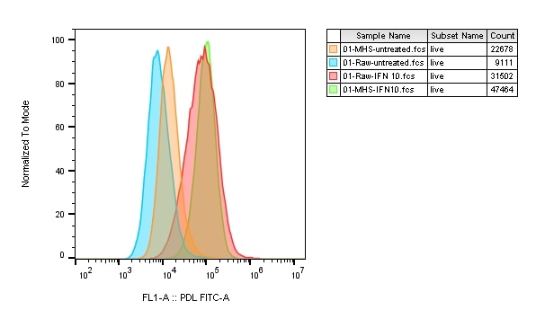
MH-S and RAW264.7 cells were treated with 10ug/ml concentration of IFN alpha. Cells were analyzed by flow cytometer (surface staining) for PD-L1 expression using a labelled secondary antibody. Protocol was followed as given with the article. Worked exactly as expected.
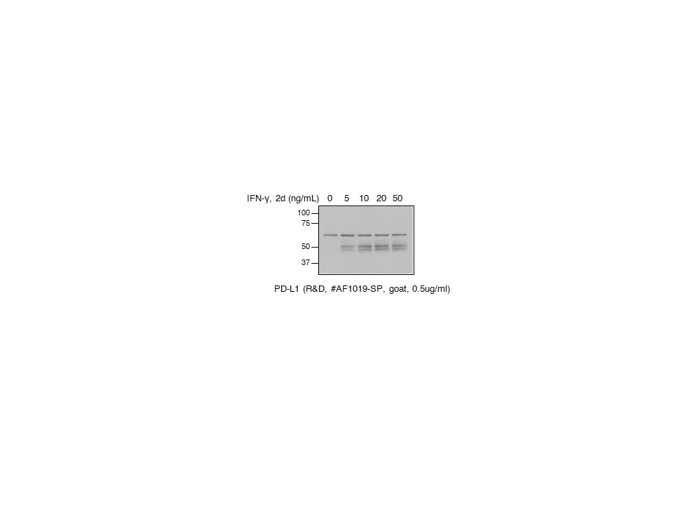
MH-S mice macrophage cell lysates. Dark bands in the middle are PD-L1
Technical Service will be following up with the customer.
Buffer: tbst
Dilution: 1/1000
