Human/Rat EGLN1/PHD2 Antibody Summary
Ala2-Phe426
Accession # Q9GZT9
Applications
Please Note: Optimal dilutions should be determined by each laboratory for each application. General Protocols are available in the Technical Information section on our website.
Scientific Data
 View Larger
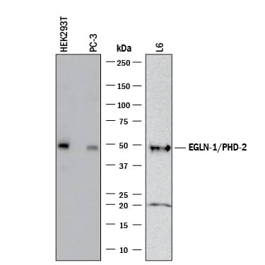
Detection of Human and Rat EGLN1/PHD2 by Western Blot. Western blot shows lysates of HEK293T human embryonic kidney cell line, PC-3 human prostate cancer cell line, and L6 rat myoblast cell line. PVDF membrane was probed with 1 µg/mL of Rabbit Anti-Human/Rat EGLN1/PHD2 Monoclonal Antibody (Catalog # MAB7680) followed by HRP-conjugated Anti-Rabbit IgG Secondary Antibody (HAF008). A specific band was detected for EGLN1/PHD2 at approximately 49 kDa (as indicated). This experiment was conducted under reducing conditions and using Immunoblot Buffer Group 1.
 View Larger
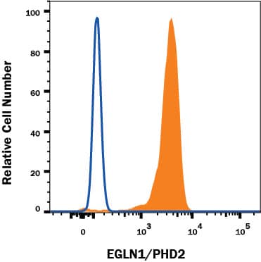
Detection of EGLN1/PHD2 in Human Jurkat cell line by Flow Cytometry. Human Jurkat T Cell Leukemia Cell Line was stained with Rabbit Anti-Human/Rat EGLN1/PHD2 Monoclonal Antibody (Catalog # MAB7680, filled histogram) or Rabbit IgG Isotype Control Antibody (MAB1050, open histogram) followed by Phycoerythrin-conjugated Anti-Rabbit IgG Secondary Antibody (Catalog # F0110). To facilitate intracellular staining, cells were fixed and permeabilized with FlowX FoxP3 Fixation & Permeabilization Buffer Kit (FC012). View our protocol for Staining Membrane-associated Proteins.
 View Larger
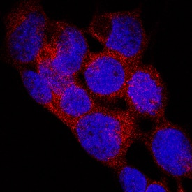
EGLN1/PHD2 in WM-115 Human Cell Lines. EGLN1/PHD2 was detected in immersion fixed WM-115 human malignant melanoma cell line using Rabbit Anti-Human/Rat EGLN1/PHD2 Monoclonal Antibody (Catalog # MAB7680) at 3 µg/mL for 3 hours at room temperature. Cells were stained using the NorthernLights™ 557-conjugated Anti-Rabbit IgG Secondary Antibody (red; NL004) and counterstained with DAPI (blue). Specific staining was localized to cell cytoplasm and nuclei. View our protocol for Fluorescent ICC Staining of Cells on Coverslips.
 View Larger
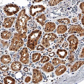
EGLN1/PHD2 in Human Kidney. EGLN1/PHD2 was detected in immersion fixed paraffin-embedded sections of human kidney using Rabbit Anti-Human/Rat EGLN1/PHD2 Monoclonal Antibody (Catalog # MAB7680) at 3 µg/mL for 1 hour at room temperature followed by incubation with the Anti-Rabbit IgG VisUCyte™ HRP Polymer Antibody (VC003). Before incubation with the primary antibody, tissue was subjected to heat-induced epitope retrieval using Antigen Retrieval Reagent-Basic (CTS013). Tissue was stained using DAB (brown) and counterstained with hematoxylin (blue). Specific staining was localized to cell cytoplasm and nuclei. View our protocol for IHC Staining with VisUCyte HRP Polymer Detection Reagents.
 View Larger
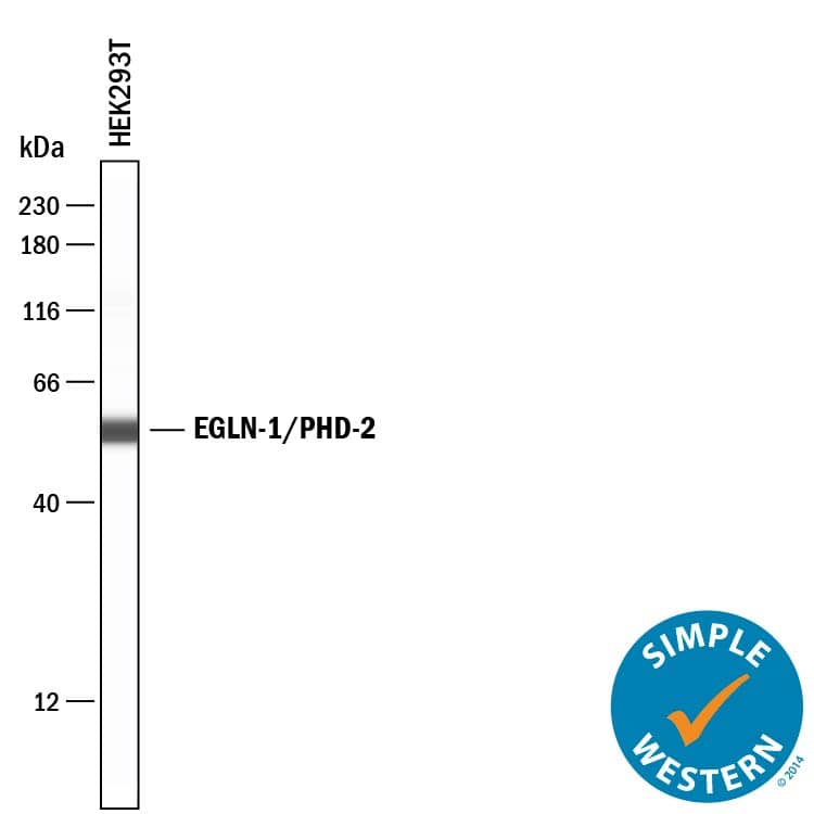
Detection of Human EGLN1/PHD2 by Simple WesternTM. Simple Western lane view shows lysates of HEK293T human embryonic kidney cell line, loaded at 0.2 mg/mL. A specific band was detected for EGLN1/PHD2 at approximately 55 kDa (as indicated) using 10 µg/mL of Rabbit Anti-Human/Rat EGLN1/PHD2 Monoclonal Antibody (Catalog # MAB7680). This experiment was conducted under reducing conditions and using the 12-230 kDa separation system.
 View Larger
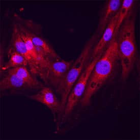
Bax in RAW264.7 cells. EGLN1/PHD2 was detected in immersion fixed L6 cells using Rabbit Anti-Human/Rat EGLN1/PHD2 Monoclonal Antibody (Catalog # MAB7680) at 15 µg/mL for 3 hours at room temperature. Cells were stained using the NorthernLights™ 557-conjugated Anti-Rabbit IgG Secondary Antibody (red; Catalog # NL004) and counterstained with DAPI (blue). Specific staining was localized to plasma membrane. View our protocol for Fluorescent ICC Staining of Non-adherent Cells.
Reconstitution Calculator
Preparation and Storage
- 12 months from date of receipt, -20 to -70 °C as supplied.
- 1 month, 2 to 8 °C under sterile conditions after reconstitution.
- 6 months, -20 to -70 °C under sterile conditions after reconstitution.
Background: EGLN1/PHD2
PHD2 (Prolyl Hydroxylase Domain-containing protein 2; also HPH2, EGLN1 and HIF-PH2) is a 45-47 kDa dioxygenase member of the PH family of enzymes. It is ubiquitously expressed, and serves to regulate the availability of the oxygen-sensitive HIF transcription factor. Active HIF1 alpha is a heterodimer of alpha - and beta -subunits and when intact, promotes VEGF and EPO production. The beta -subunit is constitutively expressed, while alpha -subunit levels are regulated by intracellular oxygen concentration. At normoxic levels, the alpha -subunit is hydroxylated on Pro by one of three PHDs, inducing its ubiquitination/degradation. The hydroxylation event requires oxygen, and thus PH activity (particularly PHD2) is a measure of a cell's oxygen concentration. Human PHD2 is 426 amino acids (aa) in length. It contains an NES (aa 6-20), a Zn-finger region (aa 21-58), and a catalytic domain (aa 291-392). There are five nitrosylated cysteines plus one acetylated alanine. Two isoform variants are known, one that shows a deletion of aa 338-359, and another that contains a 17 aa substitution for aa 58-175. Over aa 157-426, human PHD2 shares 93% aa sequence identity with mouse PHD2.
Product Datasheets
FAQs
No product specific FAQs exist for this product, however you may
View all Antibody FAQsReviews for Human/Rat EGLN1/PHD2 Antibody
There are currently no reviews for this product. Be the first to review Human/Rat EGLN1/PHD2 Antibody and earn rewards!
Have you used Human/Rat EGLN1/PHD2 Antibody?
Submit a review and receive an Amazon gift card.
$25/€18/£15/$25CAN/¥75 Yuan/¥2500 Yen for a review with an image
$10/€7/£6/$10 CAD/¥70 Yuan/¥1110 Yen for a review without an image
