Human Phospho-Akt1 (T308) Antibody Summary
Applications
Please Note: Optimal dilutions should be determined by each laboratory for each application. General Protocols are available in the Technical Information section on our website.
Scientific Data
 View Larger
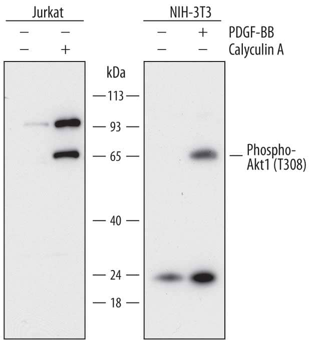
Detection of Human and Mouse Phospho-Akt1 (T308) by Western Blot. Western blot shows lysates of Jurkat human acute T cell leukemia cell line and NIH-3T3 mouse embryonic fibroblast cell line untreated (-) or treated (+) with 100 ng/mL Recombinant Human PDGF-BB (Catalog # 220-BB) for 20 minutes or 100nM Calyculin A for 30 minutes. PVDF membrane was probed with 1 µg/mL of Mouse Anti-Human Phospho-Akt1 (T308) Monoclonal Antibody (Catalog # MAB7419) followed by HRP-conjugated Anti-Mouse IgG Secondary Antibody (Catalog # HAF018). A specific band was detected for Phospho-Akt1 (T308) at approximately 65 kDa (as indicated). This experiment was conducted under reducing conditions and using Immunoblot Buffer Group 1.
 View Larger
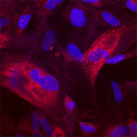
Phospho-Akt1 (T308) in CCD‑1070Sk Human Cell Line. Akt1 phosphorylated at T308 was detected in immersion fixed CCD-1070Sk human foreskin fibroblast cell line stimulated with Recombinant Human PDGF-BB (Catalog # 220-BB) using Mouse Anti-Human Phospho-Akt1 (T308) Mono-clonal Antibody (Catalog # MAB7419) at 25 µg/mL for 3 hours at room temperature. Cells were stained using the Northern-Lights™ 557-conjugated Anti-Mouse IgG Secondary Antibody (red; Catalog # NL007) and counterstained with DAPI (blue). Specific staining was localized to cytoplasm. View our protocol for Fluorescent ICC Staining of Cells on Coverslips.
 View Larger
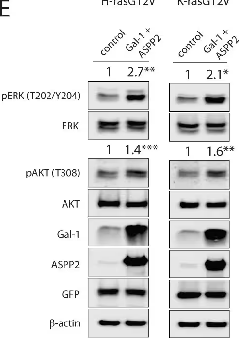
Detection of Human AKT1 by Western Blot ASPP2 blocks Gal-1 dependent nanoclustering and halts oncogenic H-ras induced transformation.Nanoclustering-FRET analysis in HEK cells coexpressing mGFP- and mCherry-tagged (A, B) H-rasG12V or (C, D) K-rasG12V. Cells were analysed after overexpression of either Gal-1 or ASPP2 plasmids, or both (1:1 ratio). Plotted are the means ± SEM, n = 3. (E) Representative Western blots from HEK cells expressing mGFP-H-rasG12V (left) or K-rasG12V (right) alone or together with Gal-1 and ASPP2. Statistical significance of differences was examined using t-test (n = 3; *, p<0.05, **, p< 0.01, ***, p<0.001). (F, G) Colony survival assay of NIH/3T3 cells stably expressing (F) H-rasG12V or (G) K-rasG12V and transiently expressing indicated constructs. Colony survival was graphed based on mean foci areas calculated from at least 4 independent biological repeats. (A-D, F-G) Statistical significance of differences between controls and treated samples was examined using one-way ANOVA (ns, not significant; *, p<0.05; **, p<0.01; ****, p<0.0001). Image collected and cropped by CiteAb from the following publication (https://dx.plos.org/10.1371/journal.pone.0159677), licensed under a CC-BY license. Not internally tested by R&D Systems.
 View Larger
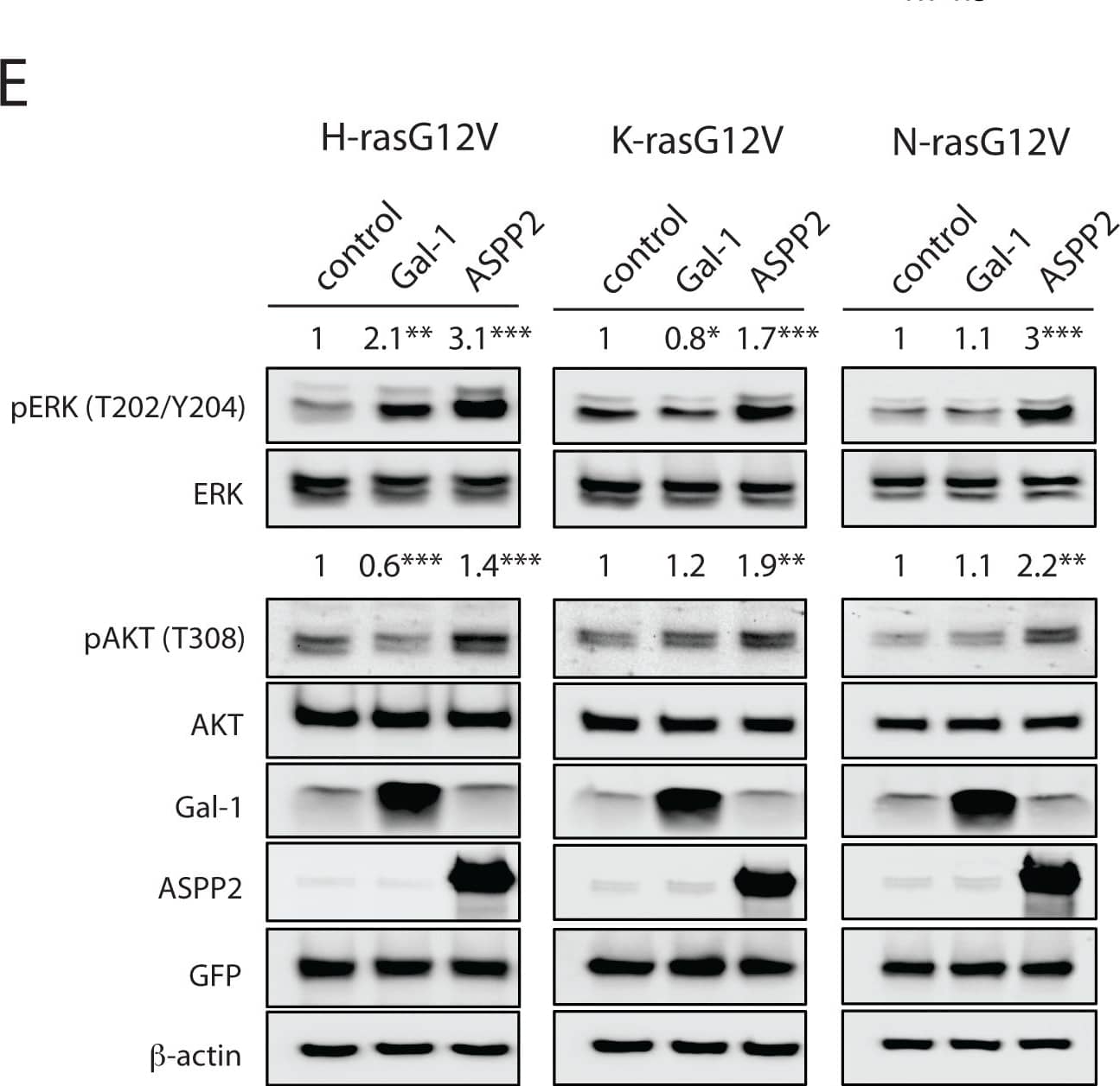
Detection of Human AKT1 by Western Blot ASPP2 increases oncogenic H-ras, K-ras and N-ras-effector-recruitment, as well as ERK- and AKT-signalling.(A) Left, scheme explaining effector-recruitment FRET analysis in HEK cells. Right, examples of FLIM-FRET images of HEK cells from the different FRET samples as indicated. (B-D) Effector-recruitment FRET analysis in HEK cells coexpressing (B) mGFP-H-rasG12V, (C) mGFP-K-rasG12V or (D) mGFP-NrasG12V and mRFP-RBD from C-Raf. The effect of Gal-1 or ASPP2 expression on effector-recruitment FRET was compared to control samples. (E) Representative Western blots from HEK cells expressing mGFP-H-rasG12V (left), K-rasG12V (middle) or N-rasG12V (right) without or with Gal-1 or ASPP2. Statistical significance of differences between controls and treated samples was examined using one-way ANOVA (mean ± SEM, n = 3; ns, not significant; *, p<0.05, **, p< 0.01, ***, p<0.001, ****, p< 0.0001). Image collected and cropped by CiteAb from the following publication (https://dx.plos.org/10.1371/journal.pone.0159677), licensed under a CC-BY license. Not internally tested by R&D Systems.
Reconstitution Calculator
Preparation and Storage
- 12 months from date of receipt, -20 to -70 °C as supplied.
- 1 month, 2 to 8 °C under sterile conditions after reconstitution.
- 6 months, -20 to -70 °C under sterile conditions after reconstitution.
Background: Akt1
Akt, also known as protein kinase B (PKB), is a central kinase in such diverse cellular processes as glucose uptake, cell cycle progression, and apoptosis. Three highly homologous members define the Akt family: Akt1 (PKB alpha ), Akt2 (PKB beta ), and Akt3 (PKB gamma ). All three Akts contain an amino-terminal pleckstrin homology domain, a central kinase domain, and a carboxyl-terminal regulatory domain. Akt1 is the most widely expressed family member and is frequently activated in a number of carcinomas, including breast, prostate, lung, pancreatic, liver, ovarian, and colorectal cancer. Akt1 is activated in a multistep process that involves the sequential phosphorylation of Thr450 by JNK kinases, Thr308 by PDK1, and Ser473 by PDK2 or mTORC2. Activated Akt1 phosphorylates a wide variety of cytosolic, nuclear, and mitochondrial substrates. Human Akt1 shares 98% aa sequence identity with mouse and rat Akt1. MAB7419 also detects mouse Phospho-Akt1 (T308) in Western Blot.
Product Datasheets
Citations for Human Phospho-Akt1 (T308) Antibody
R&D Systems personnel manually curate a database that contains references using R&D Systems products. The data collected includes not only links to publications in PubMed, but also provides information about sample types, species, and experimental conditions.
7
Citations: Showing 1 - 7
Filter your results:
Filter by:
-
A Three-Dimensional Xeno-Free Culture Condition for Wharton's Jelly-Mesenchymal Stem Cells: The Pros and Cons
Authors: B Koh, N Sulaiman, MB Fauzi, JX Law, MH Ng, TL Yuan, AGN Azurah, MH Mohd Yunus, RBH Idrus, MD Yazid
International Journal of Molecular Sciences, 2023-02-13;24(4):.
Species: Human
Sample Types: Cell Lysates
Applications: Simple Western -
Cathepsin V suppresses GATA3 protein expression in luminal A breast cancer
Authors: Naphannop Sereesongsaeng, Sara H. McDowell, James F. Burrows, Christopher J. Scott, Roberta E. Burden
Breast Cancer Research
-
Distinct molecular pathways mediate Mycn and Myc-regulated miR-17-92 microRNA action in Feingold syndrome mouse models
Authors: F Mirzamoham, A Kozlova, G Papaioanno, E Paltrinier, UM Ayturk, T Kobayashi
Nat Commun, 2018-04-10;9(1):1352.
Species: Mouse
Sample Types: Cell Lysates
Applications: Western Blot -
Opposite feedback from mTORC1 to H-ras and K-ras4B downstream of SREBP1
Authors: Itziar M. D. Posada, Benoit Lectez, Farid A. Siddiqui, Christina Oetken-Lindholm, Mukund Sharma, Daniel Abankwa
Scientific Reports
-
Rapalogs can promote cancer cell stemness in vitro in a Galectin-1 and H-ras-dependent manner
Authors: Itziar M.D. Posada, Benoit Lectez, Mukund Sharma, Christina Oetken-Lindholm, Laxman Yetukuri, Yong Zhou et al.
Oncotarget
-
ASPP2 Is a Novel Pan-Ras Nanocluster Scaffold
PLoS ONE, 2016-07-20;11(7):e0159677.
Species: Human
Sample Types: Cell Lysates
Applications: Western Blot -
Specific cancer-associated mutations in the switch III region of Ras increase tumorigenicity by nanocluster augmentation
Authors: Maja Šolman, Alessio Ligabue, Olga Blaževitš, Alok Jaiswal, Yong Zhou, Hong Liang et al.
eLife
FAQs
No product specific FAQs exist for this product, however you may
View all Antibody FAQsReviews for Human Phospho-Akt1 (T308) Antibody
There are currently no reviews for this product. Be the first to review Human Phospho-Akt1 (T308) Antibody and earn rewards!
Have you used Human Phospho-Akt1 (T308) Antibody?
Submit a review and receive an Amazon gift card.
$25/€18/£15/$25CAN/¥75 Yuan/¥2500 Yen for a review with an image
$10/€7/£6/$10 CAD/¥70 Yuan/¥1110 Yen for a review without an image


