Human/Mouse/Rat p53 Antibody Summary
Asp7-Asp393
Accession # P04637
Applications
Please Note: Optimal dilutions should be determined by each laboratory for each application. General Protocols are available in the Technical Information section on our website.
Scientific Data
 View Larger
Detection of Human p53 by Western Blot. Western blot shows lysates of CEM human T-lymphoblastoid cell line and MCF-7 human breast cancer cell line were mock-treated (-) or exposed (+) to 10 Gy ionizing radiation (IR) and harvested after 1 hour. PVDF membrane was probed with 0.5 µg/mL of Goat Anti-Human/Mouse/Rat p53 Antigen Affinity-purified Polyclonal Antibody (Catalog # AF1355), followed by HRP-conjugated Anti-Goat IgG Secondary Antibody (Catalog # HAF109). A specific band was detected for p53 at approximately 53 kDa (as indicated). This experiment was conducted under reducing conditions and using Immunoblot Buffer Group 1.
 View Larger
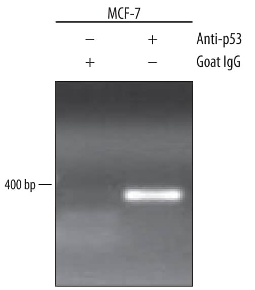
Detection of p53-regulated Genes by Chromatin Immunoprecipitation. MCF-7 human breast cancer cell line treated with 300 nM camptothecin overnight were fixed using formaldehyde, resuspended in lysis buffer, and sonicated to shear chromatin. p53/DNA complexes were immunoprecipitated using 5 µg Goat Anti-Human/Mouse/Rat p53 Antigen Affinity-purified Polyclonal Antibody (Catalog # AF1355) or control antibody (Catalog # AB-108-C) for 15 minutes in an ultrasonic bath, followed by Biotinylated Anti-Goat IgG Secondary Antibody (Catalog # BAF109). Immunocomplexes were captured using 50 µL of MagCellect Streptavidin Ferrofluid (Catalog # MAG999) and DNA was purified using chelating resin solution. The p21 promoter was detected by standard PCR.
 View Larger
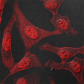
p53 in HeLa Human Cell Line. p53 was detected in immersion fixed HeLa human cervical epithelial carcinoma cell line using Goat Anti-Human/Mouse/Rat p53 Antigen Affinity-purified Polyclonal Antibody (Catalog # AF1355) at 1.7 µg/mL for 3 hours at room temperature. Cells were stained using the NorthernLights™ 557-conjugated Anti-Goat IgG Secondary Antibody (red; Catalog # NL001) and counterstained with DAPI (blue). Specific staining was localized to nuclei. View our protocol for Fluorescent ICC Staining of Cells on Coverslips.
 View Larger
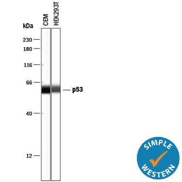
Detection of Human p53 by Simple WesternTM. Simple Western lane view shows lysates of CEM human T-lymphoblastoid cell line and HEK293T human embryonic kidney cell line, loaded at 0.2 mg/mL. A specific band was detected for p53 at approximately 59 kDa (as indicated) using 2.5 µg/mL of Goat Anti-Human/Mouse/Rat p53 Antigen Affinity-purified Polyclonal Antibody (Catalog # AF1355) followed by 1:50 dilution of HRP-conjugated Anti-Goat IgG Secondary Antibody (Catalog # HAF109). This experiment was conducted under reducing conditions and using the 12-230 kDa separation system.
 View Larger
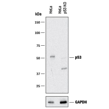
Western Blot Shows Human p53 Specificity by Using Knockout Cell Line. Western blot shows lysates of HeLa human cervical epithelial carcinoma parental cell line and p53 knockout HeLa cell line (KO). PVDF membrane was probed with 0.25 µg/mL of Goat Anti-Human/Mouse/Rat p53 Antigen Affinity-purified Polyclonal Antibody (Catalog # AF1355) followed by HRP-conjugated Anti-Goat IgG Secondary Antibody (Catalog # HAF017). A specific band was detected for p53 at approximately 53 kDa (as indicated) in the parental HeLa cell line, but is not detectable in knockout HeLa cell line. GAPDH (Catalog # AF5718) is shown as a loading control.This experiment was conducted under reducing conditions and using Immunoblot Buffer Group 1.
 View Larger
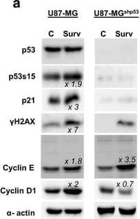
Detection of Human p53 by Knockdown Validated Overexpression of Survivin in glioma cells affects cell cycle and proliferation in a p53 dependent manner. a Western blot analysis showing cell lysates from U87-MG and U87-MGshp53 after transduction with Survivin and control vectors. The membranes were blotted with anti-p53 (53 kDa), anti-p53s15 (53 kDa), anti-p21waf/cip (21 kDa), anti-gamma H2AX (16 kDa), anti-cyclin D1 (37 kDa) and E (42 kDa singlets +50 kDa doublets) antibodies and the signals comparing control and Survivin cells were measured. The relative band density (fold increase) obtained from the densitometric analysis and normalized to the corresponding control is depicted. alpha -actin (42 kDa) was used as an internal loading control. b Representative BrdU-incorporation assays of U87-MG (left panel) and U87-MGshp53 cells (right panel) with overexpression of Survivin (bottom) compared to the empty vector-transduced controls (top). The overall amount of BrdU-positive fractions as well as the relative levels of BrdU+ and BrdU− cells in fractions with DNA contents of 2n, 4n, 8n and >8n are depicted. c Proliferation index of the U87-MG and U87-MGshp53 cells transduced with Survivin and empty vector controls. Depicted are mean values ± SD. **p < 0.01. d BrdU-incorporation in U87-MG and U87-MGshp53 cell fractions with DNA content >4n. Depicted are mean values ± SD. **p < 0.01. All data were collected 72 h after transduction of cells Image collected and cropped by CiteAb from the following publication (https://bmccancer.biomedcentral.com/articles/10.1186/s12885-017-3932-y), licensed under a CC-BY license. Not internally tested by R&D Systems.
Reconstitution Calculator
Preparation and Storage
- 12 months from date of receipt, -20 to -70 °C as supplied.
- 1 month, 2 to 8 °C under sterile conditions after reconstitution.
- 6 months, -20 to -70 °C under sterile conditions after reconstitution.
Background: p53
The p53 tumor suppressor protein is a multi-functional transcription factor that regulates cellular decisions regarding proliferation, cell cycle checkpoints, and apoptosis. The importance of p53 is underscored by its mutation in over 50% of human cancers. Mice that lack one or both copies of p53 also showed an increased incidence of tumors, which makes the p53 deficient mouse a model system for studying cancer generation and progression.
Product Datasheets
Citations for Human/Mouse/Rat p53 Antibody
R&D Systems personnel manually curate a database that contains references using R&D Systems products. The data collected includes not only links to publications in PubMed, but also provides information about sample types, species, and experimental conditions.
30
Citations: Showing 1 - 10
Filter your results:
Filter by:
-
Prediction of breast cancer risk based on flow-variant analysis of circulating peripheral blood B cells.
Authors: Syeda MM, Upadhyay K, Loke J et al.
Genet Med
-
Fanca deficiency is associated with alterations in osteoclastogenesis that are rescued by TNF?
Authors: Oppezzo, A;Monney, L;Kilian, H;Slimani, L;Maczkowiak-Chartois, F;Rosselli, F;
Cell & bioscience
Species: Mouse
Sample Types: Cell Lysates
Applications: Western Blot -
Role of p53 in Cisplatin-Induced Myotube Atrophy
Authors: Matsumoto C, Sekine H, Zhang N et al.
International Journal of Molecular Sciences
-
Calorie restriction alters the mechanisms of radiation-induced mouse thymic lymphomagenesis
Authors: T Nakayama, M Sunaoshi, Y Shang, M Takahashi, T Saito, BJ Blyth, Y Amasaki, K Daino, Y Shimada, A Tachibana, S Kakinuma
PLoS ONE, 2023-01-20;18(1):e0280560.
Species: Mouse
Sample Types: Protein
Applications: Simple Western -
Aberrant phosphorylation inactivates Numb in breast cancer causing expansion of the stem cell pool
Authors: Maria Grazia Filippone, Stefano Freddi, Silvia Zecchini, Silvia Restelli, Ivan Nicola Colaluca, Giovanni Bertalot et al.
Journal of Cell Biology
-
Non-targeting control for MISSION shRNA library silences SNRPD3 leading to cell death or permanent growth arrest
Authors: Czarnek M, Sarad K, Kara? A et al.
Molecular Therapy - Nucleic Acids
-
Accumulation of cholesterol, triglycerides and ceramides in hepatocellular carcinomas of diethylnitrosamine injected mice
Authors: EM Haberl, R Pohl, L Rein-Fisch, M Höring, S Krautbauer, G Liebisch, C Buechler
Lipids in Health and Disease, 2021-10-10;20(1):135.
Species: Mouse
Sample Types: Tissue Homogenates
Applications: Western Blot -
MYC Promotes Bone Marrow Stem Cell Dysfunction in Fanconi Anemia
Authors: A Rodríguez, K Zhang, A Färkkilä, J Filiatraul, C Yang, M Velázquez, E Furutani, DC Goldman, B García de, G Garza-Mayé, K McQueen, LA Sambel, B Molina, L Torres, M González, E Vadillo, R Pelayo, WH Fleming, M Grompe, A Shimamura, S Hautaniemi, J Greenberge, S Frías, K Parmar, AD D'Andrea
Cell Stem Cell, 2020-09-29;0(0):.
Species: Human
Sample Types: Whole Cells
Applications: Flow Cytometry -
Pseudopterosin and O-Methyltylophorinidine Suppress Cell Growth in a 3D Spheroid Co-Culture Model of Pancreatic Ductal Adenocarcinoma
Authors: Bailu Xie, Jan Hänsel, Vanessa Mundorf, Janina Betz, Irene Reimche, Mert Erkan et al.
Bioengineering (Basel)
-
Germline mutation of MDM4, a major p53 regulator, in a familial syndrome of defective telomere maintenance
Authors: E Toufektcha, V Lejour, R Durand, N Giri, I Draskovic, B Bardot, P Laplante, S Jaber, BP Alter, JA Londono-Va, SA Savage, F Toledo
Sci Adv, 2020-04-10;6(15):eaay3511.
Species: Mouse
Sample Types: Cell Culture Lysates
Applications: Western Blot -
Stress-Induced, p53-Mediated Tumor Growth Inhibition of Melanoma by Modulated Electrohyperthermia in Mouse Models without Major Immunogenic Effects
Authors: Balázs Besztercei, Tamás Vancsik, Anett Benedek, Enikő Major, Mbuotidem J. Thomas, Csaba A. Schvarcz et al.
International Journal of Molecular Sciences
-
Modulated electro‐hyperthermia induced p53 driven apoptosis and cell cycle arrest additively support doxorubicin chemotherapy of colorectal cancer in vitro
Authors: Tamas Vancsik, Gertrud Forika, Andrea Balogh, Eva Kiss, Tibor Krenacs
Cancer Medicine
-
Inflammatory responses induced by Helicobacter pylori on the carcinogenesis of gastric epithelial GES‑1 cells
Authors: Jianjun Wang, Yongliang Yao, Qinghui Zhang, Shasha Li, Lijun Tang
International Journal of Oncology
-
Progranulin protects the mouse retina under hypoxic conditions via inhibition of the Toll?like receptor?4?NADPH oxidase 4 signaling pathway
Authors: ZP You, MJ Yu, YL Zhang, K Shi
Mol Med Rep, 2018-11-08;0(0):.
Species: Mouse
Sample Types: Tissue Homogenates
Applications: Western Blot -
Oxidative stress induces p38MAPK-dependent senescence in the feto-maternal interface cells
Authors: Jin Jin, Lauren Richardson, Samantha Sheller-Miller, Nanbert Zhong, Ramkumar Menon
Placenta
-
B-cell lymphoma 2 is associated with advanced tumor grade and clinical stage, and reduced overall survival in young Chinese patients with colorectal carcinoma
Authors: J Wang, G He, Q Yang, L Bai, B Jian, Q Li, Z Li
Oncol Lett, 2018-04-13;15(6):9009-9016.
Species: Human
Sample Types: Tissue Homogenates, Whole Tissue
Applications: IHC-P, Western Blot -
Reciprocity of Action of Increasing Oct4 and Repressing p53 in Transdifferentiation of Mouse Embryonic Fibroblasts into Cardiac Myocytes
Authors: Hongran Wang, Shuying Zhao, Michelle Barton, Todd Rosengart, Austin J. Cooney
Cellular Reprogramming
-
Chromosomal instability induced by increased BIRC5/Survivin levels affects tumorigenicity of glioma cells
Authors: M Conde, S Michen, R Wiedemuth, B Klink, E Schröck, G Schackert, A Temme
BMC Cancer, 2017-12-28;17(1):889.
Species: Human
Sample Types: Cell Lysates
Applications: Western Blot -
FAS apoptotic inhibitory molecule 2 is a stress-induced intrinsic neuroprotective factor in the retina
Authors: Mercy Pawar, Boris Busov, Aaruran Chandrasekhar, Jingyu Yao, David N Zacks, Cagri G Besirli
Cell Death & Differentiation
-
Dysfunction of the MDM2/p53 axis is linked to premature aging
Authors: D Lessel, D Wu, C Trujillo, T Ramezani, I Lessel, MK Alwasiyah, B Saha, FM Hisama, K Rading, I Goebel, P Schütz, G Speit, J Högel, H Thiele, G Nürnberg, P Nürnberg, M Hammerschm, Y Zhu, DR Tong, C Katz, GM Martin, J Oshima, C Prives, C Kubisch
J. Clin. Invest., 2017-08-28;0(0):.
Species: Human
Sample Types: Cell Lysates
Applications: Western Blot -
Combining anti-miR-155 with chemotherapy for the treatment of lung cancers
Authors: Katrien Van Roosbr
Clin. Cancer Res, 2016-11-30;0(0):.
Species: Human
Sample Types: Cell Lysates
Applications: Western Blot -
Recombinant human neuregulin-1 beta is protective against radiation-induced myocardial cell injury
Authors: QIANG ZHOU, WENBING HU, XINXIONG FEI, XUQUN HUANG, XI CHEN, DEQING ZHAO et al.
Molecular Medicine Reports
-
Growth hormone is permissive for neoplastic colon growth
Proc Natl Acad Sci USA, 2016-05-25;0(0):.
Species: Human
Sample Types: Cell Lysates
Applications: Western Blot -
Survivin safeguards chromosome numbers and protects from aneuploidy independently from p53.
Authors: Wiedemuth R, Klink B, Topfer K, Schrock E, Schackert G, Tatsuka M, Temme A
Mol Cancer, 2014-05-09;13(0):107.
Species: Human
Sample Types: Cell Lysates
Applications: Western Blot -
IL-12-dependent innate immunity arrests endothelial cells in G0-G1 phase by a p21(Cip1/Waf1)-mediated mechanism
Authors: Lucia Napione, Marina Strasly, Claudia Meda, Stefania Mitola, Maria Alvaro, Gabriella Doronzo et al.
Angiogenesis
-
Optimization of Stability, Encapsulation, Release, and Cross-Priming of Tumor Antigen-Containing PLGA Nanoparticles
Authors: Shashi Prasad, Virginia Cody, Jennifer K. Saucier-Sawyer, Tarek R. Fadel, Richard L. Edelson, Martin A. Birchall et al.
Pharmaceutical Research
-
Polymer nanoparticles containing tumor lysates as antigen delivery vehicles for dendritic cell-based anti-tumor immunotherapy
Authors: Shashi Prasad, Virginia Cody, Jennifer K. Saucier-Sawyer, W. Mark Saltzman, Clarence T. Sasaki, Richard L. Edelson et al.
Nanomedicine: Nanotechnology, Biology and Medicine
-
Expression of S100A6 in cardiac myocytes limits apoptosis induced by tumor necrosis factor-alpha.
Authors: Tsoporis JN, Izhar S, Parker TG
J. Biol. Chem., 2008-08-27;283(44):30174-83.
Species: Rat
Sample Types: Cell Lysates
Applications: Western Blot -
Protection of human keratinocytes from UVB-induced inflammation using root extract of Lithospermum erythrorhizon.
Authors: Ishida T, Sakaguchi I
Biol. Pharm. Bull., 2007-05-01;30(5):928-34.
Species: Human
Sample Types: Cell Lysates
Applications: Western Blot -
TIMP-1 gene deficiency increases tumour cell sensitivity to chemotherapy-induced apoptosis.
Authors: Davidsen ML, Wurtz SØ, Romer MU, Sorensen NM, Johansen SK, Christensen IJ, Larsen JK, Offenberg H, Brunner N, Lademann U
Br. J. Cancer, 2006-10-23;95(8):1114-20.
Species: Mouse
Sample Types: Cell Lysates
Applications: Western Blot
FAQs
No product specific FAQs exist for this product, however you may
View all Antibody FAQsReviews for Human/Mouse/Rat p53 Antibody
Average Rating: 5 (Based on 3 Reviews)
Have you used Human/Mouse/Rat p53 Antibody?
Submit a review and receive an Amazon gift card.
$25/€18/£15/$25CAN/¥75 Yuan/¥2500 Yen for a review with an image
$10/€7/£6/$10 CAD/¥70 Yuan/¥1110 Yen for a review without an image
Filter by:


