Human HLA Class I Antibody Summary
*Small pack size (-SP) is supplied either lyophilized or as a 0.2 μm filtered solution in PBS.
Applications
Please Note: Optimal dilutions should be determined by each laboratory for each application. General Protocols are available in the Technical Information section on our website.
Scientific Data
 View Larger
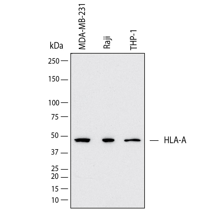
Detection of Human HLA Class I by Western Blot. Western Blot shows lysates of MDA‑MB‑231 human breast cancer cell line, Raji human Burkitt's lymphoma cell line and THP‑1 human acute monocytic leukemia cell line. PVDF membrane was probed with 2 µg/ml of Mouse Anti-Human HLA Class I Monoclonal Antibody (Catalog # MAB11502) followed by HRP-conjugated Anti-Mouse IgG Secondary Antibody (Catalog # HAF018). A specific band was detected for HLA Class I at approximately 48 kDa (as indicated). This experiment was conducted under reducing conditions and using Western Blot Buffer Group 1.
 View Larger
Detection of HLA Class I in Human Spleen. HLA Class I was detected in immersion fixed paraffin-embedded sections of human spleen using Mouse Anti-Human HLA Class I Monoclonal Antibody (Catalog # MAB11502) at 1.7 µg/ml for 1 hour at room temperature followed by incubation with the HRP-conjugated Anti-Mouse IgG Secondary Antibody (Catalog # HAF007) or the Anti-Mouse IgG VisUCyte™ HRP Polymer Antibody (Catalog # VC001). Before incubation with the primary antibody, tissue was subjected to heat-induced epitope retrieval using VisUCyte Antigen Retrieval Reagent-Basic (Catalog # VCTS021). Tissue was stained using DAB (brown) and counterstained with hematoxylin (blue). Specific staining was localized to endothelial cells. View our protocol for Chromogenic IHC Staining of Paraffin-embedded Tissue Sections.
 View Larger
Detection of HLA Class I in PBMC by Flow Cytometry PBMC were stained with Mouse Anti-Human CD19 APC‑conjugated Monoclonal Antibody (Catalog # FAB4867A) and either (A) Mouse Anti-Human HLA Class I Monoclonal Antibody (Catalog # MAB11502) or (B) isotype control antibody (Catalog # MAB004) followed by Phycoerythrin-conjugated Anti-Mouse IgG Secondary Antibody (Catalog # F0102B). View our protocol for Staining Membrane-associated Proteins.
Reconstitution Calculator
Preparation and Storage
- 12 months from date of receipt, -20 to -70 °C as supplied.
- 1 month, 2 to 8 °C under sterile conditions after reconstitution.
- 6 months, -20 to -70 °C under sterile conditions after reconstitution.
Background: HLA Class I
HLA-A, B, and C are approximately 45 kDa transmembrane glycoproteins in the major histocompatibility complex 1 (MHC I) family. They contain three alpha domains in their extracellular regions. HLA molecules are expressed on nearly all nucleated cells in association with the 12 kDa beta 2-Microglobulin. This complex binds peptides derived from pathogenic cytosolic or extracellular proteins such as viral or microbial proteins. It presents these peptides on the cell surface for recognition by the T cell receptor on CD8+ cytotoxic T cells. The activated cytotoxic T cell then kills the presenting cell. Mismatched MHC I alleles between a host and a donor lead to transplant rejection.
Product Datasheets
FAQs
No product specific FAQs exist for this product, however you may
View all Antibody FAQsReviews for Human HLA Class I Antibody
There are currently no reviews for this product. Be the first to review Human HLA Class I Antibody and earn rewards!
Have you used Human HLA Class I Antibody?
Submit a review and receive an Amazon gift card.
$25/€18/£15/$25CAN/¥75 Yuan/¥2500 Yen for a review with an image
$10/€7/£6/$10 CAD/¥70 Yuan/¥1110 Yen for a review without an image

