Human GFAP Antibody Summary
Leu292-Met432
Accession # P14136
Applications
Please Note: Optimal dilutions should be determined by each laboratory for each application. General Protocols are available in the Technical Information section on our website.
Scientific Data
 View Larger
Detection of Human GFAP by Western Blot. Western blot shows lysates of human brain (motor cortex) tissue, human brain (cerebellum) tissue, and human brain (hypothalamus) tissue. PVDF membrane was probed with 1 µg/mL of Mouse Anti-Human GFAP Monoclonal Antibody (Catalog # MAB2594) followed by HRP-conjugated Anti-Mouse IgG Secondary Antibody (Catalog # HAF018). Specific bands were detected for GFAP at approximately 35-50 kDa (as indicated). This experiment was conducted under reducing conditions and using Immunoblot Buffer Group 1.
.") View Larger
View Larger
GFAP in Rat Cortical Stem Cells. GFAP was detected in immersion fixed differentiated rat cortical stem cells using Mouse Anti-Human GFAP Monoclonal Antibody (Catalog # MAB2594) at 10 µg/mL for 3 hours at room temperature. Cells were stained using the NorthernLights™ 557-conjugated Anti-Mouse IgG Secondary Antibody (red; Catalog # NL007) and counterstained with DAPI (blue). Specific staining was localized to cytoplasm. View our protocol for Fluorescent ICC Staining of Stem Cells on Coverslips.
 View Larger
Detection of Human GFAP by Simple WesternTM. Simple Western lane view shows lysates of human brain (cerebellum) tissue and human brain (motor cortex) tissue, loaded at 0.2 mg/mL. A specific band was detected for GFAP at approximately 51-52 kDa (as indicated) using 50 µg/mL of Mouse Anti-Human GFAP Monoclonal Antibody (Catalog # MAB2594). This experiment was conducted under reducing conditions and using the 12-230 kDa separation system.
 View Larger
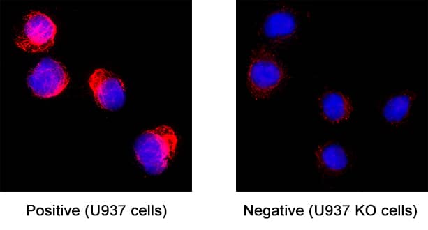
GFAP Specificity is Shown by Immunocytochemistry in Knockout Cell Line. GFAP was detected in immersion fixed U937 human histiocytic lymphoma cell line but is not detected in GFAP knockout (KO) U937 Human cell line using Mouse Anti-Human GFAP Monoclonal Antibody (Catalog # MAB2594) at 8 µg/mL for 3 hours at room temperature. Cells were stained using the NorthernLights™ 557-conjugated Anti-Mouse IgG Secondary Antibody (red; NL007) and counterstained with DAPI (blue). Specific staining was localized to cytoplasm. Staining was performed using our protocol for Fluorescent ICC Staining of Non-adherent Cells.
Reconstitution Calculator
Preparation and Storage
- 12 months from date of receipt, -20 to -70 °C as supplied.
- 1 month, 2 to 8 °C under sterile conditions after reconstitution.
- 6 months, -20 to -70 °C under sterile conditions after reconstitution.
Background: GFAP
Glial Fibrillary Acidic Protein (GFAP) is the predominant component of astrocyte intermediate filaments in the central nervous system. It has also been detected in the glial cells of the enteric nervous system and some Schwann cells in the peripheral nervous systems.
Product Datasheets
Citations for Human GFAP Antibody
R&D Systems personnel manually curate a database that contains references using R&D Systems products. The data collected includes not only links to publications in PubMed, but also provides information about sample types, species, and experimental conditions.
8
Citations: Showing 1 - 8
Filter your results:
Filter by:
-
Tissue- and cell-type-specific manifestations of heteroplasmic mtDNA 3243A>G mutation in human induced pluripotent stem cell-derived disease model.
Authors: Hamalainen RH, Manninen T, Koivumaki H et al.
Proc Natl Acad Sci U S A.
-
Open-source personal pipetting robots with live-cell incubation and microscopy compatibility
Authors: Philip Dettinger, Tobias Kull, Geethika Arekatla, Nouraiz Ahmed, Yang Zhang, Florin Schneiter et al.
Nature Communications
-
Enhanced sphingosine-1-phosphate receptor 2 expression underlies female CNS autoimmunity susceptibility.
Authors: Cruz-Orengo L, Daniels B, Dorsey D, Basak S, Grajales-Reyes J, McCandless E, Piccio L, Schmidt R, Cross A, Crosby S, Klein R
J Clin Invest, 2014-05-08;124(6):2571-84.
Species: Human
Sample Types: Whole Tissue
Applications: IHC -
MicroRNA-210 overexpression induces angiogenesis and neurogenesis in the normal adult mouse brain.
Authors: Zeng, L, He, X, Wang, Y, Tang, Y, Zheng, C, Cai, H, Liu, J, Wang, Y, Fu, Y, Yang, G-Y
Gene Ther, 2013-10-24;21(1):37-43.
Species: Mouse
Sample Types: Whole Tissue
Applications: IHC -
Gene expression profile of glioblastoma peritumoral tissue: an ex vivo study.
Authors: Mangiola, Annunzia, Saulnier, Nathalie, De Bonis, Pasquale, Orteschi, Daniela, Sica, Gigliola, Lama, Gina, Pettorini, Benedett, Sabatino, Giovanni, Zollino, Marcella, Lauriola, Libero, Colabianchi, Anna, Proietti, Gabriell, Kovacs, Gyula, Maira, Giulio, Anile, Carmelo
PLoS ONE, 2013-03-05;8(3):e57145.
Species: Human
Sample Types: Whole Tissue
Applications: IHC -
CD133 positive embryonal rhabdomyosarcoma stem-like cell population is enriched in rhabdospheres.
Authors: Walter D, Satheesha S, Albrecht P, Bornhauser BC, D'Alessandro V, Oesch SM, Rehrauer H, Leuschner I, Koscielniak E, Gengler C, Moch H, Bernasconi M, Niggli FK, Schafer BW
PLoS ONE, 2011-05-13;6(5):e19506.
Species: Human
Sample Types: Whole Cells
Applications: ICC -
Presence of pluripotent CD133+ cells correlates with malignancy of gliomas.
Authors: Thon N, Damianoff K, Hegermann J, Grau S, Krebs B, Schnell O, Tonn JC, Goldbrunner R
Mol. Cell. Neurosci., 2008-08-07;43(1):51-9.
Species: Human
Sample Types: Whole Cells
Applications: ICC -
Development of a culture system that supports adult microglial cell proliferation and maintenance in the resting state.
Authors: Ponomarev ED, Novikova M, Maresz K, Shriver LP, Dittel BN
J. Immunol. Methods, 2005-04-26;300(1):32-46.
Species: Mouse
Sample Types: Whole Cells
Applications: ICC
FAQs
No product specific FAQs exist for this product, however you may
View all Antibody FAQsReviews for Human GFAP Antibody
Average Rating: 5 (Based on 1 Review)
Have you used Human GFAP Antibody?
Submit a review and receive an Amazon gift card.
$25/€18/£15/$25CAN/¥75 Yuan/¥2500 Yen for a review with an image
$10/€7/£6/$10 CAD/¥70 Yuan/¥1110 Yen for a review without an image
Filter by:


