Human Clusterin Antibody Summary
Asp23-Arg227 (beta) & Ser228-Glu449 (alpha)
Accession # NP_001822
Applications
Please Note: Optimal dilutions should be determined by each laboratory for each application. General Protocols are available in the Technical Information section on our website.
Scientific Data
 View Larger
Detection of Human Clusterin by Western Blot. Western blot shows lysates of human liver tissue and human serum. PVDF membrane was probed with 0.5 µg/mL of Mouse Anti-Human Clusterin Monoclonal Antibody (Catalog # MAB2937) followed by HRP-conjugated Anti-Mouse IgG Secondary Antibody (Catalog # HAF018). Specific bands were detected for Clusterin Precursor at approximately 60-65 kDa and Clusterin a chain at approximately 36 kDa (as indicated). This experiment was conducted under reducing conditions and using Immunoblot Buffer Group 1.
 View Larger
Clusterin in Mouse Spleen. Clusterin was detected in immersion fixed frozen sections of mouse spleen using Mouse Anti-Human Clusterin Monoclonal Antibody (Catalog # MAB2937) at 5 µg/mL for 1 hour at room temperature followed by incubation with the Anti-Mouse IgG VisUCyte™ HRP Polymer Antibody (Catalog # VC001). Tissue was stained using DAB (brown) and counterstained with hematoxylin (blue). Specific staining was localized to cytoplasm. View our protocol for IHC Staining with VisUCyte HRP Polymer Detection Reagents.
 View Larger
Clusterin in Human Prostate Cancer Tissue. Clusterin was detected in immersion fixed paraffin-embedded sections of human prostate cancer tissue using Mouse Anti-Human Clusterin Monoclonal Antibody (Catalog # MAB2937) at 5 µg/mL for 1 hour at room temperature followed by incubation with the Anti-Mouse IgG VisUCyte™ HRP Polymer Antibody (Catalog # VC001). Tissue was stained using DAB (brown) and counterstained with hematoxylin (blue). Specific staining was localized to cytoplasm. View our protocol for IHC Staining with VisUCyte HRP Polymer Detection Reagents.
 View Larger
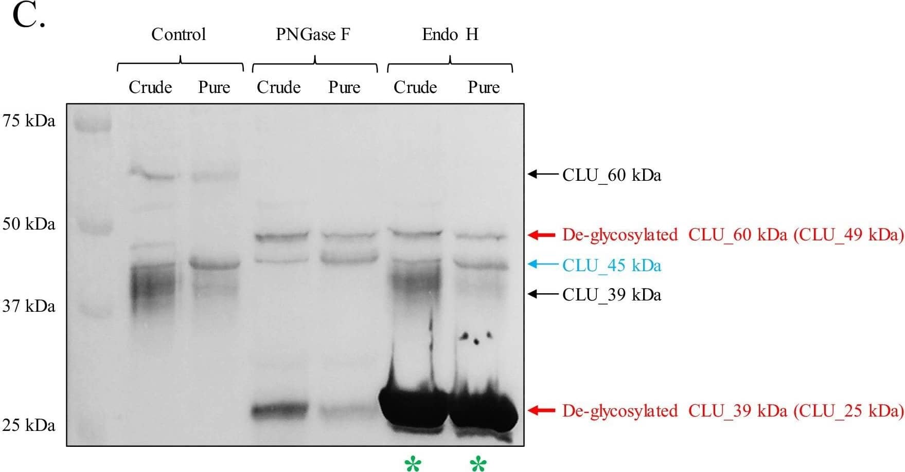
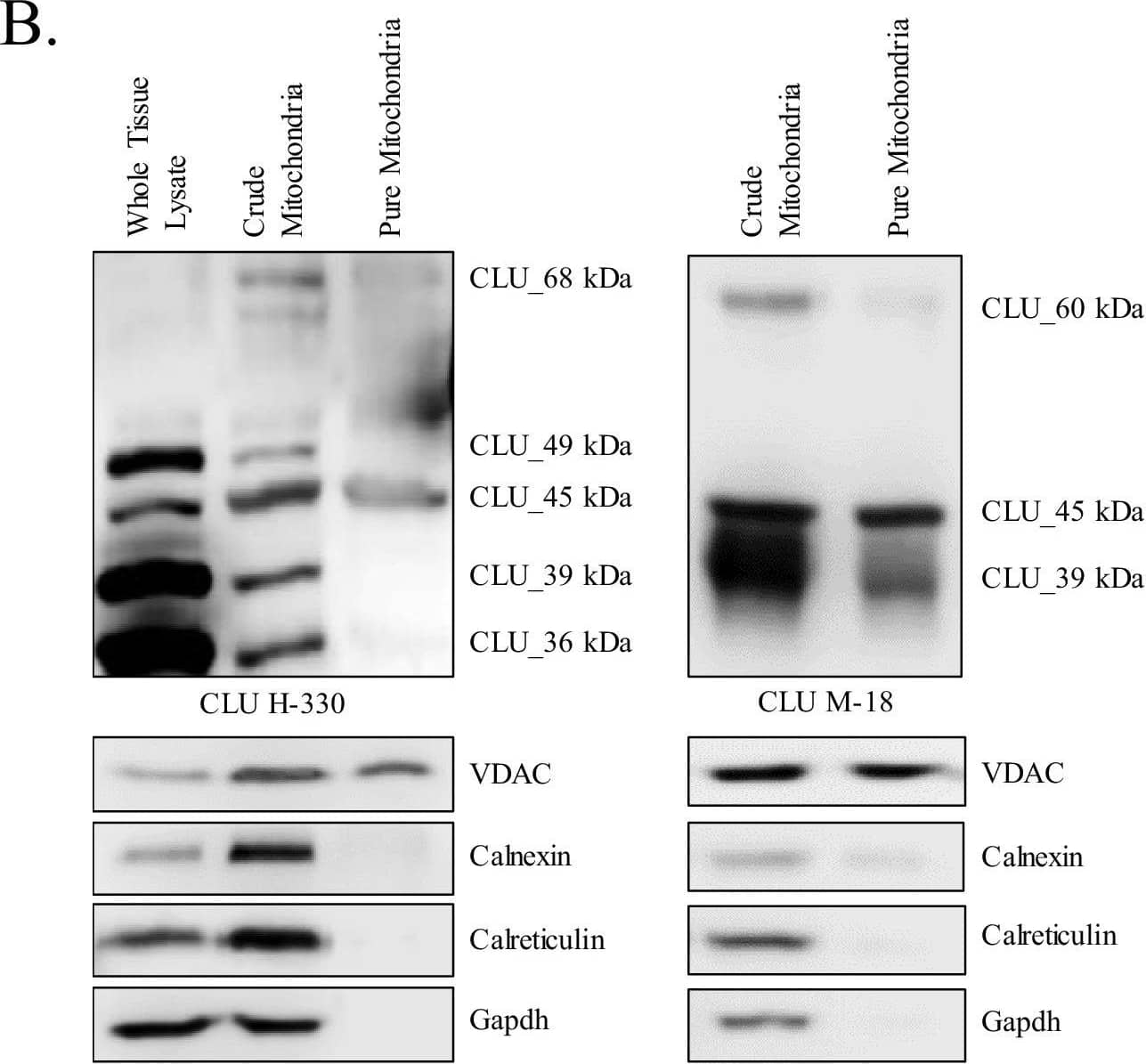
Detection of Rat Clusterin/APOJ by Western Blot Identification of a mitochondrial CLU protein isoform.(A) DIV 9 Mitotracker-stained (green) primary neurons were probed for CLU immunoreactivity using anti-CLU H-330 (red) and visualized using 40X confocal microscopy. (B) Pure cortical mitochondria were isolated as indicated. Equal concentrations of whole tissue lysate, crude mitochondria, and pure mitochondria were analyzed via SDS-PAGE and probed for CLU immunoreactivity using anti-CLU H-330 (left panel) and anti-CLU M-18 (right panel) (n = 3 independent isolations). Biochemical characterization of isolated fractions was performed using a panel of organelle-specific antibodies: voltage-dependent anion channels (VDAC) (mitochondria), calnexin and calreticulin (ER), and Gapdh (cytosol). (C) Crude and pure mitochondria were isolated and subjected to endoglycosidase treatment using PNGase F and Endo H. Deglycosylated mitochondrial lysates were then analyzed for CLU immunoreactivity using anti-CLU M-18. Red font: deglycosylated protein isoforms; blue font: isoforms that were unaffected by glycosidase treatment; green asterisk: excess Endo H enzyme.Positive controls for deglycosylation studies.RNase B, a high mannose glycoprotein, has a single N-linked glycosylation site and was used as a positive control for endoglycosidases that cleave N-linked carbohydrates. Fetuin, a glycoprotein containing sialylated N-linked and O-linked glycans, was used as a positive control for endoglycosidases that cleave both N-linked and O-linked carbohydrates. Image collected and cropped by CiteAb from the following open publication (https://pubmed.ncbi.nlm.nih.gov/31738162), licensed under a CC-BY license. Not internally tested by R&D Systems.
Reconstitution Calculator
Preparation and Storage
- 12 months from date of receipt, -20 to -70 °C as supplied.
- 1 month, 2 to 8 °C under sterile conditions after reconstitution.
- 6 months, -20 to -70 °C under sterile conditions after reconstitution.
Background: Clusterin
Clusterin, also known as Apolipoprotein J, Sulfated Glycoprotein 2 (SGP-2), TRPM-2, and SP-40, is a secreted multi-functional protein that was named for its ability to induce cellular clustering. It binds a wide range of molecules and may function as a chaperone of misfolded extracellular proteins. It also participates in the control of cell proliferation, apoptosis, and carcinogenesis (1, 2). Clusterin is predominantly expressed in adult testis, ovary, adrenal gland, liver, heart, and brain and in many epithelial tissues during embryonic development (3). Human Clusterin is synthesized as a precursor that contains two coiled coil domains, three nuclear localization signals (NLS), and one heparin binding domain (4-6). Intracellular cleavages of the precursor remove the signal peptide and generate comparably sized alpha and beta chains which are secreted as an 80 kDa N-glycosylated disulfide-linked heterodimer (7, 8). Mature human Clusterin shares 77% amino acid sequence identity with mouse and rat Clusterin. High μg/mL concentrations of Clusterin circulate predominantly as a component of high density lipoprotein particles, and these are internalized and degraded through interactions with LRP-2/Megalin (9, 10). In human, an alternately spliced 50 kDa isoform of Clusterin (nCLU) lacks the signal peptide and remains intracellular (5, 11). This molecule is neither glycosylated nor cleaved into alpha and beta chains (11). In the cytoplasm, nCLU destabilizes the actin cytoskeleton and inhibits NF kappa B activation (12, 13). Cellular exposure to ionizing radiation promotes the translocation of nCLU to the nucleus where it interacts with Ku70 and promotes apoptosis (5, 11). This function contrasts with the cytoprotective effect of secreted Clusterin (14). During colon cancer tumor progression there is a downregulation of the intracellular form and an upregulation of the glycosylated secreted form (11).
- Carver, J.A. et al. (2003) IUBMB Life 55:661.
- Shannan, B. et al. (2006) Cell Death Differ. 13:12.
- French, L.E. et al. (1993) J. Cell Biol. 122:1119.
- Kirszbaum, L. et al. (1989) EMBO J. 8:711.
- Leskov, K.S. et al. (2003) J. Biol. Chem. 278:11590.
- Pankhurst, G.J. et al. (1998) Biochemistry 37:4823.
- Burkey, B.F. et al. (1991) J. Lipid. Res. 32:1039.
- de Silva, H.V. et al. (1990) J. Biol. Chem. 265:14292.
-
Jenne, D.E. et al. (1991) J. Biol. Chem. 266:11030.
-
Kounnas, M.Z. et al. (1995) J. Biol. Chem. 270:13070.
-
Pucci, S. et al. (2004) Oncogene 23:2298.
-
Moretti, R. M. et al. (2007) Cancer Res. 67:10325.
-
Santilli, G. et al. (2003) J. Biol. Chem. 278:38214.
-
Trougakos, I.P. et al. (2004) Cancer Res. 64:1834.
Product Datasheets
Citations for Human Clusterin Antibody
R&D Systems personnel manually curate a database that contains references using R&D Systems products. The data collected includes not only links to publications in PubMed, but also provides information about sample types, species, and experimental conditions.
5
Citations: Showing 1 - 5
Filter your results:
Filter by:
-
Structural Remodeling of the Human Colonic Mesenchyme in Inflammatory Bowel Disease.
Authors: Kinchen J, Chen HH, Parikh K et al.
Cell
-
Divergent single cell transcriptome and epigenome alterations in ALS and FTD patients with C9orf72 mutation
Authors: Li J, Jaiswal MK, Chien JF et al.
Nat Commun
-
Divergent single cell transcriptome and epigenome alterations in ALS and FTD patients with C9orf72 mutation
Authors: Li J, Jaiswal MK, Chien JF et al.
Nat Commun
-
Low-dose brain irradiation normalizes TSPO and CLUSTERIN levels and promotes the non-amyloidogenic pathway in pre-symptomatic TgF344-AD rats
Authors: K Ceyzériat, T Zilli, P Millet, N Koutsouvel, G Dipasquale, C Fossey, T Cailly, F Fabis, GB Frisoni, V Garibotto, BB Tournier
Journal of Neuroinflammation, 2022-12-22;19(1):311.
Species: Rat
Sample Types: Protein
Applications: Western Blot -
Brain clusterin protein isoforms and mitochondrial localization
Authors: Sarah K Herring, Hee-Jung Moon, Punam Rawal, Anindit Chhibber, Liqin Zhao
eLife
FAQs
No product specific FAQs exist for this product, however you may
View all Antibody FAQsReviews for Human Clusterin Antibody
There are currently no reviews for this product. Be the first to review Human Clusterin Antibody and earn rewards!
Have you used Human Clusterin Antibody?
Submit a review and receive an Amazon gift card.
$25/€18/£15/$25CAN/¥75 Yuan/¥2500 Yen for a review with an image
$10/€7/£6/$10 CAD/¥70 Yuan/¥1110 Yen for a review without an image
