Human CCL19/MIP-3 beta Antibody Summary
Gly22-Ser98
Accession # Q99731.1
Applications
Please Note: Optimal dilutions should be determined by each laboratory for each application. General Protocols are available in the Technical Information section on our website.
Scientific Data
 View Larger
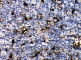
CCL19/MIP‑3 beta in Human Tonsil. CCL19/MIP-3 beta was detected in immersion fixed paraffin-embedded sections of human tonsil using 25 µg/mL Mouse Anti-Human CCL19/ MIP-3 beta Monoclonal Antibody (Catalog # MAB361) overnight at 4 °C. Tissue was stained with the Anti-Mouse HRP-DAB Cell & Tissue Staining Kit (brown; Catalog # CTS002) and counter-stained with hematoxylin (blue). View our protocol for Chromogenic IHC Staining of Paraffin-embedded Tissue Sections.
 View Larger
CCL19/MIP‑3 beta in Human PBMCs. CCL19/MIP-3 beta was detected in immersion fixed human peripheral blood mononuclear cells (PBMCs) using 10 µg/mL Mouse Anti-Human CCL19/MIP-3 beta Monoclonal Anti-body (Catalog # MAB361) for 3 hours at room temperature. Cells were stained with the NorthernLights™ 557-conjugated Anti-Mouse IgG Secondary Antibody (red; Catalog # NL007) and counter-stained with DAPI (blue). View our protocol for Fluorescent ICC Staining of Non-adherent Cells.
 View Larger
Detection of CCL19/MIP‑3 beta in Human Dendritic Cells by Flow Cytometry. Human monocyte-derived dendritic cells were stained with Mouse Anti-Human CCL19/MIP-3 beta Monoclonal Antibody (Catalog # MAB361, filled histogram) or isotype control antibody (Catalog # MAB0041, open histogram), followed by Phycoerythrin-conjugated Anti-Mouse IgG Secondary Antibody (Catalog # F0102B). To facilitate intracellular staining, cells were fixed with Flow Cytometry Fixation Buffer (Catalog # FC004) and permeabilized with Flow Cytometry Permeabilization/Wash Buffer I (Catalog # FC005).
Reconstitution Calculator
Preparation and Storage
- 12 months from date of receipt, -20 to -70 °C, as supplied.
- 1 month, 2 to 8 °C under sterile conditions after opening.
- 6 months, -20 to -70 °C under sterile conditions after opening.
Background: CCL19/MIP-3 beta
CCL19, also known as MIP-3 beta and ELC (EBI1-Ligand Chemokine), is a 77 amino acid (aa) beta chemokine that is distantly related to other beta chemokines (20-30% aa sequence identity). The gene for MIP-3 beta has been mapped to chromosome 9p13 rather than chromosome 17 where the genes for many human beta chemokines are clustered. MIP-3 beta is constitutively expressed in various lymphoid tissues (including thymus, lymph nodes, appendix and spleen). The expression of MIP-3 beta is
down‑regulated by the anti-inflammatory cytokine IL-10. Recombinant MIP-3 beta is chemotactic for cultured human lymphocytes. MIP-3 beta is a ligand for CCR7 (previously referred to as the Epstein-Barr virus-induced gene 1 (EBI1) orphan receptor), a chemokine receptor that is expressed in various lymphoid tissues and activated B and T lymphocytes. CCR7 is strongly up-regulated in B cells infected with Epstein-Barr virus and T cells infected with herpesvirus 6 or 7.
Product Datasheets
Citations for Human CCL19/MIP-3 beta Antibody
R&D Systems personnel manually curate a database that contains references using R&D Systems products. The data collected includes not only links to publications in PubMed, but also provides information about sample types, species, and experimental conditions.
14
Citations: Showing 1 - 10
Filter your results:
Filter by:
-
CC-chemokine receptor 7 and its ligand CCL19 promote mitral valve interstitial cell migration and repair
Authors: Xiaozhi Xiaozhi, Liang Liang, Liping Liping, Zhao Rong, Yanhu Yanhu, Xiangqing Xiangqing
The Journal of Biomedical Research
-
Structural Remodeling of the Human Colonic Mesenchyme in Inflammatory Bowel Disease.
Authors: Kinchen J, Chen HH, Parikh K et al.
Cell
-
The Ccr7 Ligand ELC (Ccl19) Is Transcytosed in High Endothelial Venules and Mediates T Cell Recruitment
Authors: Espen S. Baekkevold, Takeshi Yamanaka, Roger T. Palframan, Hege S. Carlsen, Finn P. Reinholt, Ulrich H. von Andrian et al.
J. Exp. Med
-
Soluble pathogenic tau enters brain vascular endothelial cells and drives cellular senescence and brain microvascular dysfunction in a mouse model of tauopathy
Authors: Hussong, SA;Banh, AQ;Van Skike, CE;Dorigatti, AO;Hernandez, SF;Hart, MJ;Ferran, B;Makhlouf, H;Gaczynska, M;Osmulski, PA;McAllen, SA;Dineley, KT;Ungvari, Z;Perez, VI;Kayed, R;Galvan, V;
Nature communications
-
A Versatile Toolkit for Semi-Automated Production of Fluorescent Chemokines to Study CCR7 Expression and Functions
Authors: M Artinger, C Matti, OJ Gerken, CT Veldkamp, DF Legler
International Journal of Molecular Sciences, 2021-04-16;22(8):.
Species: Human
Sample Types: Protein Lysate, Whole Cells
Applications: Flow Cytometry, Western Blot -
An initial investigation into endothelial CC chemokine expression in the human rheumatoid synovium.
Authors: Rump L, Mattey D, Kehoe O, Middleton J
Cytokine, 2017-09-01;97(0):133-140.
Species: Human
Sample Types: Whole Tissue
Applications: IHC-Fr -
Induction of immunoregulatory CD271+ cells by metastatic tumor cells that express human endogenous retrovirus H.
Authors: Kudo-Saito C, Yura M, Yamamoto R, Kawakami Y
Cancer Res, 2014-03-01;74(5):1361-70.
Species: Human
Sample Types: Whole Tissue
Applications: IHC-P -
Intracellular coexpression of CXC- and CC- chemokine receptors and their ligands in human melanoma cell lines and dynamic variations after xenotransplantation.
Authors: Pinto S, Martinez-Romero A, O'Connor J, Gil-Benso R, San-Miguel T, Terradez L, Monteagudo C, Callaghan R
BMC Cancer, 2014-02-22;14(0):118.
Species: Human
Sample Types: Whole Cells
Applications: Flow Cytometry, ICC -
CCL21 overexpressed on lymphatic vessels drives thymic hyperplasia in myasthenia.
Authors: Berrih-Aknin S, Ruhlmann N, Bismuth J, Cizeron-Clairac G, Zelman E, Shachar I, Dartevelle P, de Rosbo NK, Le Panse R
Ann. Neurol., 2009-10-01;66(4):521-31.
Species: Human
Sample Types: Tissue Homogenates
Applications: ELISA Development -
Cutting edge: nonproliferating mature immune cells form a novel type of organized lymphoid structure in idiopathic pulmonary fibrosis.
Authors: Marchal-Somme J, Uzunhan Y, Marchand-Adam S, Valeyre D, Soumelis V, Crestani B, Soler P
J. Immunol., 2006-05-15;176(10):5735-9.
Species: Human
Sample Types: Whole Tissue
Applications: IHC-Fr -
Expression of macrophage inflammatory protein-3 beta/CCL19 in pulmonary sarcoidosis.
Authors: Gibejova A, Mrazek F, Subrtova D, Sekerova V, Szotkowska J, Kolek V, du Bois RM, Petrek M
Am. J. Respir. Crit. Care Med., 2003-03-05;167(12):1695-703.
Species: Human
Sample Types: Whole Cells
Applications: ICC -
Single-cell and spatial transcriptomics reveal aberrant lymphoid developmental programs driving granuloma formation
Authors: Thomas Krausgruber, Anna Redl, Daniele Barreca, Konstantin Doberer, Daria Romanovskaia, Lina Dobnikar et al.
Immunity
-
The Vaccine-site Microenvironment Induced by Injection of Incomplete Freund's Adjuvant, With or Without Melanoma Peptides
Authors: Rebecca C. Harris, Kimberly A. Chianese-Bullock, Gina R. Petroni, Jochen T. Schaefer, Louis B. Brill, Kerrington R. Molhoek et al.
Journal of Immunotherapy
FAQs
No product specific FAQs exist for this product, however you may
View all Antibody FAQsReviews for Human CCL19/MIP-3 beta Antibody
Average Rating: 5 (Based on 1 Review)
Have you used Human CCL19/MIP-3 beta Antibody?
Submit a review and receive an Amazon gift card.
$25/€18/£15/$25CAN/¥75 Yuan/¥2500 Yen for a review with an image
$10/€7/£6/$10 CAD/¥70 Yuan/¥1110 Yen for a review without an image
Filter by:


