Human Axl APC-conjugated Antibody Summary
Met1-Pro440
Accession # AAA61243
Applications
Please Note: Optimal dilutions should be determined by each laboratory for each application. General Protocols are available in the Technical Information section on our website.
Scientific Data
 View Larger
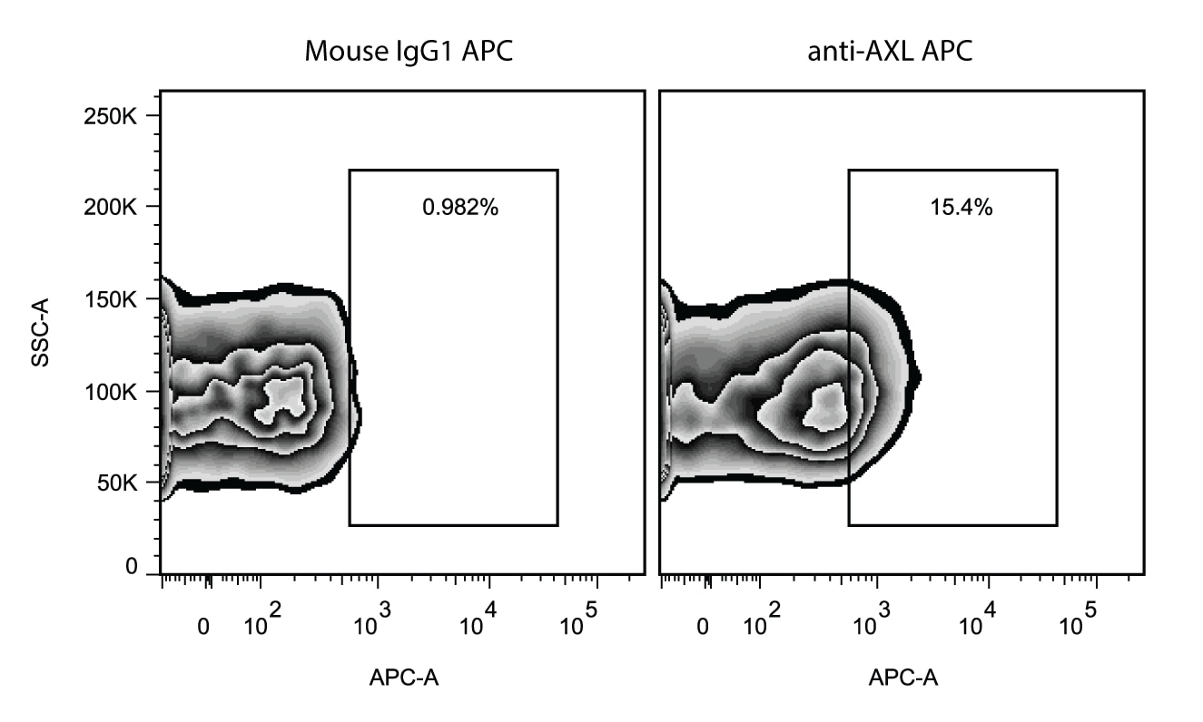
Detection of Axl in HeLa Human Cell Line by Flow Cytometry. HeLa human cervical epithelial carcinoma cell line was stained with Mouse Anti-Human Axl APC-conjugated Monoclonal Antibody (Catalog # FAB154A, filled histogram) or isotype control antibody (Catalog # IC002A, open histogram). View our protocol for Staining Membrane-associated Proteins.
 View Larger
Detection of Axl in A431 Human Cell Line by Flow Cytometry. A431 human epithelial carcinoma cell line was stained with APC-conjugated Mouse Anti-Human Axl Monoclonal Antibody (Catalog # FAB154A, filled histogram) or isotype control antibody (Catalog # IC002A, open histogram). View our protocol for Staining Membrane-associated Proteins.
 View Larger
Axl Specificity is Shown by Flow Cytometry in Knockout Cell Line. Axl knockout A431 human epithelial carcinoma cell line was stained with APC-conjugated Mouse Anti-Human Axl Monoclonal Antibody (Catalog # FAB154A, filled histogram) or isotype control antibody (Catalog # IC002A, open histogram). No staining in the Axl knockout A431 cell line was observed. View our protocol for Staining Membrane-associated Proteins.
 View Larger
Detection of Human Axl by Flow Cytometry CD1c+ DCs in LPS-BAL are likely to be tissue-recruited blood CD1c+ DCs. a Expression of surface antigens by flow cytometry on DC2/3 from HC blood (green), SS-BAL (blue) and LPS-BAL (red) relative to isotype control (gray). Antigens predicted to discriminate between blood and tissue CD1c+ DCs were tested. Representative plots from more than three experiments are shown. b Dendrogram showing hierarchical clustering of monocyte/macrophage and DC gene expression by indicated subsets isolated from HC blood and SS/LPS BAL. In vitro monocyte-derived DCs were also included. Clustering used Pearson correlation metric. Monocyte/macrophage and DC identifying genes were taken from McGovern et al.28. The 25 genes available on the NanoString Immunology v2 panel were used. c Proliferation of allogeneic peripheral blood T cells measured by CSFE dilution during 7-day co-culture with or without MP subsets isolated from LPS BAL. Flow cytometry plots are gated on CD3+ T cells from a representative experiment. Summary graph shows 2–3 replicates per subset. Bars show mean ± SEM. Means were compared by one-way ANOVA with Dunnett’s multiple comparison test of DC2/3 against other subsets. *p < 0.05. d Analysis of differentially expressed genes (DEGs) by DC2/3 from SS/LPS-BAL and HC blood. Comparisons were made by unpaired t-test with p < 0.05 and >3-fold difference in mean expression. Venn diagram shows DEGs in DC2/3 from SS-BAL, LPS-BAL, and HC blood. Circles represent DEGs between SS BAL and blood (cyan; 154 genes, 33%) and between LPS-BAL and blood (yellow; 124 genes, 39%). The overlapping circles represents DEGs shared between comparisons (100 genes, 21%). The top 10 upregulated genes (standard type) and top 5 downregulated genes (italic type) in BAL relative to blood are listed. Heatmap shows DEGs common to DC2/3 in both SS-BAL and LPS-BAL relative to HC blood. Genes with >10-fold difference in mean expression are shown. Genes discussed in the text are colored burgundy. e Heatmap showing DEGs between DC2/3 from SS-BAL and LPS-BAL. Genes with >10-fold difference in mean expression are shown. Genes discussed in the text are colored burgundy Image collected and cropped by CiteAb from the following publication (https://pubmed.ncbi.nlm.nih.gov/31040289), licensed under a CC-BY license. Not internally tested by R&D Systems.
 View Larger
Detection of Human Axl by Flow Cytometry Inflammatory stimuli associated with viral infections upregulate Axl and promote Gas6 binding to macrophages. (A–E) MDMs were stimulated for 24 h with dexamethasone (Dex), LPS, IFN‐ gamma, poly(I:C) or IFN‐ alpha. (A) MerTK and (C) Axl protein expression determined by ELISA of total cell lysates (mean+SEM, n = 4). (B) MerTK and (D) Axl protein expression analyzed by western blotting; actin was used as loading control. A representative of two to three independent experiments is shown. (E) Flow cytometric analysis of Axl expression on MDMs stimulated with poly(I:C) or IFN‐ alpha for 24 h. Representative histograms of two independent experiments are shown; dotted line/shaded: FMO control. (F) Axl and MerTK relative mRNA and (G) protein expression in MDMs differentiated by 6‐day culture in M‐CSF or GM‐CSF determined by qPCR (mean+SEM, n = 6) and by ELISA of total cell lysates (mean+SEM, n = 3), respectively. (H) Axl, MerTK and Tyro3 relative mRNA expression in MDMs stimulated as in (A–C) analyzed by qPCR (mean+SEM, n = 2–3). (I and J) Flow cytometric analysis of Gas6 binding to MDMs stimulated with poly(I:C) for 24 h. (I) Representative of four independent experiments is shown; dotted line/shaded: isotype control. (J) Ratio of Gas6 to isotype control geometric mean fluorescence intensity (MFI)+SEM in unstimulated and poly(I:C)‐stimulated MDMs (n = 4). *p < 0.01, paired t‐test. ELISA and qPCR samples were assayed in duplicate. Image collected and cropped by CiteAb from the following publication (https://pubmed.ncbi.nlm.nih.gov/29400409), licensed under a CC-BY license. Not internally tested by R&D Systems.
 View Larger
Detection of Human Axl by Flow Cytometry Neutrophils and mononuclear phagocytes are expanded in the alveolar airspace following LPS inhalation. a Schematic overview of study design. Solid arrows denote LPS inhalation (red) or saline inhalation control (blue). Black arrows denote blood sampling. Dashed arrows denote BAL. b Flow cytometry of leukocyte preparations from SS-BAL, LPS-BAL, and HC blood. The CD45 versus SSC plot was used to define CD45+SSChi AM, CD45loSSCmid neutrophils and CD45+SSClo mononuclear cells (see also Supplementary Fig. 1). Monocyte/macrophages and DCs were negative for lineage markers CD3, CD19, CD20, and CD56 and expressed HLA-DR. Monocyte/macrophages were divided into CD14++CD16−, CD14++CD16+, and CD14-CD16++ populations analogous to blood classical, intermediate and non-classical monocytes. DCs within the CD14-CD16− gate were divided into subsets: Axl+Siglec6+ DC5s, CD11cloCD123+ pDCs, CD1c+BTLAlo-mid DC2/3s, and BTLAhi DC1s. Plots are representative of n = 9 SS BAL and n = 10 LPS BAL. c Concentrations of neutrophil, monocyte/macrophage and DC subsets in SS-BAL and LPS BAL. Bars represent mean and lines SEM. p-values from unpaired t-tests of SS versus LPS are shown: “ns” p > 0.05, *p < 0.05, **p < 0.01, ***p < 0.001, ****p < 0.0001. d Monocyte/macrophage and DC frequency in HC blood, SS-BAL and LPS-BAL as a proportion of SSClo MHC class II-expressing cells (not-including CD1c- DCs). e Concentration of selected leukocyte populations in peripheral blood at 2-h intervals following inhalation of saline (blue line) or LPS (red line). Data points show mean ± SEM for 3–5 participants. p-values from one-way ANOVA are shown. p-value representation is described in c Image collected and cropped by CiteAb from the following publication (https://pubmed.ncbi.nlm.nih.gov/31040289), licensed under a CC-BY license. Not internally tested by R&D Systems.
Reconstitution Calculator
Preparation and Storage
- 12 months from date of receipt, 2 to 8 °C as supplied.
Background: Axl
Axl (Ufo, Ark), Dtk (Sky, Tyro3, Rse, Brt), and Mer (human and mouse homologues of chicken c-Eyk) constitute a subfamily of the receptor tyrosine kinases (1,2). The extracellular domains of these proteins contain two Ig-like motifs and two fibronectin type III motifs. This characteristic topology is also found in neural cell adhesion molecules and in receptor tyrosine phosphatases. The human Axl cDNA encodes an 887 amino acid (aa) precursor that includes an 18 aa signal sequence, a 426 aa extracellular domain, a 21 aa transmembrane segment, and a 422 aa cytoplasmic domain. The extracellular domains of human and mouse Axl share 81% aa sequence identity. A short alternately spliced form of human Axl is distinguished by a 9 aa deletion in the extracellular juxtamembrane region. These receptors bind the vitamin K-dependent protein growth arrest specific gene 6 (Gas6) which is structurally related to the anticoagulation factor protein S. Binding of Gas6 induces receptor autophosphorylation and downstream signaling pathways that can lead to cell proliferation, migration, or the prevention of apoptosis (3). This family of tyrosine kinase receptors is involved in hematopoiesis, embryonic development, tumorigenesis, and regulation of testicular functions.
- Yanagita, M. (2004) Curr. Opin. Nephrol. Hypertens. 13:465.
- Nagata, K. et al. (1996) J. Biol. Chem. 22:30022.
- Holland, S. et al. (2005) Canc. Res. 65:9294.
Product Datasheets
Citations for Human Axl APC-conjugated Antibody
R&D Systems personnel manually curate a database that contains references using R&D Systems products. The data collected includes not only links to publications in PubMed, but also provides information about sample types, species, and experimental conditions.
11
Citations: Showing 1 - 10
Filter your results:
Filter by:
-
Lineage-dependence of the neuroblastoma surfaceome defines tumor cell state-dependent and independent immunotherapeutic targets
Authors: Kendsersky, NM;Odrobina, M;Mabe, NW;Farrel, A;Grossmann, L;Tsang, M;Groff, D;Wolpaw, AJ;Zammarchi, F;van Berkel, PH;Dang, CV;Mossé, YP;Stegmaier, K;Maris, JM;
bioRxiv : the preprint server for biology
Species: Human
Sample Types: Whole Cells
Applications: Flow Cytometry -
AXL-Receptor Targeted 14FN3 Based Single Domain Proteins (Pronectins�) from 3 Synthetic Human Libraries as Components for Exploring Novel Bispecific Constructs against Solid Tumors
Authors: CA Hokanson, E Zacco, G Cappuccill, T Odineca, R Crea
Biomedicines, 2022-12-08;10(12):.
Species: Mouse
Sample Types: Whole Cell
Applications: Flow Cytometry -
Anti-inflammatory clearance of amyloid-beta by a chimeric Gas6 fusion protein
Authors: H Jung, SY Lee, S Lim, HR Choi, Y Choi, M Kim, S Kim, Y Lee, KH Han, WS Chung, CH Kim
Nature Medicine, 2022-08-04;0(0):.
Species: Human
Sample Types: Whole Cells
Applications: Flow Cytometry -
The Most Common VHL Point Mutation R167Q in Hereditary VHL Disease Interferes with Cell Plasticity Regulation
Authors: S Buart, S Terry, MK Diop, P Dessen, S Couvé, A Abdou, J Adam, J Thiery, P Savagner, S Chouaib
Cancers, 2021-08-02;13(15):.
Species: Human
Sample Types: Whole Cells
Applications: Flow Cytometry -
An epigenetic switch regulates the ontogeny of AXL-positive/EGFR-TKi-resistant cells by modulating miR-335 expression
Authors: Polona Safaric Tepes, Debjani Pal, Trine Lindsted, Ingrid Ibarra, Amaia Lujambio, Vilma Jimenez Sabinina et al.
eLife
-
Discriminatory Power of Combinatorial Antigen Recognition in Cancer T Cell Therapies
Authors: Ruth Dannenfelser, Gregory M. Allen, Benjamin VanderSluis, Ashley K. Koegel, Sarah Levinson, Sierra R. Stark et al.
Cell Systems
-
Differential IRF8 Transcription Factor Requirement Defines Two Pathways of Dendritic Cell Development in Humans
Authors: Urszula Cytlak, Anastasia Resteu, Sarah Pagan, Kile Green, Paul Milne, Sheetal Maisuria et al.
Immunity
-
TAM Family Receptor kinase inhibition reverses MDSC-mediated suppression and augments anti-PD-1 therapy in melanoma.
Authors: Alisha Holtzhausen, William Harris, Eric Ubil, Debra M. Hunter, Jichen Zhao, Yuewei Zhang et al.
Cancer Immunology Research
-
Lipopolysaccharide inhalation recruits monocytes and dendritic cell subsets to the alveolar airspace
Authors: L Jardine, S Wiscombe, G Reynolds, D McDonald, A Fuller, K Green, A Filby, I Forrest, MH Ruchaud-Sp, J Scott, M Collin, M Haniffa, AJ Simpson
Nat Commun, 2019-04-30;10(1):1999.
Species: Human
Sample Types: Whole Cells
Applications: Flow Cytometry -
Characterization of the Filovirus-Resistant Cell Line SH-SY5Y Reveals Redundant Role of Cell Surface Entry Factors
Authors: FJ Zapatero-B, E Dietzel, O Dolnik, K Döhner, R Costa, B Hertel, B Veselkova, J Kirui, A Klintworth, MP Manns, S Pöhlmann, T Pietschman, T Krey, S Ciesek, G Gerold, B Sodeik, S Becker, T von Hahn
Viruses, 2019-03-19;11(3):.
Species: Human
Sample Types: Whole Cells
Applications: Flow Cytometry -
Normal breast-derived epithelial cells with luminal and intrinsic subtype-enriched gene expression document inter-individual differences in their differentiation cascade
Authors: B Kumar, MS Prasad, P Bhat-Naksh, M Anjanappa, M Kalra, N Marino, AMV Storniolo, X Rao, S Liu, J Wan, Y Liu, H Nakshatri
Cancer Res., 2018-07-11;0(0):.
Species: Human
Sample Types: Whole Cells
Applications: Flow Cytometry
FAQs
No product specific FAQs exist for this product, however you may
View all Antibody FAQsReviews for Human Axl APC-conjugated Antibody
Average Rating: 4.3 (Based on 3 Reviews)
Have you used Human Axl APC-conjugated Antibody?
Submit a review and receive an Amazon gift card.
$25/€18/£15/$25CAN/¥75 Yuan/¥2500 Yen for a review with an image
$10/€7/£6/$10 CAD/¥70 Yuan/¥1110 Yen for a review without an image
Filter by:
the gating was based on FMO control. In my hand, Axl antibody with APC works better than PE.


