Endothelial Cell Growth Media
Endothelial Cell Growth Media Summary
Media for the low-serum expansion of primary endothelial cells.
Key Benefits
- Unique and optimized formulation that promotes endothelial cell growth.
- Reduces cell senescence at higher passages
- Includes a single supplement featuring key growth factors and small molecules
- 250 mL bottle results in less wasted media
Why Use Pre-optimized, Low-serum Media to Grow Endothelial Cells?
Primary endothelial cells are commonly used for the study of developmental and disease mechanisms involving the blood vessels, such as angiogenesis. Endothelial cells are phenotypically characterized as CD31+, KDR+, and vWF+ cells.
R&D Systems® Endothelial Cell Growth Media is formulated to support the expansion of human primary endothelial cells under low-serum conditions. Culturing endothelial cells under serum-free conditions allows for the researcher to have more controlled and defined system. This media has been tested for its ability to support CD31+, KDR+, and vWF+ human umbilical vein endothelial cells growth in vitro.
This kit contains the following reagents to maintain and expand human and mouse endothelial cells in culture:
- Endothelial Cell Growth Base Media (250 mL)
- Endothelial Cell Growth Supplement (50X)
Containing optimized concentrations of:
- Human FGF basic
- Human LR3 IGF-1
- Human VEGF165
- Human EGF
- Heparin
- Hydrocortisone
- L-Ascorbic Acid
- Fetal Bovine Serum
Note: The components for this kit require different storage/shipping temperatures and will arrive in separate packaging.
Stability and Storage
Store in the dark at < -20 °C in a manual defrost freezer. Do not use past the expiration date.
Precaution
When handling biohazardous materials such as human cells, safe laboratory procedures should be followed and protective clothing should be worn.
Limitations
- FOR LABORATORY RESEARCH USE ONLY. NOT FOR USE IN DIAGNOSTIC PROCEDURES.
- The safety and efficacy of this product in diagnostic or other clinical uses has not been established.
- This reagent should not be used beyond the expiration date indicated on the label.
- Results may vary due to variations among cells derived from different donors.
Specifications
Product Datasheets
Scientific Data
 View Larger
Endothelial Cell Growth Media Supports Cell Proliferation of Human Umbilical Cord Endothelial Cells (HUVECs). One T25 flask of HUVECs (0.17 x 10⁶cells) were expanded over 10 passages using Complete Endothelial Cell Growth Media. Cells were passaged every 4-6 days. The histogram shows the cumulative number of HUVECs expanded over 10 passages if all cells were replated at each passage.
 View Larger
Human Umbilical Cord Endothelial Cells (HUVECs) Express Endothelial Cell Markers CD31, KDR, and vWF. HUVECs were expanded in Complete Endothelial Cell Growth Media for 5 passages prior to staining for marker expression. (A,B) Flow cytometry analysis of endothelial cell marker expression. Filled histograms indicate cells stained with PE-conjugated Mouse Anti-CD31 (A; R&D Systems®, Catalog # FAB3567P) or PE-conjugated Mouse Anti-KDR (B; R&D Systems®, Catalog # FAB357P). Open histograms in A and B indicate staining with PE-conjugated Mouse IgG1 isotype control (R&D Systems®, Catalog # IC002P). (C) Immunocytochemistry analysis of endothelial cell marker expression. Cells were double stained with Mouse Anti-CD31 (red; R&D Systems®, Catalog # BBA7) and Rabbit Anti-Von Willebrand Factor (vWF) (green; Novus Biologicals®, Catalog # NB600-586). The nuclei were counterstained with DAPI (blue)
 View Larger
Increased HUVEC Survival and CD31 Expression Compared to Competitor Endothelial Cell Media. HUVECs were expanded for 14 passages in either R&D Systems Endothelial Cell Growth Media (Catalog # CCM027) or competitor endothelial cell media. Cells were stained for CD31 expression using the Mouse Anti-Human CD31 Antibody (yellow; R&D Systems; Catalog # BBA7) and counterstained with DAPI (Catalog # 5748).A) Representative images of CD31 expression in p14 HUVECs grown in R&D Systems Media or competitor media. There is a greater density of cells and a higher expression of CD31 in HUVECs grown in R&D Systems Media.B) Histogram showing that at p14, there is a higher percentage of surviving cells that express CD31 in HUVECs grown in R&D Systems Media compared to competitor.
 View Larger
Improved Yield of Murine CD31+PDGFa- Lung Cells using Endothelial Cell Growth Media A single cell suspension of mouse lung cells was prepared and seeded onto gelatin coated plates in 3 different media, R&D Systems®Endothelial Cell Growth Media (Catalog # CCM027), competitor media, or 20% FBS/DMEM. After one week in culture, cells were analyzed for expression of CD31 (APC) and PDGFRα (PE). The percentage of CD31+PDGFRα- cells obtained from R&D Systems Endothelial Cell Growth Media was superior (13.2%) compared to competitor (3.5%) and 20% FBS/DMEM (4.5%) media. Data courtesy of the laboratory of Dr. Michael Kyba at the University of Minnesota.
 View Larger
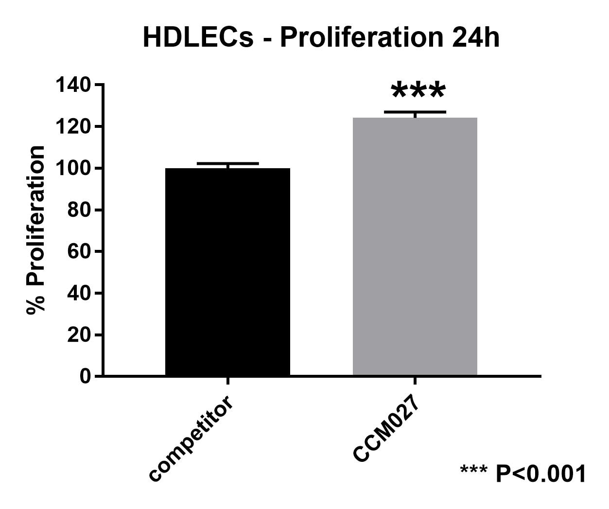
Improved Proliferation of Primary Human Dermal Lymphatic Endothelial Cells. Human dermal lymphatic endothelial cells (HDLECs) were cultured in either Endothelial Cell Growth Media (R&D Systems, Catalog # CCM027) or a competitor endothelial cell media. After 48 hours, HDLEC proliferation was assessed using a MTT proliferation assay. HDLECs cultured in R&D Systems Endothelial Cell Growth Medium showed a 24% increase in proliferation compared to competitor media. The P-value was calculated using an unpaired t-test. Data provided courtesy of the laboratory of Dr. Constantinos Mikelis at Texas Tech University Health Sciences Center.
 View Larger
Improved Yield of Murine CD31+PDGF alpha - Lung Cells using Endothelial Cell Growth Media. A single cell suspension of mouse lung cells was prepared and seeded onto gelatin coated plates in 3 different media, R&D Systems Endothelial Cell Growth Media (Catalog # CCM027), competitor media, or 20% FBS/DMEM. After one week in culture, cells were analyzed for expression of CD31 (APC) and PDGFR alpha (PE). The percentage of CD31+PDGFR alpha - cells obtained from R&D Systems Endothelial Cell Growth Media was superior (13.2%) compared to competitor (3.5%) and 20% FBS/DMEM (4.5%) media. Data courtesy of the laboratory of Dr. Michael Kyba at the University of Minnesota.
Assay Procedure
Refer to the product datasheet for complete product details.
Briefly, Complete Endothelial Cell Growth Media is prepared using the following procedure:
- Add Endothelial Growth Media Supplement to Endothelial Cell Growth Base Media
- Store completed media at 2 - 8 °C
Reagents provided with the Endothelial Cell Growth Media (Catalog # CCM027):
- Endothelial Cell Growth Base Media Human
- Endothelial Cell Growth Supplement (50X)
Note: The components of this list require different storage/shipping temperatures and will arrive in separate packaging.
Reagents
- HUVECs
- Penicillin-Streptomycin (100X), optional
- TrypLE™ Express
- Phosphate-Buffered Saline (PBS) (Tocris; Catalog # 3156)
Materials
- 75 cm2 tissue culture (T75) flasks
- 15 mL centrifuge tubes
- Serological pipettes
- Pipette and pipette tips
Equipment
- 37°C and 5% CO2 humidified incubator
- Centrifuge (low speed clinical or equivalent)
- Hemocytometer
- Inverted microscope
- Water bath
Reagent Preparation
Endothelial Cell Growth Supplement - Thaw the Endothelial Cell Growth Supplement (50X) at 2-8 °C or room temperature.
Complete Endothelial Cell Growth Media - Add 5 mL of Endothelial Cell Growth Supplement to 250 mL of Endothelial Cell Growth Base Media. Add Penicillin-Streptomycin at a 1:100 dilution. Store under sterile conditions at 2-8 °C for up to 2 weeks.
Note: If Penicillin-Streptomycin is not needed for the experiment, it can be omitted.
Culturing of HUVECs
Warm Complete Endothelial Cell Growth Media to 37 °C.
Determine the size and number of flasks needed for plating.

Warm the frozen vial of HUVECs until just thawed.

Transfer cells immediately and gently into a 15 mL centrifuge tube containing > 5 mL of prewarmed Complete Endothelial Cell Growth Media.
Centrifuge at 200 x g for 5 minutes.

Resuspend the pellet in prewarmed Complete Endothelial Cell Growth Media
Transfer HUVEC suspension into a T75 flask(s).

Change media the day following thaw.
Monitor cells daily and change media every other day.
Passage the cells at 80-90% confluency.

Subculturing of HUVECs
Warm Complete Endothelial Cell Growth Media to 37 °C.

Remove and discard the media from the flasks.
Rinse cells once with sterile 1X PBS.
Digest cells with TrypLE Express for 1-2 minutes at 37 °C.

Tap the side of the flask to aid the detachment of the cells.
Transfer the cell suspension to a 15 mL centrifuge tube containing 8 mL of prewarmed Complete Endothelial Cell Growth
Centrifuge at 200 x g for 5 minutes.

Plate cells (approximately 6.7 x 10³ cells/cm2).
Change media the day following thaw.
Monitor cells daily and change media every other day.
Passage the cells at 80-90% confluency.

Citations for Endothelial Cell Growth Media
R&D Systems personnel manually curate a database that contains references using R&D Systems products. The data collected includes not only links to publications in PubMed, but also provides information about sample types, species, and experimental conditions.
9
Citations: Showing 1 - 9
Filter your results:
Filter by:
-
First Application of a Mixed Porcine-Human Repopulated Bioengineered Liver in a Preclinical Model of Post-Resection Liver Failure
Authors: Felgendreff, P;Hosseiniasl, SM;Minshew, A;Amiot, BP;Wilken, S;Ahmadzada, B;Huebert, RC;Sakrikar, NJ;Engles, NG;Halsten, P;Mariakis, K;Barry, J;Riesgraf, S;Fecteau, C;Ross, JJ;Nyberg, SL;
Biomedicines 2024-06-07
-
Enhanced Angiogenesis in HUVECs Preconditioned with Media from Adipocytes Differentiated from Lipedema Adipose Stem Cells In Vitro
Authors: Al-Ghadban, S;Walczak, SG;Isern, SU;Martin, EC;Herbst, KL;Bunnell, BA;
International journal of molecular sciences 2023-09-01
-
Lung Inflammation Resolution by RvD1 and RvD2 in a Receptor-Dependent Manner
Authors: Gao, J;Su, Y;Wang, Z;
Pharmaceutics 2023-05-18
-
Low- and high-grade glioma endothelial cells differentially regulate tumor growth
Authors: Muthukrishnan, S;Qi, H;Wang, D;Elahi, L;Pham, A;Alvarado, A;Li, T;Gao, F;Kawaguchi, R;Lai, A;Kornblum, H;
bioRxiv 2023-01-01
-
Amino Acid Nanofibers Improve Glycemia and Confer Cognitive Therapeutic Efficacy to Bound Insulin
Authors: A Lee, ML Mason, T Lin, SB Kumar, D Kowdley, JH Leung, D Muhanna, Y Sun, J Ortega-Ana, L Yu, J Fitzgerald, AC DeVries, RJ Nelson, ZM Weil, R Jiménez-Fl, JR Parquette, O Ziouzenkov
Pharmaceutics, 2021-12-29;14(1):. 2021-12-29
-
Circulating microRNA profile in humans and mice with congenital GH deficiency
Authors: TD Saccon, A Schneider, CG Marinho, ADC Nunes, S Noureddine, J Dhahbi, YO Nunez Lope, G LeMunyan, R Salvatori, CRP Oliveira, AA Oliveira-S, N Musi, A Bartke, MH Aguiar-Oli, MM Masternak
Aging Cell, 2021-06-12;0(0):e13420. 2021-06-12
-
Induction of CXCL10-Mediated Cell Migration by Different Types of Galectins
Authors: DB AbuSamra, N Panjwani, P Argüeso
Cells, 2021-01-30;10(2):. 2021-01-30
-
Mitofusin 2 regulates neutrophil adhesive migration and the actin cytoskeleton
Authors: W Zhou, AY Hsu, Y Wang, R Syahirah, T Wang, J Jeffries, X Wang, H Mohammad, MN Seleem, D Umulis, Q Deng
J. Cell. Sci., 2020-09-04;133(17):. 2020-09-04
-
Imaging specific cellular glycan structures using glycosyltransferases via click chemistry.
Authors: Wu Z, Person A, Anderson M, Burroughs B, Tatge T, Khatri K, Zou Y, Wang L, Geders T, Zaia J, Sackstein R
Glycobiology, 2018-02-01;0(0):. 2018-02-01
FAQs
No product specific FAQs exist for this product, however you may
View all Cell Culture Product FAQsReviews for Endothelial Cell Growth Media
Average Rating: 5 (Based on 9 Reviews)
Have you used Endothelial Cell Growth Media?
Submit a review and receive an Amazon gift card.
$25/€18/£15/$25CAN/¥75 Yuan/¥2500 Yen for a review with an image
$10/€7/£6/$10 CAD/¥70 Yuan/¥1110 Yen for a review without an image
Filter by:
Tube assay using culture media with supplemented growth factors
Im working with HUVEC cells for the first time. I was very confused about the medium, so I ordered this medium. It was delivered on time and had needed growth factor supplements for HUVEC. I will highly recommend this medium.
Our primary retina endothelial cells grow fast and nice with this media.


