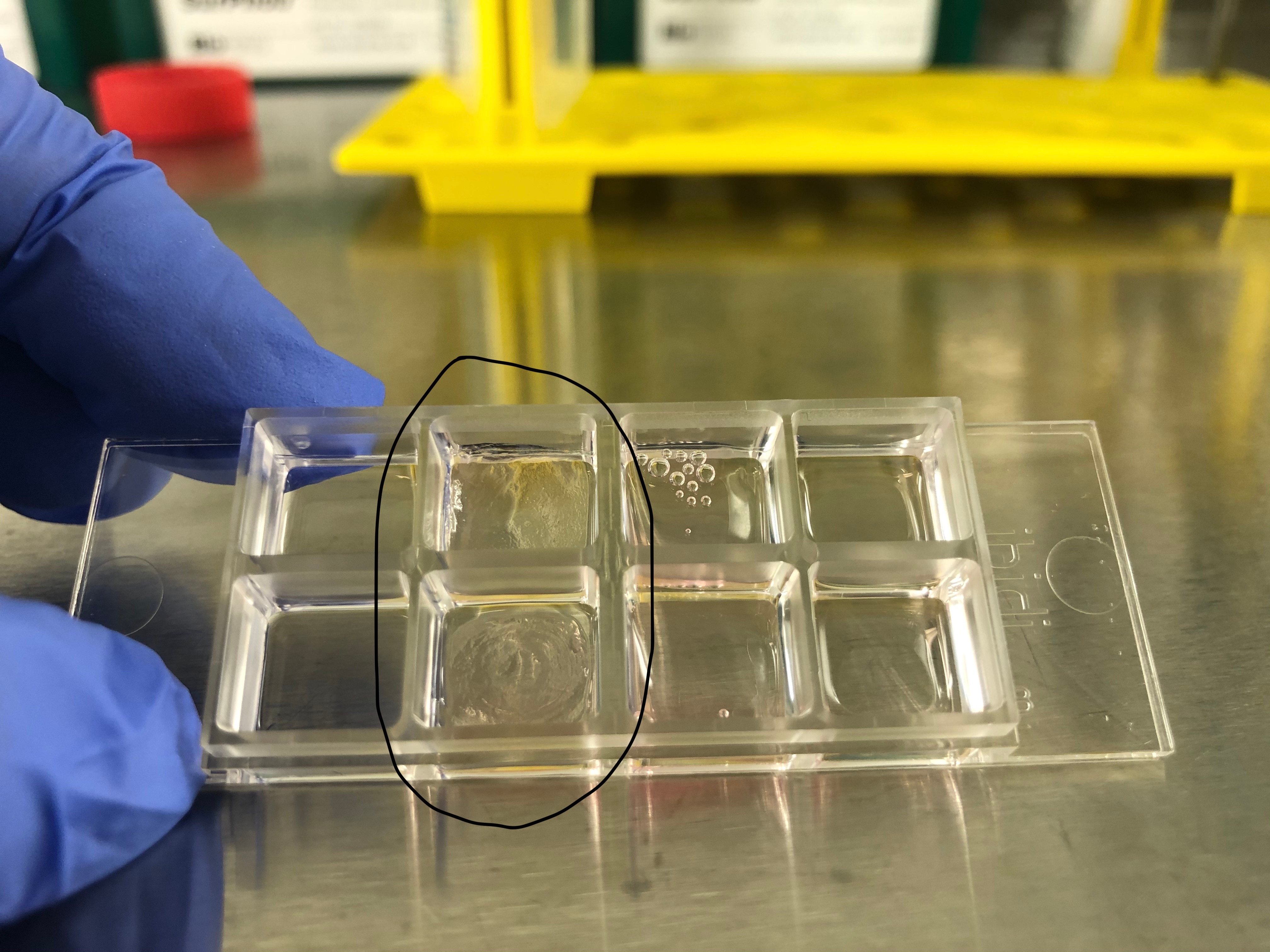
Cultrex 3-D Culture Matrix Reduced Growth Factor Basement Membrane Extract, Pathclear
Try it on your cultures! Request a Sample of Cultrex 3-D Culture Matrix RGF BME
Cultrex 3-D Culture Matrix Reduced Growth Factor Basement Membrane Extract, Pathclear Summary
Cultrex 3-D Culture Matrix RGF BME is an extracellular matrix hydrogel that directs cells to grow in three dimensions and assemble into organotypic structures in vitro.Key Benefits
• Qualified to support 3-D cell culture applications
• Reduced growth factor formulation provides a more defined culture system
• Quality controlled for performance consistency
Why Use Cultrex 3-D Culture Matrix RGF BME ?
Cultrex 3-D Culture Matrix Reduced Factor Basement Membrane Extract (RGF BME) is a soluble form of basement membrane purified from Engelbreth-Holm-Swarm (EHS) tumor. This extract provides a natural extracellular matrix hydrogel that polymerizes at 37°C to form a reconstituted basement membrane. Cultrex 3-D Culture Matrix RGF BME is produced and qualified specifically for use in 3-D culture studies. It provides the foundation for cells to grow in three dimensions allowing for the formation of complex in vitro structures. To provide the most standardized basement membrane extract for use in 3-D cultures, a special process is employed to reduce growth factors and manufacture matrix at a standard and consistent concentration. This product is then evaluated in 3-D culture assays to validate efficacy. Basement membranes are continuous sheets of specialized extracellular matrix that form an interface between endothelial, epithelial, muscle, or neuronal cells and their adjacent stroma and that play an essential role in tissue organization by influencing cell adhesion, migration, proliferation, and differentiation. The major components of BME include laminin, collagen IV, entactin, and heparin sulfate proteoglycans.
Specifications
Tube Formation Assay - Cultrex 3-D Culture Matrix RGF BME Supports formation of capillary-like structures by human (HBMVEC; HUVEC) or mouse (SVEC4-10) endothelial cells.
Gelling Assay - Cultrex 3-D Culture Matrix RGF BME gels in less than 30 minutes at 37 °C, and maintains the gelled form in culture medium for a minimum of 7 days at 37 °C.
Limitations
For research use only. Not for diagnostic use.
Product Datasheets
Citations for Cultrex 3-D Culture Matrix Reduced Growth Factor Basement Membrane Extract, Pathclear
R&D Systems personnel manually curate a database that contains references using R&D Systems products. The data collected includes not only links to publications in PubMed, but also provides information about sample types, species, and experimental conditions.
21
Citations: Showing 1 - 10
Filter your results:
Filter by:
-
Therapeutic effect and transcriptome-methylome characteristics of METTL3 inhibition in liver hepatocellular carcinoma
Authors: Liu, Q;Qi, J;Li, W;Tian, X;Zhang, J;Liu, F;Lu, X;Zang, H;Liu, C;Ma, C;Yu, Y;Jiang, S;
Cancer cell international 2023-11-27
-
LKB1 signaling is altered in skeletal muscle of a Duchenne muscular dystrophy mouse model
Authors: Boccanegra, B;Mantuano, P;Conte, E;Cerchiara, AG;Tulimiero, L;Quarta, R;Caputo, E;Sanarica, F;Forino, M;Spadotto, V;Cappellari, O;Fossati, G;Steinkühler, C;De Luca, A;
Disease models & mechanisms 2023-07-01
-
A distal lung organoid model to study interstitial lung disease, viral infection and human lung development
Authors: Matkovic Leko, I;Schneider, RT;Thimraj, TA;Schrode, N;Beitler, D;Liu, HY;Beaumont, K;Chen, YW;Snoeck, HW;
Nature protocols 2023-05-10
-
Advanced Cell Culture Models Illuminate the Interplay between Mammary Tumor Cells and Activated Fibroblasts
Authors: Del Nero, M;Colombo, A;Garbujo, S;Baioni, C;Barbieri, L;Innocenti, M;Prosperi, D;Colombo, M;Fiandra, L;
Cancers 2023-04-26
-
Designing Organoid Models to Monitor Cancer Progression, Plasticity and Resistance: The Right Set Up for the Right Question
Authors: F Doffe, F Bonini, E Lakis, S Terry, S Chouaib, P Savagner
Cancers, 2022-07-22;14(15):. 2022-07-22
-
Invasive phenotype in triple negative breast cancer is inhibited by blocking SIN3A-PF1 interaction through KLF9 mediated repression of ITGA6 and ITGB1
Authors: R Kadamb, BA Leibovitch, EF Farias, N Dahiya, H Suryawansh, N Bansal, S Waxman
Translational Oncology, 2021-12-27;16(0):101320. 2021-12-27
-
Extensive transcriptional and chromatin changes underlie astrocyte maturation in vivo and in culture
Authors: M Lattke, R Goldstone, JK Ellis, S Boeing, J Jurado-Arj, N Marichal, JI MacRae, B Berninger, F Guillemot
Nature Communications, 2021-07-15;12(1):4335. 2021-07-15
-
T cell exosome-derived miR-142-3p impairs glandular cell function in Sj�gren's syndrome
Authors: J Cortes-Tro, SI Jang, P Perez, J Hidalgo, T Ikeuchi, T Greenwell-, BM Warner, NM Moutsopoul, I Alevizos
JCI Insight, 2020-05-07;5(9):. 2020-05-07
-
CDK4/6 inhibitors target SMARCA4-determined cyclin D1 deficiency in hypercalcemic small cell carcinoma of the ovary
Authors: Y Xue, B Meehan, E Macdonald, S Venneti, XQD Wang, L Witkowski, P Jelinic, T Kong, D Martinez, G Morin, M Firlit, A Abedini, RM Johnson, R Cencic, J Patibandla, H Chen, AI Papadakis, A Auguste, I de Rink, RM Kerkhoven, N Bertos, WH Gotlieb, BA Clarke, A Leary, M Witcher, MC Guiot, J Pelletier, J Dostie, M Park, AR Judkins, R Hass, DA Levine, J Rak, B Vanderhyde, WD Foulkes, S Huang
Nat Commun, 2019-02-04;10(1):558. 2019-02-04
-
Impairment of an Endothelial NAD+-H2S Signaling Network Is a Reversible Cause of Vascular Aging
Authors: A Das, GX Huang, MS Bonkowski, A Longchamp, C Li, MB Schultz, LJ Kim, B Osborne, S Joshi, Y Lu, JH Treviño-Vi, MJ Kang, TT Hung, B Lee, EO Williams, M Igarashi, JR Mitchell, LE Wu, N Turner, Z Arany, L Guarente, DA Sinclair
Cell, 2018-03-22;173(1):74-89.e20. 2018-03-22
-
Human mesenchymal stem cells secrete hyaluronan-coated extracellular vesicles.
Authors: Arasu U, Karna R, Harkonen K, Oikari S, Koistinen A, Kroger H, Qu C, Lammi M, Rilla K
Matrix Biol, 2017-05-05;64(0):54-68. 2017-05-05
-
PEDF increases the tumoricidal activity of macrophages towards prostate cancer cells in vitro
Authors: D Martinez-M, C Jarvis, T Nelius, W de Riese, OV Volpert, S Filleur
PLoS ONE, 2017-04-12;12(4):e0174968. 2017-04-12
-
Live-Cell Imaging of Protease Activity: Assays to Screen Therapeutic Approaches.
Authors: Chalasani A, Ji K, Sameni M, Mazumder S, Xu Y, Moin K, Sloane B
Methods Mol Biol, 2017-01-01;1574(0):215-225. 2017-01-01
-
In vitro differentiation of human amniotic epithelial cells into insulin-producing 3D spheroids.
Authors: Okere B, Alviano F, Costa R, Quaglino D, Ricci F, Dominici M, Paolucci P, Bonsi L, Iughetti L
Int J Immunopathol Pharmacol, 2015-07-27;28(3):390-402. 2015-07-27
-
Organotypic culture of untransformed and tumorigenic primary mammary epithelial cells.
Authors: Jechlinger M
Cold Spring Harb Protoc, 2015-05-01;2015(5):457-61. 2015-05-01
-
Silencing beta3 Integrin by Targeted ECO/siRNA Nanoparticles Inhibits EMT and Metastasis of Triple-Negative Breast Cancer.
Authors: Parvani J, Gujrati M, Mack M, Schiemann W, Lu Z
Cancer Res, 2015-04-09;75(11):2316-2325. 2015-04-09
-
Tumor-induced pressure in the bone microenvironment causes osteocytes to promote the growth of prostate cancer bone metastases.
Authors: Sottnik J, Dai J, Zhang H, Campbell B, Keller E
Cancer Res, 2015-04-08;75(11):2151-8. 2015-04-08
-
Survival Outcome and EMT Suppression Mediated by a Lectin Domain Interaction of Endo180 and CD147.
Authors: Rodriguez-Teja M, Gronau J, Minamidate A, Darby S, Gaughan L, Robson C, Mauri F, Waxman J, Sturge J
Mol Cancer Res, 2014-11-07;13(3):538-47. 2014-11-07
-
AMPK reverses the mesenchymal phenotype of cancer cells by targeting the Akt-MDM2-Foxo3a signaling axis.
Authors: Chou C, Lee K, Lai I, Wang D, Mo X, Kulp S, Shapiro C, Chen C
Cancer Res, 2014-07-03;74(17):4783-95. 2014-07-03
-
Tumor-associated macrophages promote invasion while retaining Fc-dependent anti-tumor function.
Authors: Grugan K, McCabe F, Kinder M, Greenplate A, Harman B, Ekert J, Van Rooijen N, Anderson G, Nemeth J, Strohl W, Jordan R, Brezski R
J Immunol, 2012-10-26;189(11):5457-66. 2012-10-26
-
SnoN regulates mammary gland alveologenesis and onset of lactation by promoting prolactin/Stat5 signaling.
Authors: Jahchan N, Wang D, Bissell M, Luo K
Development, 2012-07-25;139(17):3147-56. 2012-07-25
FAQs
-
How does Cultrex® Basement Membrane Extract (BME) promote cell differentiation?
All epithelial and endothelial cells are in contact with a basement membrane matrix on at least one of their surfaces. By providing them with their natural matrix in vitro as a substrate for the cells that provides biological cues, the cells can assume a more physiological morphology (i.e. correct shape) and begin expression of cell-lineage specific proteins. Two-dimensional plastic surfaces, in combination with serum-containing media, cause cells to flatten, proliferate and de-differentiate.
-
How should Cultrex Basement Membrane Extract (BME) be stored and handled?
For best practices for handling our BME products, please refer to our helpful guide at the following link: https://www.bio-techne.com/reagents/cell-culture-reagents/cultrex-bme-and-ecm-proteins/cell-culture-matrix-for-your-research
-
What is the difference between Cultrex® Basement Membrane Extract (BME) and Cultrex® 3D Culture Matrix?
Cultrex® 3D Culture Matrix was developed to provide the most standardized basement membrane extract for use in 3D Cultures. A special process is employed to reduce growth factors. This material is then incorporated in a 3D culture to validate efficacy. 3D Culture Basement Membrane Extract promotes differentiation of a human epithelial cell line derived from mammary gland (MCF-10A) and human prostate (PC-3) into acinar structures. The Cultrex® 3D Culture Matrix is essentially the same as our standard Cultrex® BME, but has been additionally qualified with the functional 3D assay as described above.
-
What is the Tube Formation Assay?
The Tube Formation Assay is based on the ability of endothelial cells to form three-dimensional capillary-like tubular structures when cultured on a hydrogel of reconstituted basement membrane, such as Cultrex Basement Membrane Extract (BME).
-
What are the advantages of the Tube Formation Assay?
The Tube Formation Assay is the most widely used in vitro angiogenesis assay. The assay is rapid, inexpensive and quantifiable. It can be used to identify potentially angiogenic and anti-angiogenic factors, to determine endothelial cell phenotype, and to study pathways and mechanisms involved in angiogenesis. It can be performed in a high throughput mode to screen for a large number of compounds.
-
What cell types can be used in the Tube Formation Assay?
The Tube Formation Assay is specific for endothelial cells, either primary cells or immortalized cell lines. Only endothelial cells form capillary-like structures with a lumen inside. Other endothelial cell types form other structures.
-
What are the variables associated with the Tube Formation Assay?
The major variables associated with tube formation are composition of the Cultrex Basement Membrane Extract (BME) hydrogel, thickness of the hydrogel, cell density, composition of angiogenic factors in the assay medium, and assay period.
-
Which Cultrex Basement Membrane Extract (BME) should I use for the Tube Formation Assay?
Cultrex Reduced Growth Factor BME (RGF BME) is generally used for testing compounds that promote angiogenesis because formation of capillary-like structures (tubes) is significantly less compared to non-growth factor reduced varieties of Cultrex BME. The Cultrex In Vitro Angiogeneis Assay (Tube Formation) includes a qualified production lot of Cultrex RGF BME that exhibits reduced background tube formation in the absence of angiogenic factors.
-
How do I reduce spontaneous formation of tubular structures on Cultrex BME in the absence of angiogenic factors?
Primary endothelial cells, such as Human Umbilical Vein Endothelial Cells (HUVECs) form capillary-like structures in the absence of added angiogenic factors less often than immortalized endothelial cells. Generally, reducing the number of cells per cm2 plated onto Cultrex BME will result in less background or spontaneous tube formation. Titrate the number of cells and find optimal conditions for your specific cell line. When endothelial cells fully form capillary structures in response to angiogenic activators, but not in their absence, you may proceed.
-
What type of analysis is typically applied for organoid or 3-D cell cultures?
Within the organoid, spheroid, or 3-D culture, cells may be assessed for morphology, apical/basal polarity, protein localization, and relative proliferation. In addition, cells may be isolated from the 3-D culture and evaluated for levels of RNA and protein expression, as well as modifications to DNA.
-
What are 3-D cultures?
3-D cultures are in vitro cultures where immortalized cell lines, primary cell lines, stem cells, or tissue explants are placed within hydrogel matrices, such as Cultrex Basement Membrane Extract, that mimic in vivo cell environments and allow cells to proliferate in three dimensions.
-
What is the advantage of 3-D culture over traditional 2-D culture?
While 2-D culture has been used for studying many aspects of cell function and behavior, the tissue-culture treated plastic environment is unlike anything found within living organisms. As a result, cells in 2-D culture exhibit altered morphology, function, proliferation, and gene expression when compared to their emanating tissues. By placing these cells in a 3-D environment, they assume biological and biochemical characteristics similar to what is observed in vivo.
-
What are the variables associated with 3-D culture?
The major variables associated with 3-D culture are cell type, cell seeding density, composition of hydrogel, thickness of hydrogel, stiffness of hydrogel, composition of cell culture medium, and time of culture.
-
What are the different types of 3-D culture?
The two principal methods for performing 3-D culture are the top assay and embedded assay. For the top assay, cells are seeded on a thick gel of Cultrex Basement Membrane Extract (BME) or Extracellular Matrix Protein. A thin overlay of cell culture medium is then applied to the cells. For the embedded assay, cells are resuspended within a thick gel of Cultrex BME or ECM and the culture media is applied on top. The top assay is easier to setup, to control seeding densities, and to keep cells within one focal plane for analysis.
-
How should cells be cultured prior to setting up the 3-D culture?
Cells need to be healthy and actively dividing in 2-D culture. Cells should be passaged two or three times after resuspension from cryopreservation, and they should never surpass 80% confluency during each passage. Cells should also be assessed for viability using trypan blue, and they should exhibit less than 5% staining.
-
What type of plates are recommended?
In order for the Cultrex 3-D Culture Matrix RGF BME to stick to the plate, it must be tissue-culture treated. However, if the application is spheroids or when using BME diluted in media as a slurry to form floating 3-D structures, then low attachment plates are recommended.
Reviews for Cultrex 3-D Culture Matrix Reduced Growth Factor Basement Membrane Extract, Pathclear
Average Rating: 5 (Based on 6 Reviews)
Have you used Cultrex 3-D Culture Matrix Reduced Growth Factor Basement Membrane Extract, Pathclear?
Submit a review and receive an Amazon gift card.
$25/€18/£15/$25CAN/¥75 Yuan/¥2500 Yen for a review with an image
$10/€7/£6/$10 CAD/¥70 Yuan/¥1110 Yen for a review without an image
Filter by:
Thick three-dimensional matrix that allows great cell growth!
Application: Tumor 3D Proliferation assay
Reason for Rating: We used the 3-D culture Cultrex RGF for our Tumor Spheroids 3D Proliferation assay.


